Review
. 2020 Oct;228(2):445-458.
doi: 10.1111/nph.16650. Epub 2020 Jun 19.

Devastating intimacy: the cell biology of plant-Phytophthora interactions

Affiliations
Review

Devastating intimacy: the cell biology of plant-Phytophthora interactions

Petra C Boevink et al. New Phytol. 2020 Oct.

Abstract

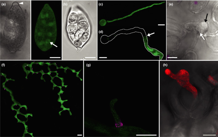
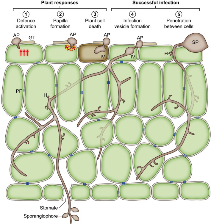
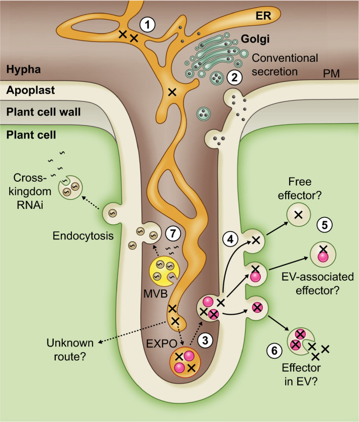
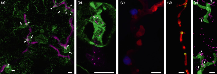
An understanding of the cell biology underlying the burgeoning molecular genetic and genomic knowledge of oomycete pathogenicity is essential to gain the full context of how these pathogens cause disease on plants. An intense research focus on secreted Phytophthora effector proteins, especially those containing a conserved N-terminal RXLR motif, has meant that most cell biological studies into Phytophthora diseases have focussed on the effectors and their host target proteins. While these effector studies have provided novel insights into effector secretion and host defence mechanisms, there remain many unanswered questions about fundamental processes involved in spore biology, host penetration and haustorium formation and function.

Keywords: Phytophthora; RXLR; effector; haustoria; plant defence; plant-pathogen interactions.


Figures

Fig. 1
Fig. 1
Confocal images of Phytophthora infestans growth stages. (a) A single optical section of the papillate sporangium of a GFP‐expressing P. infestans transformant is shown in false transmission and fluorescence modes. The papilla is indicated by the arrowhead. The nuclei are visible in the latter as circles of GFP fluorescence (green); one is arrowed. (b) A sporangium that has undergone zooporogenesis shown in false transmission. (c) A projection image of a germinated cyst of a GFP‐expressing transformant. (d) A germinated cyst in which the germ tube cytoplasm has concentrated towards the growing tip, presumably as a result of the formation of a plug at the point indicated with an arrow. The outline of the whole germinated cyst is traced in white. (e) A germ tube that has penetrated a cell adjacent to a stomate to form an infection vesicle (black arrow). The swollen end of the germ tube is indicated with the white arrow. Chloroplast autofluorescence is overlaid onto the false transmission image in magenta. (f) A projection image of infectious hyphae of a transformant expressing GFP in the cytoplasm and mRFP fused to the effector Avr3a (magenta), which is mainly secreted at haustoria. The image shows the stepwise pattern of growth that occurs between the upper leaf epidermis and the palisade mesophyll. (g) A magnified, deconvoluted image of a haustorium expressing Pi04314‐mRFP that has the most intense red fluorescence around the base, indicating potentially the highest level of secretion in this zone. (h) A projection image of a sporangiophore of a transformant expressing tdTomato fluorescent protein (red) emerging from an open stomate which is shown in the false transmission overlay. Bars, 10 µm.
Fig. 2
Fig. 2
Phytophthora infection processes; schematic drawing of a section of a leaf invaded by Phytophthora. Germination of a sporangium (SP) or cyst on the leaf surface produces a germ tube (GT), which may form an appressorium (AP) (1–4) or may penetrate between anticlinal walls (5). Host penetration triggers plant defence responses (1). These may result in the formation of a papilla (2) or the death of the initially infected cell (3) which can prevent further infection. Host penetration may involve the formation of an infection vesicle (IV) (3, 4), an expanded intracellular structure, from which infectious intercellular hyphae extend to ramify through the plant tissue. Intercellular growth patterns suggest that Phytophthora avoids disrupting pit fields (PF); groups of plasmodesmata that connect plant cells. During biotrophic growth haustoria (H) are formed in cells contacted by the hyphae. Sporangiophores commonly emerge through stomata in leaf infections.
Fig. 3
Fig. 3
Confocal image showing the translocation of a nuclear‐targeted RXLR effector. Infection of a transgenic plant expressing a CFP‐histone 2B fusion (cyan) by a Phytophthora infestans transformant secreting RXLR effector Pi22926‐mRFP fusion protein from haustoria. mRFP fluorescence (magenta) is detectable in the nucleus and nucleolus (arrow) of haustoriated cells indicating translocation has occurred. Chloroplast autofluorescence is shown in yellow. Bar, 10 µm.
Fig. 4
Fig. 4
Schematic drawing of a plant cell invaded by a haustorium illustrating routes for pathogenicity factor delivery. Apoplastic proteins (black circles) are secreted via the conventional secretory pathway (1). This involves entry into the endoplasmic reticulum (ER) followed by passage through the Golgi apparatus and sorting into secretory vesicles which fuse to the Phytophthora plasma membrane (PM) (2). The sequences of RXLR effectors (crosses) include signal peptides and thus they also enter the ER. Evidence from two RXLR effectors indicates that they are then secreted by a different, unknown route and are translocated into host cells. One possibility might be that they are sorted into a distinct domain of the ER (3) which may then cleave off from the rest of the ER, perhaps as an exosome positive organelle (EXPO). This is an autophagosome‐like body that is thought to form by the ER extending, curling back on itself, and engulfing cytoplasm (Wang et al., 2010). The EXPO may then fuse with the PM. In what form the RXLRs exist in the apoplast is unknown. They may be free proteins, in protein complexes, or associated with extracellular vesicles (EVs). They may be associated with the exterior, presumably proteinaceous coat, of the EVs or inside them. How the latter might occur is unknown. Once released into the apoplast the RXLR effectors could be taken into host cells by endocytosis (4). If the RXLR effectors are taken up as proteins or associated with the exterior of EVs, how they then exit the endosomes to access the plant cell interior is unknown (5). If they were inside EVs then the EVs could potentially fuse with the endosome membranes and thereby release their contents (6). Cross‐kingdom delivery of RNA molecules has been shown to involve EVs (7) (Buck et al., 2014; Cai et al., 2018). The RNAs could be taken up from the cytoplasm of the pathogen by invagination of the outer membranes of multivesicular bodies (MVBs). The MVBs then fuse with the PM to release RNA‐carrying EVs which can be endocytosed by the host cells. As suggested for RXLR effectors the RNA could be released into the host cell by fusion of the endocytosed EVs with the endosome membrane.
Fig. 5
Fig. 5
Confocal projection images showing the accumulation of cell components around P. infestans haustoria. (a) An image of the infection of a transgenic plant expressing GFP in the endoplasmic reticulum (ER; green) by a P. infestans transformant expressing the tdTomato fluorescent protein (magenta). Haustoria can be seen outlined in GFP‐labelled ER (arrowheads). Several of the haustoria have nuclei adjacent to them (arrows). (b) A higher magnification image of a haustorium from a tagRFP‐Lifeact transformant (in which actin plaques are evident as bright magenta spots) surrounded by GFP‐labelled host ER. (c) The association of nuclei with haustoria is more clearly visible in this image of a transgenic plant expressing a CFP‐histone 2B fusion (blue) infected with the same P. infestans transformant (red). Endosomes, such as those labelled by a YFP‐tagged exocyst subunit, Sec5 (d), and peroxisomes, labelled by mRFP‐SRL (in magenta in (e)) are also observed to cluster around haustoria. Haustoria are indicated by arrowheads in (e). Bars, 10 µm.

References

    1. Ah‐Fong AMV, Shrivastava J, Judelson HS. 2017. Lifestyle, gene gain and loss, and transcriptional remodeling cause divergence in the transcriptomes of Phytophthora infestans and Pythium ultimum during potato tuber colonization. BMC Genomics 18: 764. - PMC - PubMed
    1. Aram K, Rizzo DM. 2018. Distinct trophic specializations affect how Phytophthora ramorum and Clade 6 Phytophthora spp. colonize and persist on Umbellularia californica leaves in streams. Phytopathology 108: 858–869. - PubMed
    1. Armstrong MR, Whisson SC, Pritchard L, Bos JIB, Venter E, Avrova AO, Rehmany AP, Böhme U, Brooks K, Cherevach I et al 2005. An ancestral oomycete locus contains late blight avirulence gene Avr3a, encoding a protein that is recognized in the host cytoplasm. Proceedings of the National Academy of Sciences, USA 102: 7766–7771. - PMC - PubMed
    1. Attard A, Evangelisti E, Kebdani‐Minet N, Panabières F, Deleury E, Maggio C, Ponchet M, Gourges M. 2014. Transcriptome dynamics of Arabidopsis thaliana root penetration by the oomycete pathogen Phytophthora parasitica . BMC Genomics 15: 538. - PMC - PubMed
    1. Avrova AO, Boevink PC, Young V, Grenville‐Briggs LJ, van West P, Birch PRJ, Whisson SC. 2008. A novel Phytophthora infestans haustorium‐specific membrane protein is required for infection of potato. Cellular Microbiology 10: 2271–2284. - PubMed

Publication types

LinkOut - more resources