. 2020 Jun;24(12):6869-6882.
doi: 10.1111/jcmm.15344. Epub 2020 May 12.

Aberrant methylation of WD-repeat protein 41 contributes to tumour progression in triple-negative breast cancer

Affiliations

Aberrant methylation of WD-repeat protein 41 contributes to tumour progression in triple-negative breast cancer

Han Wang et al. J Cell Mol Med. 2020 Jun.

Abstract

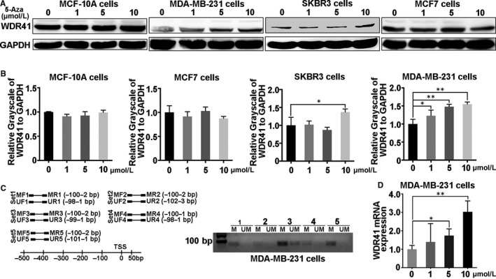
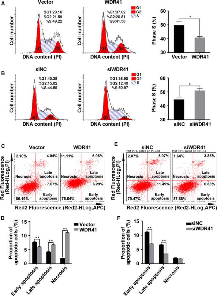
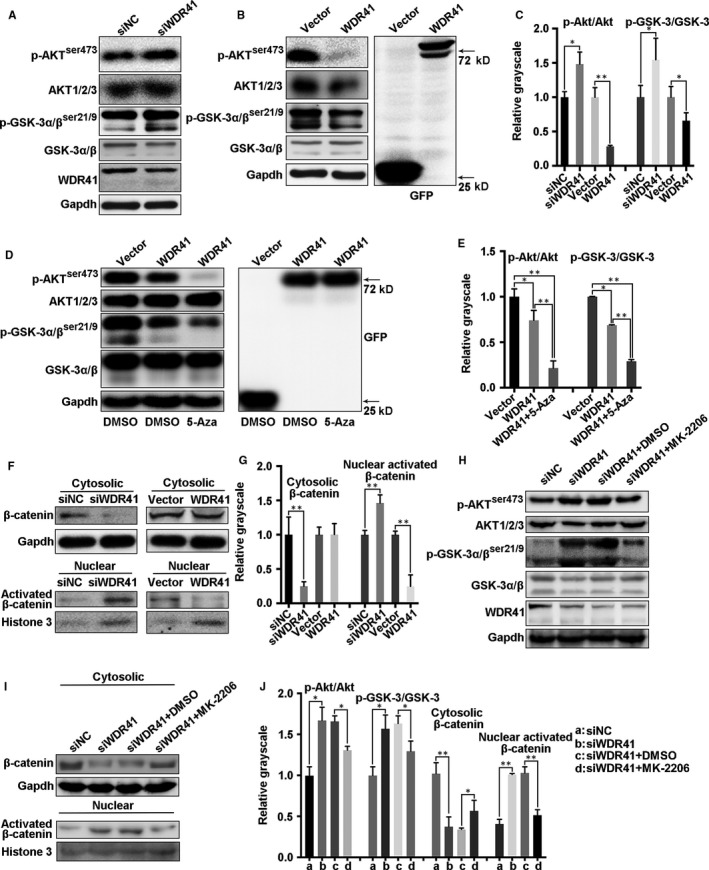
WD-repeat proteins are implicated in a variety of biological functions, most recently in oncogenesis. However, the underlying function of WD-repeat protein 41 (WDR41) in tumorigenesis remains elusive. The present study was aimed to explore the role of WDR41 in breast cancer. Combined with Western blotting and immunohistochemistry, the results showed that WDR41 was expressed at low levels in breast cancer, especially in triple-negative breast cancer (TNBC). Using methylation-specific PCR (MSP), we observed that WDR41 presented hypermethylation in MDA-MB-231 cells. Methylation inhibitor 5-aza-2'-deoxycytidine (5-aza-dC) management increased the expression of WDR41 in MDA-MB-231 cells, but not in MCF-10A (normal mammary epithelial cells) or oestrogen receptor-positive MCF-7 breast cancer cells. WDR41-down-regulation promoted, while WDR41-up-regulation inhibited the tumour characteristics of TNBC cells including cell viability, cell cycle and migration. Further, WDR41-up-regulation dramatically suppressed tumour growth in vivo. Mechanistically, WDR41 protein ablation activated, while WDR41-up-regulation repressed the AKT/GSK-3β pathway and the subsequent nuclear activation of β-catenin in MDA-MB-231 cells, and 5-aza-dC treatment enhanced this effect. After treatment with the AKT inhibitor MK-2206, WDR41-down-regulation-mediated activation of the GSK-3β/β-catenin signalling was robustly abolished. Collectively, methylated WDR41 in MDA-MB-231 cells promotes tumorigenesis through positively regulating the AKT/GSK-3β/β-catenin pathway, thus providing an important foundation for treating TNBC.

Keywords: AKT/GSK-3β/β-catenin pathway; WDR41; methylation; triple-negative breast cancer.


Conflict of interest statement

No competing interests declared.

Figures

FIGURE 1
FIGURE 1
Expression pattern of WDR41 in human breast cancer cells and tissues (A) Expression of WDR41 in breast cancer cell lines was evaluated by Western blotting. MDA‐MB‐231, BT549, MDA‐MB‐468 and MDA‐MB‐453 were the TNBC cell lines; MCF‐10A and MCF‐7 were the normal breast cell line and low invasive capability cells, respectively. The SKBR3 cell line was ER‐ and PR‐negative, but Her2‐positive. B, The mRNA level of WDR41 was determined by qRT‐PCR in normal mammary epithelial cells (MCF‐10A) and breast tumour cells (MCF‐7, MDA‐MB‐231 and SKBR3). C, Immunohistochemical staining of WDR41 in human breast cancer samples and corresponding adjacent tissues. Scale bar: 20 μm. D, Protein expression of WDR41 in human breast cancer tissues and paired para‐carcinoma tissues was measured by Western blotting. E, Relative protein expression of WBP2 in panel D. F, The mRNA level of WDR41 in human breast cancer tissues and its paired para‐carcinoma tissues was measured by qRT‐PCR. G, Immunohistochemical staining with WDR41‐specific antibody in different pathological grades of human breast cancer tissue chips. Scale bar: 50 μm. P, para‐carcinoma, T, tumour. *P < .05, **P < .01. ER, oestrogen receptor; PR, progesterone receptor; qRT‐PCR, quantitative real‐time PCR; TNBC, triple‐negative breast cancer; WDR41, WD‐repeat protein 41
FIGURE 2
FIGURE 2
Methylation level of the WDR41 promoter region in MDA‐MB‐231 cells. A, Protein expression of WDR41 was evaluated in MCF‐10A, MDA‐MB‐231, SKBR3 and MCF‐7 cells after treatment with different doses of 5‐aza‐dC. B, Relative quantification of WDR41 protein expression in MCF‐10A, MDA‐MB‐231, SKBR3 and MCF‐7 cells after stimulation with different doses of 5‐aza‐dC. C, Methylation‐specific PCR was performed to measure the methylation level of WDR41 in MDA‐MB‐231 cells. D, The mRNA level of WDR41 in MDA‐MB‐231 cells after pre‐treatment with increasing doses of 5‐aza‐dC. M, methylation primer, UM, unmethylated primer. *P < .05, **P < .01. WDR41, WD‐repeat protein 41
FIGURE 3
FIGURE 3
Effect of WDR41 on the growth and metastatic ability of MDA‐MB‐231 cells (A and B) Validation of WDR41 or WDR41‐siRNA transfection efficiency in MDA‐MB‐231 cells. Here, 0.5. 1.0 and 2.0 indicate the mass of the plasmid, and S1, S2 and S3 indicate three different WDR‐siRNA oligos. C and D, MTT assay was performed to evaluate cell growth in the four different groups. Scale bar: 200 μm. E and F, Images of wound healing and healing rates in the vector and WDR41 groups after seeding for 24 and 48 h. Scale bar: 200 μm. G and H, Images of wound healing and healing rates in siNC and siWDR41 groups after seeding for 24 and 48 h. I, The Transwell assay results have been presented here for the four different groups. Scale bar: 50 μm. J, The average number of migrated cells in 5 high‐power fields in the vector, WDR41, siNC, and siWDR41 groups. Vector, EGFP group (control); WDR41, EGFP‐WDR41 group; siNC, negative control group; siWDR41, WDR41‐siRNA group. *P < .05, **P < .01. MTT, methyl thiazolyl tetrazolium; WDR41, WD‐repeat protein 41
FIGURE 4
FIGURE 4
The impact of WDR41 on cell cycle and apoptosis of MDA‐MB‐231 cells. Cell cycle progression was analysed in MDA‐MB‐231 cells transfected with vector and WDR41 plasmids (A) or negative control and WDR41‐siRNA (B). C, The effect of WDR41‐up‐regulation on cell apoptosis was analysed by flow cytometry. D, The proportion of apoptotic cells was measured in the vector and WDR41 groups. E, The effect of WDR41‐down‐regulation on cell apoptosis was analysed by flow cytometry. F, The proportion of apoptotic cells was measured in the siNC and siWDR41 groups. Vector, EGFP group (control); WDR41, EGFP‐WDR41 group; siNC, negative control group; siWDR41, WDR41‐siRNA group. *P < .05, **P < .01. WDR41, WD‐repeat protein 41
FIGURE 5
FIGURE 5
Effect of WDR41 on the tumorigenic ability of MDA‐MB‐231 cells (A) Images of a xenograft tumour model of vector and WDR41 groups. n = 5 per group. B, Images of the size of tumours in the vector and WDR41 groups. C, Tumour growth curves of the vector and WDR41 groups. D, Tumour weights of the two groups. E, Protein levels of WDR41, caspase 3 and cleaved caspase 3 in the mice tumours. Tubulin served as the internal control. F, Relative quantification of the cleaved caspase 3 in panel E. Vector, EGFP group (control); WDR41, EGFP‐WDR41 group. Scale bar: 1 cm. *P < .05, **P < .01. WDR41, WD‐repeat protein 41
FIGURE 6
FIGURE 6
Regulatory mechanism of WDR41‐mediated tumour inhibition. A, The activation of AKT and GSK‐3α/β was detected by Western blotting after knockdown of WDR41 in MDA‐MB‐231 cells. B, The activation of AKT and GSK‐3α/β was detected by Western blotting after up‐regulation of WDR41 in MDA‐MB‐231 cells. C, Relative quantification of phosphorylated AKT and GSK‐3α/β in the siNC, siWDR41, vector and WDR41 groups. Protein levels of phosphorylated AKT and GSK‐3α/β (D) and their relative quantification (E) in DMSO‐treated vector and WDR41 groups and 5‐aza‐dC‐treated WDR41 group. F, The distribution of β‐catenin in the cytoplasm and nucleus of MDA‐MB‐231 cells was measured by extracting the nuclear and cytoplasmic protein fractions, followed by Western blotting. G, Relative quantification of cytoplasmic β‐catenin and activated nuclear β‐catenin. H, After transfection with siWDR41, the cells were exposed to DMSO or MK‐2206. The activation of AKT and GSK‐3α/β was detected by Western blotting in MDA‐MB‐231 cells. I, After treatment with siWDR41 and MK‐2206, the nuclear and cytoplasmic protein fractions were extracted and the distribution of β‐catenin was measured in MDA‐MB‐231 cells by Western blotting. J, Relative quantification of the phosphorylated AKT and GSK‐3α/β in panel H and nuclear and cytoplasmic β‐catenin in panel I. *P < .05, **P < .01. DMSO, dimethyl sulfoxide; WDR41, WD‐repeat protein 41
FIGURE 7
FIGURE 7
The proposed model of WDR41‐mediated cellular processes in TNBC cells. In the tumour microenvironment, high methylation across the WDR41 promoter region mediates the down‐regulation of WDR41, resulting in an excessive activation of the AKT/GSK‐3β signalling pathway and subsequent activation of the nuclear β‐catenin, leading to promotion of tumour progression in TNBC. TNBC, triple‐negative breast cancer; WDR41, WD‐repeat protein 41

References

    1. Crozier JA, Cornell LF, Rawal B, Perez EA. Breast cancer brain metastases: molecular subtype, treatment and survival. Breast Dis. 2016;36:133‐141. - PubMed
    1. Nagini S. Breast cancer: current molecular therapeutic targets and new players. Anti‐Cancer Agent Med. 2017;17:152‐163. - PubMed
    1. Fan L, Strasser‐Weippl K, Li JJ, et al. Breast cancer in China. Lancet Oncol. 2014;15:E279‐E289. - PubMed
    1. Akdi A, Gimenez EM, Garcia‐Quispes W, et al. WDR3 gene haplotype is associated with thyroid cancer risk in a Spanish population. Thyroid. 2010;20:803‐809. - PubMed
    1. Sato N, Koinuma J, Fujita M, et al. Activation of WD repeat and high‐mobility group box DNA binding protein 1 in pulmonary and esophageal carcinogenesis. Clin Cancer Res. 2010;16:226‐239. - PubMed

Publication types

MeSH terms