Review
. 2020 Jun 23;50(4):664-678.
doi: 10.3906/sag-2004-331.

Chest CT features of the novel coronavirus disease (COVID-19)

Affiliations
Review

Chest CT features of the novel coronavirus disease (COVID-19)

Furkan Ufuk et al. Turk J Med Sci. .

Abstract

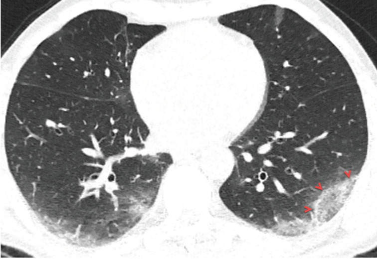
A new type of coronavirus (2019-nCoV) is rapidly spreading worldwide and causes pneumonia, respiratory distress, thromboembolic events, and death. Chest computed tomography (CT) plays an essential role in the diagnosis of viral pneumonia, monitoring disease progression, determination of disease severity, and evaluating therapeutic efficacy. Chest CT can show important clues of 2019-nCoV disease (also known as COVID-19) in patients with an appropriate clinic. Prompt diagnosis of COVID-19 is essential to prevent disease transmission and provides close clinical observation of patients with clinically severe disease. Therefore, radiologists and clinicians should be familiar with the CT imaging findings of COVID-19 pneumonia. Herein, we aimed to review the imaging findings of COVID-19 pneumonia and examine the critical points to be considered for imaging in cases with COVID-19 suspicion.

Keywords: COVID-19; chest computed tomography; diagnosis; pneumonia; radiation.


Conflict of interest statement

Authors declare that there is no conflict of interest.

Figures

Figure 1
Figure 1
a) A 48-year-old man with COVID-19 presented with cough, fever, myalgia, and malaise for 3 days. Axial chest CT image shows multifocal, peripheral-peribronchovascular ground-glass opacities (red frames). b) The magnified image of the right lower lobe shows ground-glass opacity superimposed with interlobular septal thickening and prominent intralobular lines (red arrowheads), which indicates a crazy-paving pattern.
Figure 2
Figure 2
A 66-year-old symptomatic man with COVID-19 pneumonia. Axial CT image at the carina level shows peripheral ill-defined consolidation areas in the both middle-lower lung zones with subpleural involvement (red arrowheads). Note the “air bronchogram sign” within the consolidation areas.
Figure 3
Figure 3
A33-year-old woman with COVID-19 presented with cough, fever, and pleuritic chest pain for 2 days. Axial chest CT image shows a focal consolidation with air bronchogram sign in the right lower lobe (a, red arrows). The sagittal reconstructed CT image of the right lower lobe shows air bronchogram sign inside the consolidation (b, red arrow).
Figure 4
Figure 4
A 46-year-old woman with COVID-19. Axial chest CT image shows peripheral and peribronchovascular distributed focal ground-glass opacities in both lungs, and focal vascular enlargement is seen inside the ground-glass opacity in the right lower lobe (a, red frame). The magnified image of the red frame shows vascular enlargement inside the ground-glass opacity (b, arrows).
Figure 5
Figure 5
a) A 66-year-old symptomatic man with COVID-19. Axial CT image shows subpleural fibrous stripes with ground-glass opacities in both lower lobes (red frames). b) A 53-year-old man with COVID-19. Axial CT image shows subpleural curvilinear lines, interlobular septal thickening, and fibrous stripes with ground-glass opacities in both lower lobes (red frames).
Figure 6
Figure 6
A 40-year-old female patient was presented with complaints of fever that had been ongoing for 5 days, and newly emerged severe dyspnoea. The patient’s D-dimer level was found to be very high and contrast-enhanced pulmonary CT angiography was obtained. a) Axial CT image at the level of the origin of the middle lobe bronchus with mediastinum window settings shows bilateral diffuse pulmonary embolism (red arrows), bilateral mild pleural effusion, and subpleural triangular-shaped opacity with a reverse halo sign in the left lung compatible with pulmonary infarction (red frame). b, c) Axial CT images of the same patient with lung window settings show multiple subpleural consolidation and ground-glass opacity areas (red frames). The nasopharyngeal swab test of the patient was positive for 2019-nCoV.
Figure 7
Figure 7
A 56-year-old man with COVID-19. Axial chest CT image with lung window settings shows bilateral focal ground-glass opacities in both lungs (a, red frame). Chest CT image with mediastinum window settings shows focal pleural thickening in the left lower lobe, near the focal ground-glass opacity (b, arrowheads).
Figure 8
Figure 8
A 75-year-old symptomatic man with COVID-19. Axial chest CT image shows mixed opacity (ground-glass opacity with consolidation) area in the right middle lobe (red frame), and that includes cystic air space (air bubble, red arrow).
Figure 9
Figure 9
A 36-year-old woman presented with complaints of cough and fever for 2 days. Axial chest CT image shows a nodule with halo sign in the right upper lobe (red arrow). The nasopharyngeal swab test of the patient was positive for 2019-nCoV, and the nodule disappeared at follow-up.
Figure 10
Figure 10
A 58-year-old man with COVID-19. Axial chest CT image with lung window settings shows bilateral multifocal ground-glass opacities in both lungs. The ground-glass opacity in the left lower lobe surrounded by denser ring-like (crescentic shape) consolidation, which is compatible with a reversed halo sign (arrowheads).

References

    1. Zhu N Zhang D Wang W Li X Yang B A novel coronavirus from patients with pneumonia in China, 2019. New England Journal of Medicine. 2020;382:727. - PMC - PubMed
    1. Coronavirus Disease 2019 (COVID-19) Situation Report–96. 2020.
    1. Xu Z Shi L Wang Y Zhang J Huang L Pathological findings of COVID-19 associated with acute respiratory distress syndrome. Lancet Respiratory Medicine. 2020;8:420. - PMC - PubMed
    1. Poyiadji N Shahin G Noujaim D Stone M Patel S -associated acute hemorrhagic necrotizing encephalopathy: CT and MRI features. Radiology. 2020;201187 - PMC - PubMed
    1. Zhou F Yu T Du R Fan G Liu Y Clinical course and risk factors for mortality of adult inpatients with COVID-19 in Wuhan, China: a retrospective cohort study. Lancet. 2020;395:1054. - PMC - PubMed