Comparative Study
. 2020 May 18;9(10):e014257.
doi: 10.1161/JAHA.119.014257. Epub 2020 May 12.

Natriuretic Peptide Receptor 2 Locus Contributes to Carotid Remodeling

Affiliations
Comparative Study

Natriuretic Peptide Receptor 2 Locus Contributes to Carotid Remodeling

Vyacheslav A Korshunov et al. J Am Heart Assoc. .

Abstract

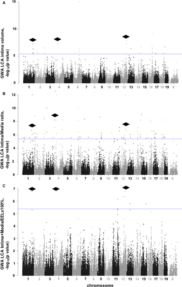
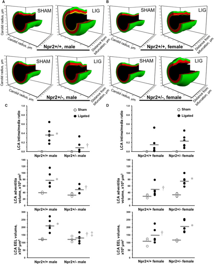
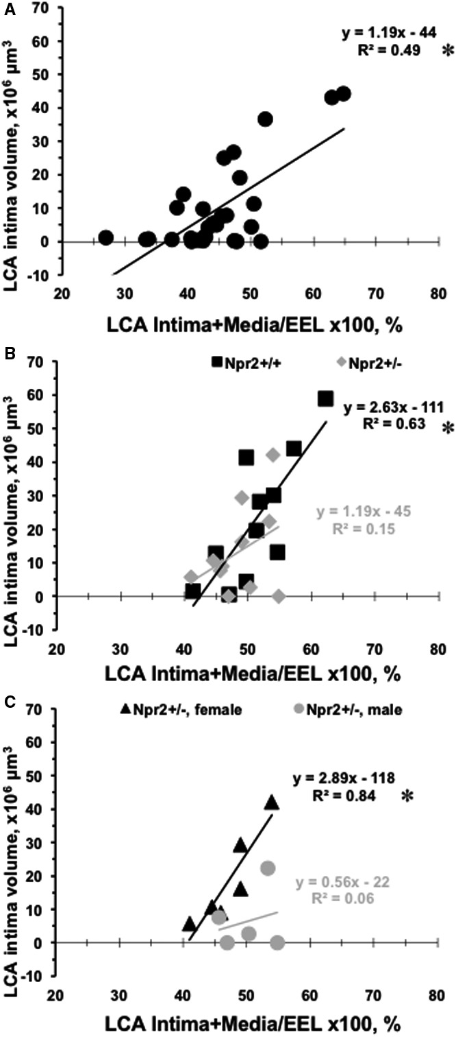
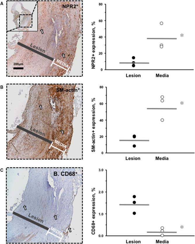
Background Carotid artery intima/media thickness (IMT) is a hallmark trait associated with future cardiovascular events. The goal of this study was to map new genes that regulate carotid IMT by genome-wide association. Methods and Results We induced IMT by ligation procedure of the left carotid artery in 30 inbred mouse strains. Histologic reconstruction revealed significant variation in left carotid artery intima, media, adventitia, external elastic lamina volumes, intima-to-media ratio, and (intima+media)/external elastic lamina percent ratio in inbred mice. The carotid remodeling trait was regulated by distinct genomic signatures with a dozen common single-nucleotide polymorphisms associated with left carotid artery intima volume, intima-to-media ratio, and (intima+media)/external elastic lamina percent ratio. Among genetic loci on mouse chromosomes 1, 4, and 12, there was natriuretic peptide receptor 2 (Npr2), a strong candidate gene. We observed that only male, not female, mice heterozygous for a targeted Npr2 deletion (Npr2+/-) exhibited defective carotid artery remodeling compared with Npr2 wild-type (Npr2+/+) littermates. Fibrosis in carotid IMT was significantly increased in Npr2+/- males compared with Npr2+/- females or Npr2+/+ mice. We also detected decreased Npr2 expression in human atherosclerotic plaques, similar to that seen in studies in Npr2+/- mice. Conclusions We found that components of carotid IMT were regulated by distinct genetic factors. We also showed a critical role for Npr2 in genetic regulation of vascular fibrosis associated with defective carotid remodeling.

Keywords: Npr2; carotid artery; genome‐wide association; inbred mice; vascular remodeling.


Figures

Figure 1
Figure 1. Variation in carotid remodeling in 30 mouse strains.
A, Left carotid artery (LCA) intima volume, ×10−6 μm3. B, LCA intima/media ratio. C, LCA (intima+media)/EEL×100%. Open circles indicate controls. Black circles indicate ligated mice. Values are mean±SEM; *P<0.05 vs control or other mouse strains. n=4 to 6 per group. D, Representative 3‐dimensional (3D) reconstructions of the 2 mm‐length from the bifurcation of the ligated LCA in mice with low values of LCA (intima+media)/EEL×100%. E, Representative 3D reconstructions of mouse strains with a greater percentage of LCA (intima+media)/EEL×100%. Black color shows lumen, yellow indicates intima, red indicates media, and green indicates adventitia volume. EEL indicates external elastic lamina.
Figure 2
Figure 2. GWA of carotid remodeling traits in 30 mouse strains.
A, GWA of LCA intima volume, ×10−6  μm3. B, GWA of LCA intima/media ratio. C, GWA of LCA (intima+media)/EEL×100%. Each circle represents an SNP. Mouse chromosomes are presented on the X‐axis. Blue lines show significance threshold. Black rhombi point to common regions of significant SNPs associated with LCA intima, intima/media ratio, and (intima+media)/EEL×100% traits in 30 inbred mouse strains. EEL indicates external elastic lamina; GWA, genome‐wide association; LCA, left carotid artery; and SNP, single‐nucleotide polymorphism.
Figure 3
Figure 3. Flow‐dependent carotid remodeling in Npr2 mice.
A, Representative 3‐dimensional (3D) reconstructions of the 2‐mm length from the bifurcation of the LCA after sham or ligation operation in males of Npr2 wild‐type (Npr2 +/+) and Npr2 heterozygous (Npr2 +/−) mice. B, Representative 3D reconstructions of LCA after sham or ligation operation in females of Npr2 +/+ and Npr2 +/− mice. Black indicates lumen, yellow indicates intima, red indicates media, and green indicates adventitia volume. C, Quantifications of LCA intima/media ratio, adventitia, and EEL volume in males of Npr2 +/+ and Npr2 +/− mice. D, Quantifications of LCA intima/media ratio, adventitia, and EEL volume in females of Npr2 +/+ and Npr2 +/− mice. Open circles indicate individual sham LCAs. Black circles indicate ligated LCAs. Gray lines indicate mean values. EEL indicates external elastic lamina; and LCA, left carotid artery. *P<0.05 vs sham; P<0.05 vs Npr2 +/+ males; P<0.05 vs Npr2 +/− females. n=3 to 6 per group.
Figure 4
Figure 4. Relationships between carotid remodeling traits in mice.
A, Ligated LCA (intima+media)/EEL×100% on X‐axis are plotted vs LCA intima volumes on Y‐axis for each of 30 inbred mouse strains (black circles). B, Ligated LCA (intima+media)/EEL×100% on the X‐axis are plotted vs LCA intima volumes on Y‐axis for Npr2 wild‐type (black squares: Npr2 +/+) and Npr2 heterozygous (gray diamonds: Npr2 +/−) mice. C, Ligated LCA (intima+media)/EEL×100% on X‐axis are plotted vs LCA intima volumes on the Y‐axis for Npr2 +/− females (black triangles) and Npr2 +/− males (gray circles). *P<0.001, P<0.01, and P<0.05 indicate level of significance of correlation between LCA (intima+media)/EEL×100% and LCA intima volume. EEL indicates external elastic lamina; and LCA, left carotid artery.
Figure 5
Figure 5. Differences in carotid fibrosis in Npr2 mice.
A, Representative image of PicroSirius Red–stained ligated LCA in male Npr2 wild‐type (Npr2 +/+) mouse. B, Representative image of Alcian Blue–stained ligated LCA in male Npr2 heterozygous (Npr2 +/−) mouse. Insets show corresponding females. Scale bar=100 μm. Black brackets indicate intima/media area. C, Quantification of fibrosis (blue color) in intima/media area of the LCA (%). Black circles indicate individual Npr2 +/+ mice. Open circles indicate Npr2 +/− mice. Gray lines indicate mean values. *P<0.001 vs Npr2 +/+ males; P<0.001 vs Npr2 +/− females. n=3 animals per group. LCA indicates left carotid artery.
Figure 6
Figure 6. Decreased NPR2 expression in smooth muscle cells in human carotid lesions.
A, NPR2 immunostaining. Inset shows lower power of the carotid endarterectomy sample. B, α1‐SM‐actin immunostaining. C, CD68 immunostaining. Positive cells are stained brown (open arrows). Black bars indicate lesion area. White brackets indicate media area. Magnification bar=200 μm. For quantification, black circles show relative expression in the lesion of the carotid endarterectomy samples and open circles indicate media of the carotid endarterectomy samples. Gray lines indicate mean values. *P<0.05 vs media; P<0.01 vs media. n=3 animals per group. SM indicates smooth muscle.

References

    1. Lusis AJ. Atherosclerosis. Nature. 2000;407:233–241. - PMC - PubMed
    1. O'Donnell CJ, Cupples LA, D'Agostino RB, Fox CS, Hoffmann U, Hwang SJ, Ingellson E, Liu C, Murabito JM, Polak JF, et al. Genome‐wide association study for subclinical atherosclerosis in major arterial territories in the NHLBI's Framingham Heart Study. BMC Med Genet. 2007;8(suppl 1):S4. - PMC - PubMed
    1. Xie G, Myint PK, Voora D, Laskowitz DT, Shi P, Ren F, Wang H, Yang Y, Huo Y, Gao W, et al. Genome‐wide association study on progression of carotid artery intima media thickness over 10 years in a Chinese cohort. Atherosclerosis. 2015;243:30–37. - PubMed
    1. O'Leary DH, Polak JF, Kronmal RA, Manolio TA, Burke GL, Wolfson SK Jr. Carotid‐artery intima and media thickness as a risk factor for myocardial infarction and stroke in older adults. Cardiovascular Health Study Collaborative Research Group. N Engl J Med. 1999;340:14–22. - PubMed
    1. Fox CS, Cupples LA, Chazaro I, Polak JF, Wolf PA, D'Agostino RB, Ordovas JM, O'Donnell CJ. Genomewide linkage analysis for internal carotid artery intimal medial thickness: evidence for linkage to chromosome 12. Am J Hum Genet. 2004;74:253–261. - PMC - PubMed

Publication types

MeSH terms

Substances