. 2020 Nov;15(11):2162-2168.
doi: 10.4103/1673-5374.282260.

Muscovite nanoparticles mitigate neuropathic pain by modulating the inflammatory response and neuroglial activation in the spinal cord

Affiliations

Muscovite nanoparticles mitigate neuropathic pain by modulating the inflammatory response and neuroglial activation in the spinal cord

Ju-Young Oh et al. Neural Regen Res. 2020 Nov.

Abstract

Despite numerous efforts to overcome neuropathic pain, various pharmacological drugs often fail to meet the needs and have many side effects. Muscovite is an aluminosilicate mineral that has been reported to have an anti-inflammatory effect, but the efficacy of muscovite for neuropathic pain has not been investigated. Here, we assessed whether muscovite nanoparticles can reduce the symptoms of pain by controlling the inflammatory process observed in neuropathic pain. The analgesic effects of muscovite nanoparticles were explored using partial sciatic nerve ligation model of neuropathic pain, in which one-third to one-half of the nerve trifurcation of the sciatic nerve was tightly tied to the dorsal side. Muscovite nanoparticles (4 mg/100 μL) was given intramuscularly to evaluate its effects on neuropathic pain (3 days per week for 4 weeks). The results showed that the muscovite nanoparticle injections significantly alleviated partial sciatic nerve ligation-induced mechanical and cold allodynia. In the spinal cord, the muscovite nanoparticle injections exhibited inhibitory effects on astrocyte and microglia activation and reduced the expression of pro-inflammatory cytokines, such as interleukin-1β, tumor necrosis factor-α, interleiukin-6 and monocyte chemoattractant protein-1, which were upregulated in the partial sciatic nerve ligation model. Moreover, the muscovite nanoparticle injections resulted in a decrease in activating transcription factor 3, a neuronal injury marker, in the sciatic nerve. These results suggest that the analgesic effects of muscovite nanoparticle on partial sciatic nerve ligation-induced neuropathic pain may result from inhibiting activation of astrocytes and microglia as well as pro-inflammatory cytokines. We propose that muscovite nanoparticle is a potential anti-nociceptive candidate for neuropathic pain. All experimental protocols in this study were approved by the Institutional Animal Ethics Committee (IACUC) at Dongguk University, South Korea (approval No. 2017-022-1) on September 28, 2017.

Keywords: astrocyte; microglia; muscovite; nanoparticle; neuropathic pain; partial sciatic nerve ligation; pharmacopuncture; pro-inflammatory cytokine; spinal cord.


Conflict of interest statement

None

Figures
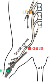
Figure 1
Figure 1
Schematic diagram of surgical ligation and muscovite injection points. Ligature was tied around the sciatic nerve in the PSNL mice. Muscovite was injected at GB30, HL, and LB. L4: Lumbar 4; L5: lumbar 5; L6: lumbar 6; LB: lumbar (GB34); HL: hindlimb (BL25); PSNL: partial sciatic nerve ligation.
Figure 2
Figure 2
Mechanical allodynia changes for 4 weeks after muscovite injections following PSNL. (A) Experimental schedule of the muscovite injections and von Frey test. (B–E) Anti-allodynic effects of muscovite was measured on the ipsilateral (B, D) and contralateral (C, E) plantar surfaces 2 hours after the muscovite injection for 0, 1, 3, 7, 14, 21, and 28 days. Data are expressed as the mean ± SEM (n = 6/group). ***P < 0.001 vs. sham group; #P < 0.05, ###P < 0.001, vs. PSNL group (two-way repeated measures analysis of variance followed by Bonferroni post hoc tests). HL: Hindlimb (BL25); LB: lumbar (GB34); PSNL: partial sciatic nerve ligation.
Figure 3
Figure 3
Cold allodynia changes for 4 weeks after muscovite injections following PSNL. (A) Experimental schedule of the muscovite injections and acetone test. (B–E) Cold allodynia in the acetone test in the ipsilateral (B, D) and contralateral paw (C, E) surfaces 2 hours after the muscovite injection for 0, 1, 2, 3, and 4 weeks. Data are expressed as the mean ± SEM (n = 6/group). ***P < 0.001 vs. sham group; #P < 0.05, ##P < 0.01, ###P < 0.001, vs. PSNL group (two-way repeated measures analysis of variance (ANOVA) followed by Bonferroni post-hoc tests). HL: Hindlimb (BL25); LB: lumbar (GB34); PSNL: partial sciatic nerve ligation.
Figure 4
Figure 4
Expression levels of pro-inflammatory cytokines after muscovite injection. (A) IL-6, (B) IL-1β, and (C) TNF-α were detected by western blot assay in the spinal cord tissue. (D) ATF-3 was detected in the sciatic nerve. Data are expressed as the mean ± SEM (n = 3/group). **P < 0.01, ***P < 0.001, vs. sham group; #P < 0.05, ##P < 0.01, ###P < 0.001, vs. PSNL group. n = 3/group (one-way analysis of variance followed by the Student-Newman-Keuls test). ATF-3: Activating transcription factor 3; HL: hindlimb (BL25); IL-6 interleukin-6; IL-1β: interleukin-1beta; LB: lumbar (GB34); PSNL: partial sciatic nerve ligation; TNF-α: tumor necrosis factor-alpha.
Figure 5
Figure 5
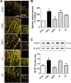
Regulation of muscovite in activated astrocytes. (A, B) Immunofluorescence staining demonstrates that GFAP is regulated for muscovite injection. (C) Western blot assay confirming GFAP expression after muscovite injection. β-Actin was used as the loading control. Scale bar: 100 µm. Data are expressed as the mean ± SEM (n = 3/group). *P < 0.05, **P < 0.01, vs. sham group; #P < 0.05, ##P < 0.01, vs. PSNL group (one-way analysis of variance followed by the Student-Newman-Keuls test). DAPI: 4′,6-Diamidino-2-phenylindole; GFAP: glial fibrillary acidic protein; HL: hindlimb (BL25); LB: lumbar (GB34); PSNL: partial sciatic nerve ligation.
Figure 6
Figure 6
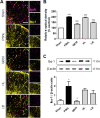
Regulation of muscovite in activated microglia. (A, B) Immunofluorescence staining demonstrates that Iba-1 is regulated for muscovite injection. (C) Western blot assay confirming Iba-1 expression after muscovite injection. β-Actin was used as the loading control. Scale bar: 100 µm. Data are expressed as the mean ± SEM (n = 3/group). *P < 0.05, ***P < 0.001, vs. sham group; #P < 0.05, ###P < 0.001, vs. PSNL group (one-way analysis of variance followed by the Student-Newman-Keuls test). DAPI: 4′,6-Diamidino-2-phenylindole; HL: hindlimb (BL25); LB: lumbar (GB34); PSNL: partial sciatic nerve ligation.

Similar articles

Cited by

References

    1. Andreev N, Urban L, Dray A. Opioids suppress spontaneous activity of polymodal nociceptors in rat paw skin induced by ultraviolet irradiation. Neuroscience. 1994;58:793–798. - PubMed
    1. Averill S, Michael GJ, Shortland PJ, Leavesley RC, King VR, Bradbury EJ, McMahon SB, Priestley JV. NGF and GDNF ameliorate the increase in ATF3 expression which occurs in dorsal root ganglion cells in response to peripheral nerve injury. Eur J Neurosci. 2004;19:1437–1445. - PubMed
    1. Benyamin R, Trescot AM, Datta S, Buenaventura R, Adlaka R, Sehgal N, Glaser SE, Vallejo R. Opioid complications and side effects. Pain Physician. 2008;11(2 Suppl):S105–120. - PubMed
    1. Breivik H, Eisenberg E, O’Brien T OPENMinds. The individual and societal burden of chronic pain in Europe: the case for strategic prioritisation and action to improve knowledge and availability of appropriate care. BMC Public Health. 2013;13:1229. - PMC - PubMed
    1. Calvo M, Dawes JM, Bennett DL. The role of the immune system in the generation of neuropathic pain. Lancet Neurol. 2012;11:629–642. - PubMed