Review
. 2020 Apr;12(4):1728-1739.
doi: 10.21037/jtd.2019.12.137.

New developments in transcatheter therapy of mitral valve disease

Affiliations
Review

New developments in transcatheter therapy of mitral valve disease

Claudia Walther et al. J Thorac Dis. 2020 Apr.

Abstract

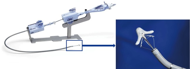
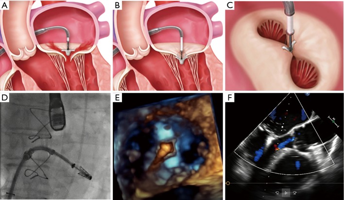
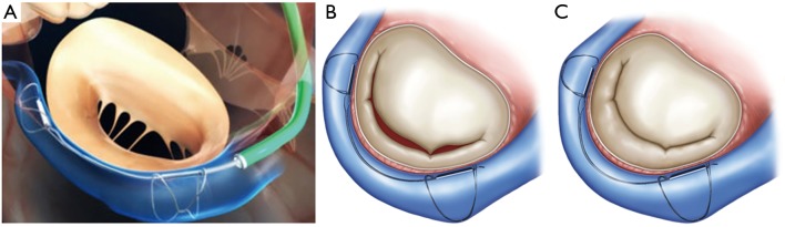
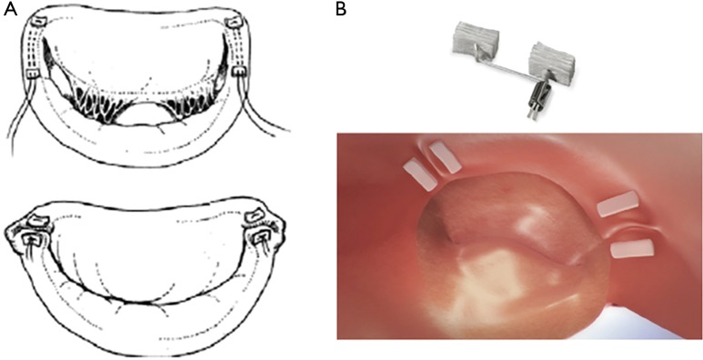
Mitral valve regurgitation (MR) belongs to one of the most common acquired valve diseases in western countries with increasing prevalence in older age. For patients with high perioperative risk and older age prohibitive for valve surgery, the development of transcatheter mitral valve therapies offers new options. Assessment of the severity and etiology of MR and thorough imaging of the mitral valve anatomy and pathology are necessary prerequisites for appropriate decision making in the field of transcatheter mitral valve therapies. Different transcatheter repair and replacement techniques are on the market, most of them mimicking surgical techniques. With some techniques (e.g., the MitraClip device), there is good clinical experience (>80,000 devices implanted worldwide), and evidence (three randomized studies), whereas for newer procedures, safety and efficacy data are still very limited. Transcatheter mitral repair and replacement techniques have to be considered as complementary treatment options for high-risk patients indicated by the Heart Teams. The different techniques and devices will be introduced and discussed in the following paper.

Keywords: MitraClip device; Mitral valve regurgitation (MR); degenerative mitral regurgitation; functional mitral regurgitation; heart failure; percutaneous mitral repair; transcatheter mitral valve repair.


Conflict of interest statement

Conflicts of Interest: All authors have completed the ICMJE uniform disclosure form (available at http://dx.doi.org/10.21037/jtd.2019.12.137). The series “Interventional Cardiology” was commissioned by the editorial office without any funding or sponsorship. CW received honoraria from Abbot Vascular. TW received honoraria from Abbot Vascular and Edwards Lifesciences. SF received honoraria from Abbot Vascular and Edwards Lifesciences. Mariuca Vasa-Nicotera received honoraria from Abbot Vascular and Edwards Lifesciences. TH and MA have no other conflicts of interest.

Figures

Figure 1
Figure 1
The MitraClip system with steerable console outside the body and the amplified MitraClip (with copyright permission from Abbot).
Figure 2
Figure 2
Standard steps of the MitraClip procedure. (A) Perpendicular positioning of the MitraClip above the mitral valve. (B) Grasping of the leaflets and closing of the MitraClip. (C) The typical “double orifice” after MitraClip implantation. (D) Fluoroscopic control of the correct position of the MitraClip. (E) 3D echocardiographic control of stable perpendicular position after advancing the MitraClip in the left ventricle. (F) The echocardiographic result after successful MitraClip implantation [figure with copyright permission from (16)].
Figure 3
Figure 3
Scetch of the Carillon device procedure. (A) Positioning of the Carillon device with the two anchors in the great cardiac vein and the ostium of the coronary sinus. Deployed Carillon device before (B) and after “cinching” (C) with the restoration of leaflet coaptation [figure with copyright permission from (16)].
Figure 4
Figure 4
Procedural steps of the Cardioband system. (A) The Cardioband is anchored to the hinge of the posterior mitral annulus starting on the anterolateral commissure and reaching the posteromedial commissure. After deployment of the Cardioband (B) cinching of the Cardioband is performed to reduce annular diameters and enhance leaflet coaptation. (C) Fluoroscopic and angiographic control is performed to confirm the correct position of the device and to exclude any damage of the left circumflex artery. (D) 3D echocardiographic imaging (“en face view”) after deployment of the Cardioband.
Figure 5
Figure 5
The Mitralign device mimicking a modified Kay Wooler annuloplasty. (A) Schematic imaging of the Mitralign device. (B) Plication of the posterior annulus with pledges.

References

    1. Nkomo VT, Gardin JM, Skelton TN, et al. Burden of valvular heart diseases: a population-based study. Lancet 2006;368:1005-11. 10.1016/S0140-6736(06)69208-8 - DOI - PubMed
    1. Grigioni F, Enriquez-Sarano M, Zehr KJ, et al. Ischemic mitral regurgitation: long-term outcome and prognostic implications with quantitative Doppler assessment. Circulation 2001;103:1759-64. 10.1161/01.CIR.103.13.1759 - DOI - PubMed
    1. Koelling TM, Aaronson KD, Cody RJ, et al. Prognostic significance of mitral regurgitation and tricuspid regurgitation in patients with left ventricular systolic dysfunction. Am Heart J 2002;144:524-9. 10.1067/mhj.2002.123575 - DOI - PubMed
    1. Enriquez-Sarano M, Avierinos JF, Messika-Zeitoun D, et al. Quantitative determinants of the outcome of asymptomatic mitral regurgitation. N Engl J Med 2005;352:875-83. 10.1056/NEJMoa041451 - DOI - PubMed
    1. Mirabel M, Iung B, Baron G, et al. What are the characteristics of patients with severe, symptomatic, mitral regurgitation who are denied surgery? Eur Heart J 2007;28:1358-65. 10.1093/eurheartj/ehm001 - DOI - PubMed