. 2019;1(1):fcz024.
doi: 10.1093/braincomms/fcz024. Epub 2019 Oct 14.

Amyloid precursor protein processing in human neurons with an allelic series of the PSEN1 intron 4 deletion mutation and total presenilin-1 knockout

Affiliations

Amyloid precursor protein processing in human neurons with an allelic series of the PSEN1 intron 4 deletion mutation and total presenilin-1 knockout

Charles Arber et al. Brain Commun. 2019.

Abstract

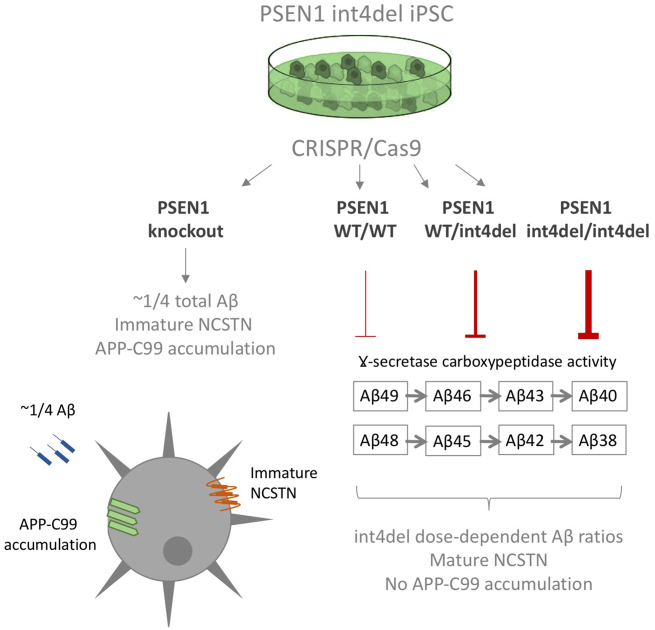
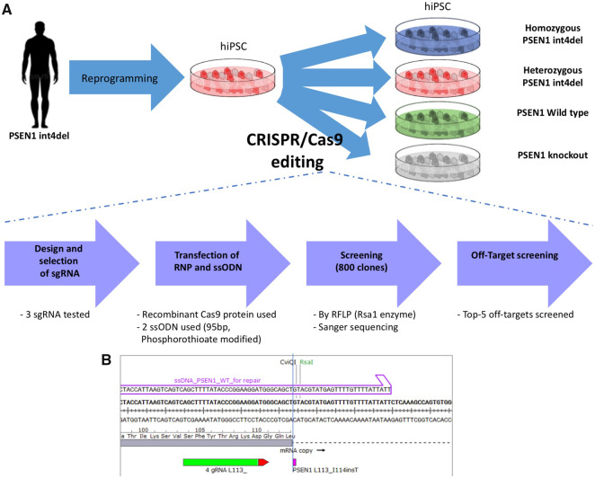
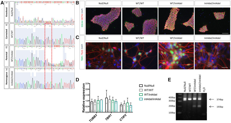
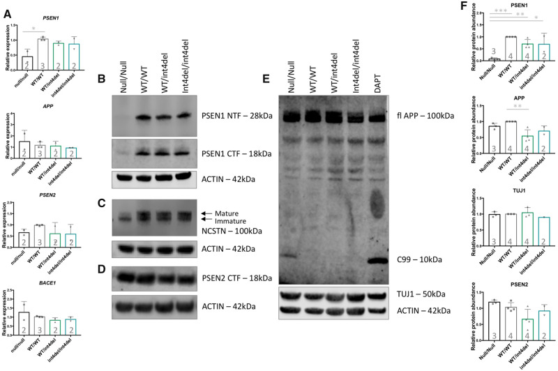
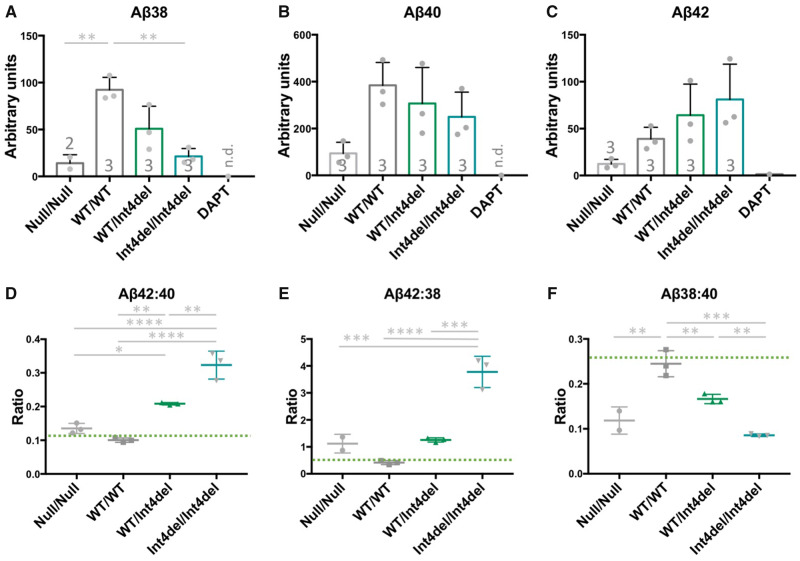
Mutations in presenilin-1 (PSEN1), encoding the catalytic subunit of the amyloid precursor protein-processing enzyme γ-secretase, cause familial Alzheimer's disease. However, the mechanism of disease is yet to be fully understood and it remains contentious whether mutations exert their effects predominantly through gain or loss of function. To address this question, we generated an isogenic allelic series for the PSEN1 mutation intron 4 deletion; represented by control, heterozygous and homozygous mutant induced pluripotent stem cells in addition to a presenilin-1 knockout line. Induced pluripotent stem cell-derived cortical neurons reveal reduced, yet detectable amyloid-beta levels in the presenilin-1 knockout line, and a mutant gene dosage-dependent defect in amyloid precursor protein processing in PSEN1 intron 4 deletion lines, consistent with reduced processivity of γ-secretase. The different effects of presenilin-1 knockout and the PSEN1 intron 4 deletion mutation on amyloid precursor protein-C99 fragment accumulation, nicastrin maturation and amyloid-beta peptide generation support distinct consequences of familial Alzheimer's diseaseassociated mutations and knockout of presenilin-1 on the function of γ-secretase.

Keywords: amyloid beta; Alzheimer’s disease; CRISPR/Cas9; iPSCs.


Conflict of interest statement

Competing interests The authors report no competing interests.

Figures

Graphical Abstract
Graphical Abstract
Figure 1
Figure 1
Scheme for the gene editing of patient-derived PSEN1 int4del iPSCs. (A) Strategy for the generation of isogenic cells using CRISPR/Cas9-editing of an iPSC line from an individual carrying the PSEN1 int4del mutation. (B) Genomic positioning of the editing site, showing ssODN repair arm (purple), mutation site (purple) and sgRNA (green) with PAM site (red). sgRNA (single guide RNA, for CRISPR/Cas9 targeting); RFLP (used for screening); ssODN (single-stranded oligodeoxynucleotide, for homology-directed repair).
Figure 2
Figure 2
Characterization of gene-edited iPSC and neurons. (A) Sanger sequencing was used to confirm the generation of a PSEN1 knockout (i), a corrected wild-type PSEN1 line (ii), an unedited heterozygous mutant line (iii) and a homozygous PSEN1 int4del mutation line (iv). (B) Immunocytochemistry was performed on all iPSC lines following CRISPR/Cas9 editing to confirm pluripotency with the pluripotency markers OCT4 (red) and SSEA4 (green). Scale bar 100 μm. (C) Successful differentiation of iPSC into neurons was characterized 50 days post-induction by immunocytochemistry for the neuronal marker TUJ1 (red) and deep-layer cortical neuronal marker TBR1 (green). Scale bar 25μm. (D) Further characterization of cortical differentiation was performed using qPCR analysis 100 days post-neural induction to assess expression of neuronal marker TUBB3 and cortical layer markers TBR1 and CTIP2. Numbers within histogram represent the number of independent neural inductions. (E) PCR analysis of PSEN1 splicing in cDNA from day 100 neurons using primers recognizing exons 3 and 5 of PSEN1 mRNA. The full-length transcript is depicted at 374 bp and one short transcript caused by aberrant splicing is evident at 193 bp in mutation-bearing neurons (we do not see evidence of a second aberrantly spliced band at 111 bp).
Figure 3
Figure 3
Analysis of γ-secretase and APP processing in iPSC-derived neurons. (A) PSEN1, PSEN2, APP and BACE1 expression in iPSC-derived neurons 100 days post-induction was assessed by qPCR in neurons from PSEN1 knockout, PSEN1 wild type, PSEN1 int4del heterozygous and PSEN1 int4del homozygous lines. PSEN1 expression was significantly reduced in the PSEN1 knockout neurons. No significant differences in PSEN2, APP and BACE1 were observed. (B–E) Western blot on whole cell lysates of day 100 neurons was used to analyse protein levels of PSEN1 N-terminal fragments (28 kDa), PSEN1 C-terminal fragments (18 kDa), NCSTN (100 kDa), PSEN2 C-terminal fragments (18 kDa), APP (100 kDa) and neuronal marker TUJ1 (50 kDa). The DAPT sample represents an unrelated control line treated with the γ-secretase inhibitor DAPT at 10 μM for 48 h. (F) Quantification of western blot band intensities in B–E. PSEN1 protein abundance is significantly reduced in PSEN1 knockout lysates and APP is significantly increased in the corrected wild-type neurons compared with parental PSEN1 int4del. *P > 0.05, **P > 0.01, ***P > 0.001 by one-way ANOVA with Tukey’s post hoc analysis. Numbers within histograms represent the number of independent neural inductions.
Figure 4
Figure 4
Aβ analysis in iPSC-neuronal-conditioned media. (A–C) Quantification of Aβ38, Aβ40 and Aβ42 in 48 h-conditioned media from neurons at day 100, measured by electrochemiluminescence and normalized to total protein content from the cell pellet. The DAPT sample is a representative unrelated control line treated with 10 μM of the γ-secretase inhibitor DAPT for 48 h; n.d., not detected. Numbers within histograms represent the number of independent neural inductions. (D–F) Aβ ratios to depict the disease-associated Aβ42:40 ratio, γ-secretase carboxypeptidase-like activity Aβ42:38 and endopeptidase cleavage position choice Aβ38:40. n=3 for each sample apart from where Aβ38 was below detection limit for one null/null sample. Green bars represent non-Alzheimer’s disease neuronal ratios from Arber et al. (2019). *P > 0.05, **P > 0.01, ***P > 0.001 and ****P > 0.0001 by one-way ANOVA with Tukey’s post hoc analysis.

References

    1. Arber C, Toombs J, Lovejoy C, Ryan NS, Paterson RW, Willumsen N, et al.Familial Alzheimer’s disease patient-derived neurons reveal distinct mutation-specific effects on amyloid beta. Mol Psychiatry 2019; 1–13. doi:10.1038/s41380-019-0410-8. - PMC - PubMed
    1. Chávez-Gutiérrez L, Bammens L, Benilova I, Vandersteen A, Benurwar M, Borgers M, et al.The mechanism of γ-secretase dysfunction in familial Alzheimer disease. EMBO J 2012; 31: 2261–74. - PMC - PubMed
    1. De Jonghe C, Cruts M, Rogaeva EA, Tysoe C, Singleton A, Vanderstichele H, et al.Aberrant splicing in the presenilin-1 intron 4 mutation causes presenile Alzheimer’s disease by increased Abeta42 secretion. Hum Mol Genet 1999; 8: 1529–40. - PubMed
    1. De Strooper B, Saftig P, Craessaerts K, Vanderstichele H, Guhde G, Annaert W, et al.Deficiency of presenilin-1 inhibits the normal cleavage of amyloid precursor protein. Nature 1998; 391: 387. - PubMed
    1. De Strooper B. Aph-1, Pen-2, and nicastrin with presenilin generate an active γ-secretase complex. Neuron 2003; 38: 9.. - PubMed

LinkOut - more resources