. 2020 Jun 17;31(6):1624-1634.
doi: 10.1021/acs.bioconjchem.0c00163. Epub 2020 May 27.

Kinetics and Optimization of the Lysine-Isopeptide Bond Forming Sortase Enzyme from Corynebacterium diphtheriae

Kinetics and Optimization of the Lysine-Isopeptide Bond Forming Sortase Enzyme from Corynebacterium diphtheriae

Christopher K Sue et al. Bioconjug Chem. .

Abstract

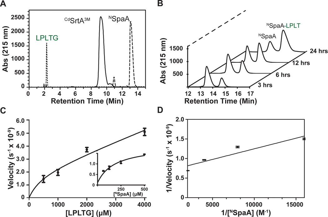
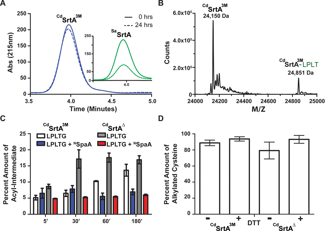
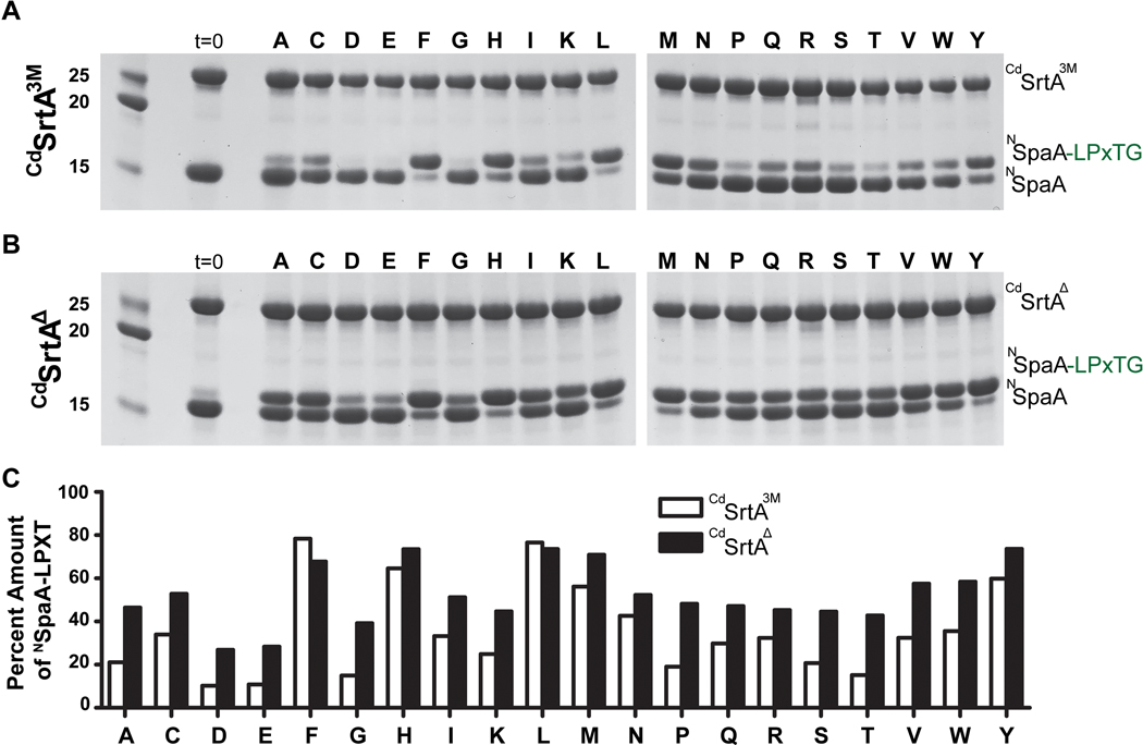
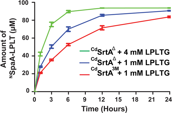
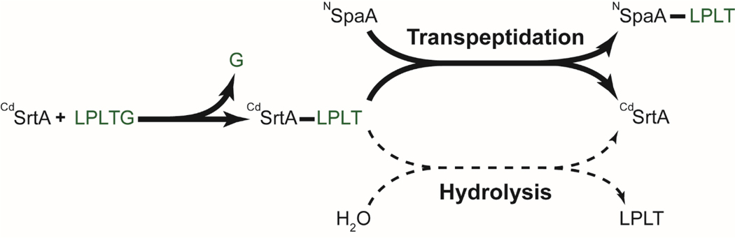
Site-specifically modified protein bioconjugates have important applications in biology, chemistry, and medicine. Functionalizing specific protein side chains with enzymes using mild reaction conditions is of significant interest, but remains challenging. Recently, the lysine-isopeptide bond forming activity of the sortase enzyme that builds surface pili in Corynebacterium diphtheriae (CdSrtA) has been reconstituted in vitro. A mutationally activated form of CdSrtA was shown to be a promising bioconjugating enzyme that can attach Leu-Pro-Leu-Thr-Gly peptide fluorophores to a specific lysine residue within the N-terminal domain of the SpaA protein (NSpaA), enabling the labeling of target proteins that are fused to NSpaA. Here we present a detailed analysis of the CdSrtA catalyzed protein labeling reaction. We show that the first step in catalysis is rate limiting, which is the formation of the CdSrtA-peptide thioacyl intermediate that subsequently reacts with a lysine ε-amine in NSpaA. This intermediate is surprisingly stable, limiting spurious proteolysis of the peptide substrate. We report the discovery of a new enzyme variant (CdSrtAΔ) that has significantly improved transpeptidation activity, because it completely lacks an inhibitory polypeptide appendage ("lid") that normally masks the active site. We show that the presence of the lid primarily impairs formation of the thioacyl intermediate and not the recognition of the NSpaA substrate. Quantitative measurements reveal that CdSrtAΔ generates its cross-linked product with a catalytic turnover number of 1.4 ± 0.004 h-1 and that it has apparent KM values of 0.16 ± 0.04 and 1.6 ± 0.3 mM for its NSpaA and peptide substrates, respectively. CdSrtAΔ is 7-fold more active than previously studied variants, labeling >90% of NSpaA with peptide within 6 h. The results of this study further improve the utility of CdSrtA as a protein labeling tool and provide insight into the enzyme catalyzed reaction that underpins protein labeling and pilus biogenesis.


Figures

Figure 1.
Figure 1.
The C. diphtheriae CdSrtA pilin sortase catalyzes lysine isopeptide bond formation. (A) Schematic showing the pilin polymerization reaction catalyzed by CdSrtA. The enzyme creates the SpaA pilus by polymerizing SpaA pilin proteins. In the reaction it recognizes lysine (K190) side chain nucleophile within the N-terminal domain of SpaA (NSpaA) and joins it the backbone threonine carbonyl carbon atom located in the C-terminal LPLTG sorting signal located within another SpaA protein. This reaction is repeated to construct the SpaA pilus that mediates bacterial adhesion. (B) Schematic of the reaction used to monitor lysine isopeptide bond formation. In this assay the CdSrtA enzyme ligates the isolated NSpaA domain to the peptide containing the LPLTG sorting signal (FELPLTGGSG). (C) The structure of CdSrtA showing H160, C222 and R231 active site residues. The “Lid” is highlighted in red (residues P77 to S89).
Figure 2.
Figure 2.
CdSrtA transpeptidation assay (A) Three separate reversed phase HPLC traces on a Waters C4 column separated the sorting signal peptide (LPLTG), CdSrtA3M, and NSpaA. (B) Several representative HPLC traces showing the decrease in NSpaA and the increase of Peptide-NSpaA. The reaction (100 μM of enzyme, 200 μM of NSpaA, and 1 mM of LPLT-10 Peptide) was sampled at 3,6,12, and 24 h. (C) A velocity vs substrate graph for both LPLTG (the sorting signal) and NSpaA from a range of concentrations of 500 μM to 4 mM and 62.5 μM to 500 μM, respectively. (n=3) Reactions were frozen with liquid N2 after 3 hours then subsequently. Initial velocities were calculated from the linear portion of each assay and peak assay was converted to concentration described in the experimental methods to obtain the initial velocity. (D) A Lineweaver-Burk graph graphing the initial velocities of CdSrtA3M versus the increasing concentration of NSpaA. Kcat and KM were estimated from a linear trend line fit.
Figure 3.
Figure 3.
The characterization of CdSrtA3M and CdSrtAΔ (A) A representative hydrolysis HPLC trace showing the hydrolysis by CdSrtA3M and SaSrtAWT at 25°C for 0 hours (solid line) and 24 hours (Dashed line). (B) A mass deconvolution of LC-MS data of CdSrtA3M. The acyl-intermediate at 24,851 Da is approximately 700 Da higher than where the enzyme is at 24,150 Da. Masses are 88 Da higher than expected due to the presence of a formic acid, acetonitrile and proton mass adduct (C) A comparison of the amounts of acyl-intermediate with and without the presence of NSpaA over a period of three hours. CdSrtA3M without NSpaA (white) in comparison to CdSrtAΔ without NSpaA (gray) shows a must faster formation of acyl-intermediate within the first 30 minutes of mixing the reaction. Upon the introduction of NSpaA to CdSrtA3M (blue) and CdSrtAΔ (red), there are similar amounts of acyl-intermediate formed over a period of three hours. (D) Fresh samples of protein were expressed and either treated with excess amount of DTT or additional buffer for one hour. The proteins were then digested with trypsin and then iodoacetamide was added to alkylate the reduced cysteines. The digests were then run on an on-line EASY-Spray HPLC (Pepmap C18 column, 25 cm x75 μm) to a Q-Exactive Orbitrap mass spectrometer (Thermo). The data was analyzed with MASCOT and the percent amount of alkylated cysteine was compared to total amount of cysteine peptides present. (n=3)
Figure 4.
Figure 4.
A) SDS-PAGE gel of the reactions of CdSrtA3M where the X position varied. Reactions (200 μM enzyme, 200 μM NSpaA, 5 mM DTT, and 1 mM Peptide) were measured after 24 hours. The majority of the more active sorting signals were non-polar while the polar residues tended to decrease the activity relative to the original sorting signal (LPLTG). B) SDS-PAGE gel of the reactions of CdSrtAΔ where the X position varied in the LPLTG Peptide. Reactions (200 μM enzyme, 200 μM NSpaA, 5 mM DTT, and 1 mM Peptide) were measured after 24 hours. C) Images were analyzed with Image J which estimated the amount of product produced for each peptide in the library.
Figure 5.
Figure 5.
A comparative study between CdSrtA3M and CdSrtAΔ at identical conditions of 1 mM LPLTG peptide and CdSrtAΔ at its ideal conditions of 4 mM of LPLTG peptide with 100 μM of enzyme and 100 μM of NSpaA. (n=3) After 6 hours, CdSrtAΔ with 4 mM peptide ligated 90% of NSpaA compared to 50% ligation by CdSrtA3M.
Scheme 1
Scheme 1

References

    1. Agarwal P; Bertozzi CR, Site-Specific Antibody Drug Conjugates: The Nexus of Bioorthogonal Chemistry, Protein Engineering, and Drug Development. Bioconjugate Chemistry 2014, 26, (2), 176–192. - PMC - PubMed
    1. Chudasama V; Maruani A; Caddick S, Recent advances in the construction of antibody drug conjugates. Nature Chemistry 2015, 8, (2), 114–119. - PubMed
    1. Hoyt EA; Cal PMSD; Oliveira BL; Bernardes G. a. J. L., Contemporary approaches to site-selective protein modification. Nature Reviews Chemistry 2019, 3, (3), 147–171.
    1. Lagasse HAD; Alexaki A; Simhadri VL; Katagiri NH; Jankowski W; Sauna ZE; Kimchi-Sarfaty C, Recent advances in (therapeutic protein) drug development. F1000Research 2017, 6, 113–113. - PMC - PubMed
    1. Elizabeth AS; Esther B; Amy EP, A Critical and Comparative Review of Fluorescent Tools for Live-Cell Imaging. Annual Review of Physiology 2016, 79, (1), 93–117. - PubMed

Publication types