. 2020 Sep;585(7824):268-272.
doi: 10.1038/s41586-020-2324-7. Epub 2020 May 12.

Respiratory disease in rhesus macaques inoculated with SARS-CoV-2

Affiliations

Respiratory disease in rhesus macaques inoculated with SARS-CoV-2

Vincent J Munster et al. Nature. 2020 Sep.

Abstract

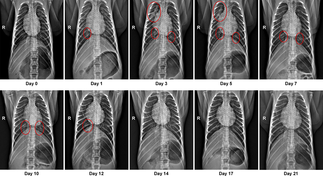
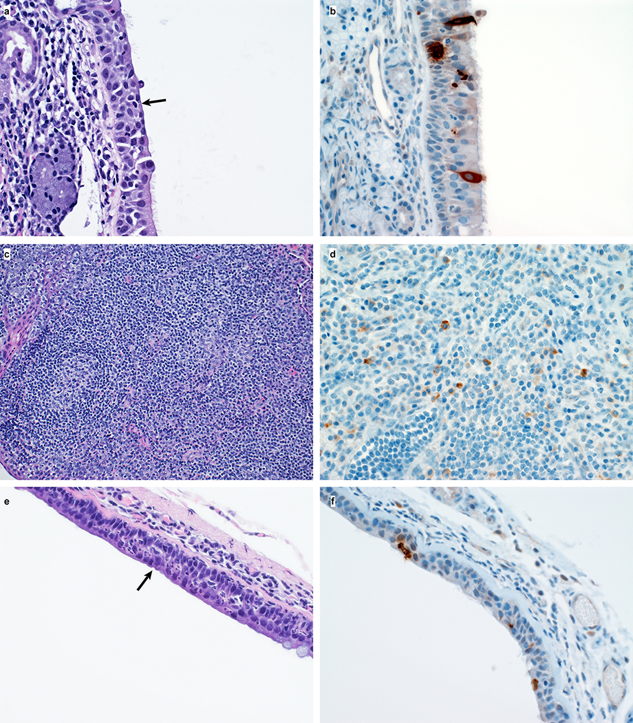
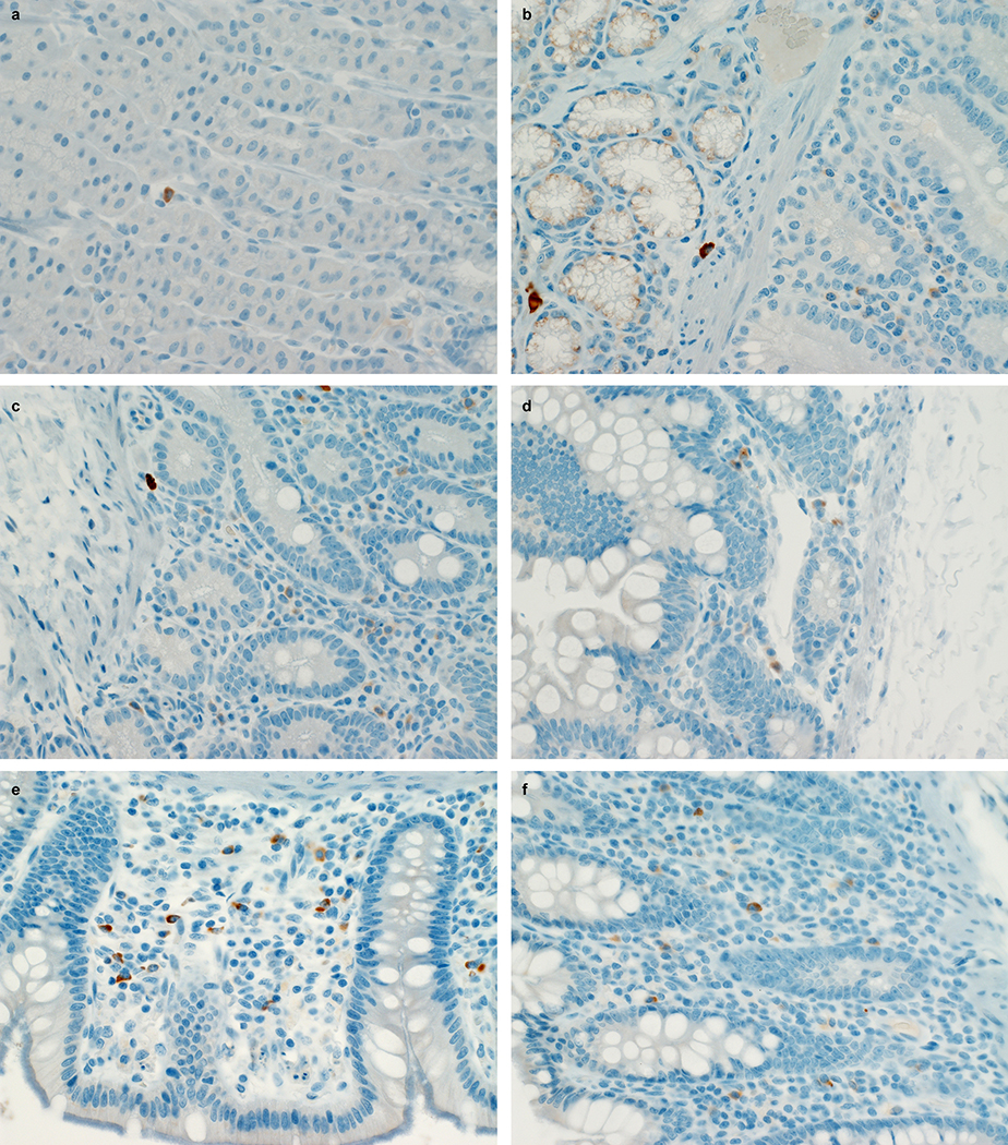
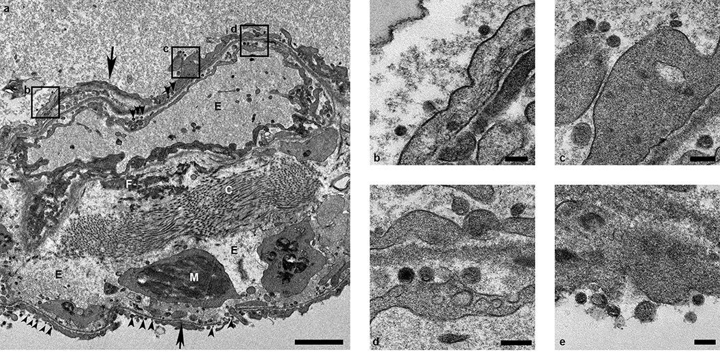
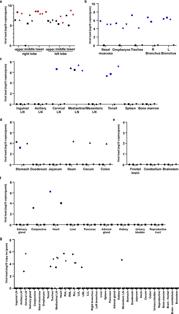
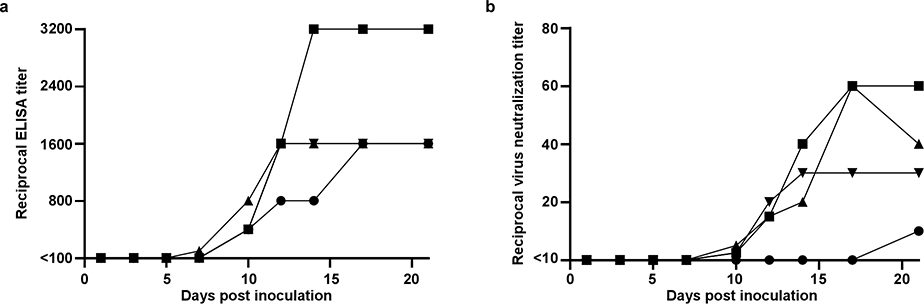
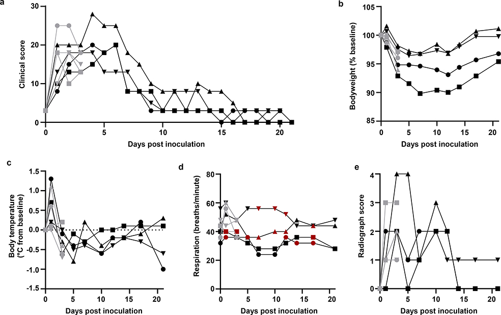
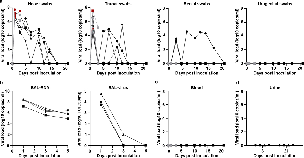
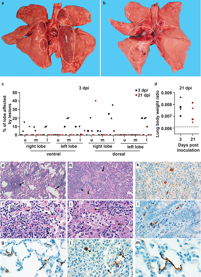
An outbreak of coronavirus disease 2019 (COVID-19), which is caused by a novel coronavirus (named SARS-CoV-2) and has a case fatality rate of approximately 2%, started in Wuhan (China) in December 20191,2. Following an unprecedented global spread3, the World Health Organization declared COVID-19 a pandemic on 11 March 2020. Although data on COVID-19 in humans are emerging at a steady pace, some aspects of the pathogenesis of SARS-CoV-2 can be studied in detail only in animal models, in which repeated sampling and tissue collection is possible. Here we show that SARS-CoV-2 causes a respiratory disease in rhesus macaques that lasts between 8 and 16 days. Pulmonary infiltrates, which are a hallmark of COVID-19 in humans, were visible in lung radiographs. We detected high viral loads in swabs from the nose and throat of all of the macaques, as well as in bronchoalveolar lavages; in one macaque, we observed prolonged rectal shedding. Together, the rhesus macaque recapitulates the moderate disease that has been observed in the majority of human cases of COVID-19. The establishment of the rhesus macaque as a model of COVID-19 will increase our understanding of the pathogenesis of this disease, and aid in the development and testing of medical countermeasures.


Conflict of interest statement

Competing interests

The authors declare no competing interests

Figures

Extended Data Figure 1.
Extended Data Figure 1.. Pulmonary infiltrates in a rhesus macaque after inoculation.
Radiographs show the progression of pulmonary infiltrates throughout the study in a single animal. Of note, this animal is denoted with a black triangle throughout the manuscript. Circles indicate areas of mild to moderate pulmonary infiltrates. A marker ‘R’ indicates right side of the animal. Three chest radiographs were taken at each timepoint: right-lateral, left-lateral and ventro-dorsal; only the ventro-dorsal radiograph is shown.
Extended Data Figure 2.
Extended Data Figure 2.. Hematological changes in rhesus macaques infected with SARS-CoV-2.
Identical symbols have been used to denote identical animals throughout the figures in this manuscript. n= 8 animals on 0, 1, and 3 dpi and n=4 animals thereafter.
Extended Data Figure 3.
Extended Data Figure 3.. Cytokine and chemokine levels in serum of rhesus macaques infected with SARS-CoV-2.
The levels of 23 cytokines and chemokines were determined in serum at different timepoints after inoculation. Levels are displayed only for those cytokines and chemokines where statistically significant (1-way ANOVA) were observed compared to levels on day of inoculation. Identical symbols have been used to denote identical animals throughout the figures in this manuscript. The lower limit of detection is indicated with a dotted line. Serum samples were analyzed in duplicate from each animal for each timepoint; n= 8 animals on 0, 1, and 3 dpi and n=4 animals thereafter.
Extended Data Figure 4.
Extended Data Figure 4.. Histological lesions in lungs of a rhesus macaque infected with SARS-CoV-2.
(a) This low magnification figure displays the focal nature of SARS-CoV-2 lesions in the lungs of animals euthanized on 3 dpi. The circle indicates the lung affected by lesion; the remaining lung tissue is healthy. (b) Lymphocytes surround pulmonary vessels. Magnification 500x. Tissue sections were collected from the same anatomical location for each animal; three tissue sections were prepared from each of the 6 lung lobes. In total, 18 lung sections were evaluated for each animal (n=4); representative images are displayed.
Extended Data Figure 5.
Extended Data Figure 5.. Histological changes in the respiratory tract of rhesus macaques infected with SARS-CoV-2.
(a) Squamous metaplasia of nasal turbinate respiratory epithelium (arrow). Magnification 400x. (b) SARS-CoV-2 antigen is detected by immunohistochemistry in respiratory epithelium of the nasal turbinate. Magnification 400x. (c) Essentially normal tonsil. Magnification 400x. (d) SARS-CoV-2 antigen is detected by immunohistochemistry in tonsillar macrophages. Magnification 400x. (e) Squamous metaplasia of tracheal columnar epithelium (arrow). Magnification 400x. (f) SARS-CoV-2 antigen is detected by immunohistochemistry in tracheal columnar epithelium. Magnification 400x. Tissue sections were collected from the same anatomical location for each animal (n=4) and organ; one tissue section was evaluated of the nasal turbinates of each animal; three tissue sections were evaluated from tonsil and trachea.
Extended Data Figure 6.
Extended Data Figure 6.. SARS-CoV-2 antigen in the gastrointestinal tract of a rhesus macaque infected with SARS-CoV-2.
Mononuclear cells staining positive for SARS-CoV-2 antigen in the lamina propria of stomach (a), duodenum (b), jejunum (c), ileum (d), cecum (e) and colon (f) of an animal infected with SARS-CoV-2 and euthanized on 3 dpi. Tissue sections were collected from the same anatomical location for each animal (n=4) and organ; three tissue sections were evaluated from each animal and organ.
Extended Data Figure 7.
Extended Data Figure 7.. Ultrastructural analysis of lungs of rhesus macaques infected with SARS-CoV-2.
Lung tissue collected on 3 dpi was analyzed by transmission electron microscopy. The alveolar interstitium is expanded by edema (E), fibrin (F) and mononuclear (M) inflammatory cells (a). Normal collagen fibers (c) and multiple virions (arrowheads) line type I pneumocytes (arrows). Boxes in (a) indicate areas enlarged in (b-d). Scale bar in (a) represents 2μm, scale bars in (b-e) represent 0.2 μm. Three tissue samples were collected from each animal (n=4) and cut into 6 samples for analysis; a minimum of 2 samples were analyzed per animal (n=4).
Extended Data Figure 8.
Extended Data Figure 8.. Viral loads in tissues collected from rhesus macaques infected with SARS-CoV-2.
Eight adult rhesus macaques were inoculated with SARS-CoV-2 isolate nCoV-WA1–2020 and euthanized on 3 (n=4) and 21 (n=4) dpi. Thirty-seven tissues were collected at necropsy and analyzed for the presence of viral RNA by qRT-PCR. Tissues are grouped by lung lobes collected on 3 dpi (a), with red symbols indicating tissues from which virus could be isolated in Vero E6 cells; other tissues from the respiratory tract on 3dpi (b); lymphoid tissues on 3 dpi (c); gastrointestinal tissueson 3 dpi (d); the central nervous system on 3 dpi (e); remaining tissues on 3 dpi (f); and all tissues collected on 21 dpi (g). Blue symbols in b-g indicate that viral mRNA was also detected in these tissues. Identical symbols have been used to denote identical animals throughout the figures in this manuscript. LN: lymph node; RUL: right upper lung lobe; RML: right middle lung lobe; RLL: right lower lung lobe; LUL: left upper lung lobe; LML: left middle lung lobe; LLL: left lower lung lobe; R: right; L: left.
Extended Data Figure 9.
Extended Data Figure 9.. Antibody response in rhesus macaques infected with SARS-CoV-2.
Sera collected after inoculation were tested for the presence of IgG against SARS-CoV-2 spike in ELISA (a) and for the presence of neutralizing antibodies in a microneutralization assay (b). All sera were analyzed in duplicate. Identical symbols have been used to denote identical animals throughout the figures in this manuscript.
Figure 1.
Figure 1.. Rhesus macaques infected with SARS-CoV-2 develop respiratory disease.
After inoculation with SARS-CoV-2, animals were observed for disease signs and scored according to a pre-established clinical scoring sheet (a). On clinical exams, body weight (b), and body temperature (c) were measured. Respiration rate was measured, and breathing pattern was recorded, with irregular respiration patterns indicated in red (d). Ventro-dorsal and lateral radiographs were taken on clinical exam days and scored for the presence of pulmonary infiltrates (0: normal; 1: mild interstitial pulmonary infiltrates; 2: moderate pulmonary infiltrates perhaps with partial cardiac border effacement and small areas of pulmonary consolidation; 3: severe interstitial infiltrates, large areas of pulmonary consolidation, alveolar patterns and air bronchograms). Individual lobes were scored and scores per animal per day totaled (e). Grey: animals euthanized 3 dpi; black: animals euthanized 21 dpi. Identical symbols have been used to denote identical animals throughout this manuscript.
Figure 2.
Figure 2.. Viral loads in respiratory samples and bodily fluids.
After inoculation, nose, throat, rectal and urogenital swabs were collected; viral loads in these samples were determined by qRT-PCR (a). On 1, 3, and 5 dpi, bronchoalveolar lavages were performed on the 4 animals remaining in the study through 21 dpi; viral loads and virus titers were determined in these samples. Viral loads were determined in blood collected during clinical exams (c) and urine collected at necropsy on 3 and 21 (d). Grey: animals euthanized 3 dpi; black: animals euthanized 21 dpi; red: virus was isolated from these samples. Identical symbols have been used to denote identical animals this manuscript.
Figure 3.
Figure 3.. Pathological changes in rhesus macaques infected with SARS-CoV-2.
Four rhesus macaques were euthanized on 3 and 21 dpi. Grossly, lungs showed focal areas of hilar consolidation and hyperemia (circles) on 3 dpi (a) and multifocal, random consolidation and hyperemia (circles) on 21 dpi (b). The percentage of the area of the lungs affected by gross lesions was estimated (c), and lung weight to bodyweight ratio was calculated. (d). The dotted line represents baseline ratio calculated from an in-house collection of rhesus macaque lung and bodyweights from animals with grossly normal lungs. Histological analysis was performed on tissues collected at 3 dpi (e-i). Tissue sections were collected from the same anatomical location for each animal; three tissue sections were prepared from each of the 6 lung lobes. In total, 18 lung sections were evaluated for each animal; representative images are displayed. (e) Pulmonary vessels surrounded by moderate numbers of lymphocytes and fewer macrophages (arrows). (f) Alveoli filled with small to moderate numbers of macrophages and neutrophils (asterisks). Adjacent alveolar interstitium (arrows) is thickened by edema, fibrin, neutrophils, lymphocytes and macrophages. (g) SARS-CoV-2 antigen detected by immunohistochemistry in type I pneumocytes. (h) Pulmonary vessels bounded by lymphocytes (arrowhead) and hyaline membranes (arrows) line alveolar spaces. (i) Hyaline membranes line alveoli (arrows). (j) SARS-CoV-2 antigen detected by immunohistochemistry in type I pneumocytes (asterisk) and type II pneumocytes (arrow) as well as alveolar macrophages (arrowheads). (k) SARS-CoV-2 antigen detected by immunohistochemistry in macrophages in a mediastinal lymph node. (l) SARS-CoV-2 antigen detected by immunohistochemistry in macrophages and lymphocytes in the lamina propria of the cecum. (m) SARS-CoV-2 detected by immunohistochemistry in type I pneumocytes. Magnification: e, h 100x; f, g, I, j, k, l 400x; m: 1000x. u: upper; m: middle; l: lower

Update of

References

    1. Wu F et al. A new coronavirus associated with human respiratory disease in China. Nature, doi:10.1038/s41586-020-2008-3 (2020). - DOI - PMC - PubMed
    1. Zhu N et al. A Novel Coronavirus from Patients with Pneumonia in China, 2019. N Engl J Med 382, 727–733, doi:10.1056/NEJMoa2001017 (2020). - DOI - PMC - PubMed
    1. Organization, W. H. Coronavirus disease (COVID-2019) situation reports, <https://www.who.int/emergencies/diseases/novel-coronavirus-2019/situatio...> (2020).
    1. Yang W et al. Clinical characteristics and imaging manifestations of the 2019 novel coronavirus disease (COVID-19):A multi-center study in Wenzhou city, Zhejiang, China. J Infect, doi:10.1016/j.jinf.2020.02.016 (2020). - DOI - PMC - PubMed
    1. Yang X et al. Clinical course and outcomes of critically ill patients with SARS-CoV-2 pneumonia in Wuhan, China: a single-centered, retrospective, observational study. Lancet Respir Med, doi:10.1016/S2213-2600(20)30079-5 (2020). - DOI - PMC - PubMed

Publication types

MeSH terms