Review
. 2020 Jul;75(7):1564-1581.
doi: 10.1111/all.14364.

Immune response to SARS-CoV-2 and mechanisms of immunopathological changes in COVID-19

Affiliations
Review

Immune response to SARS-CoV-2 and mechanisms of immunopathological changes in COVID-19

Ahmet Kursat Azkur et al. Allergy. 2020 Jul.

Abstract

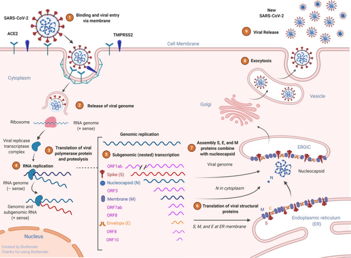
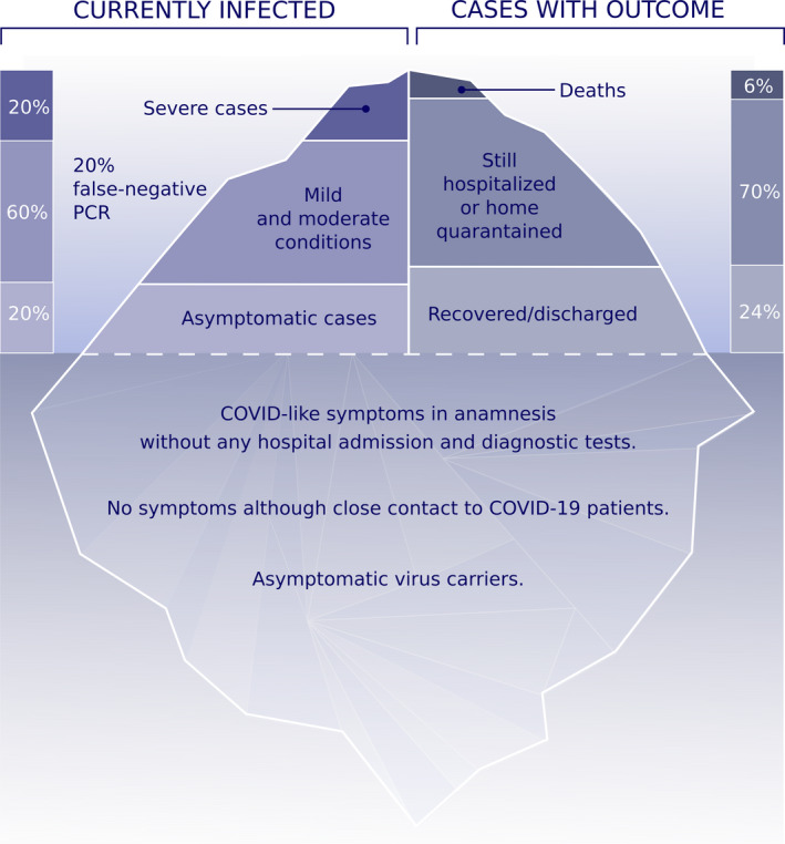
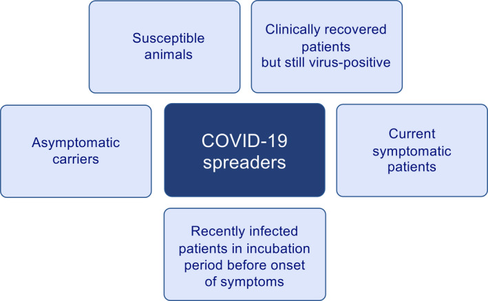
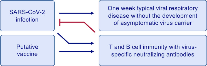
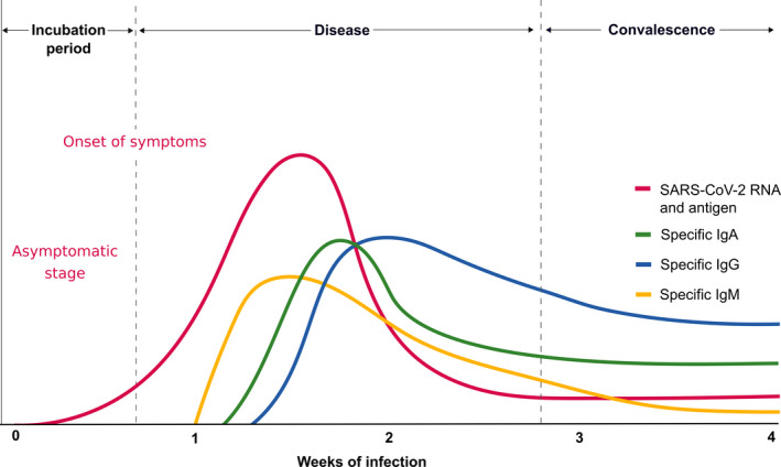
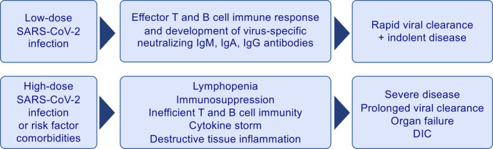
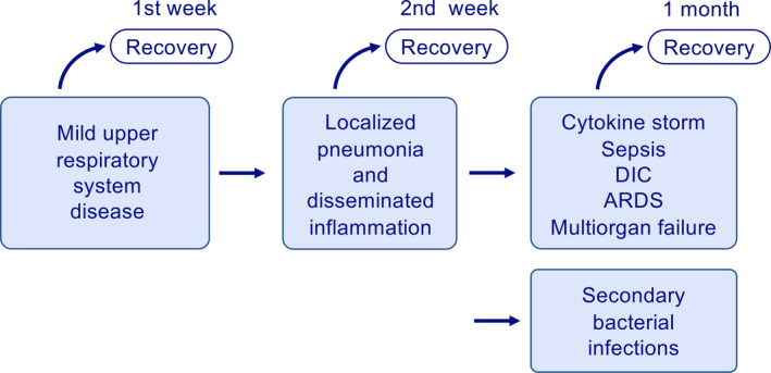
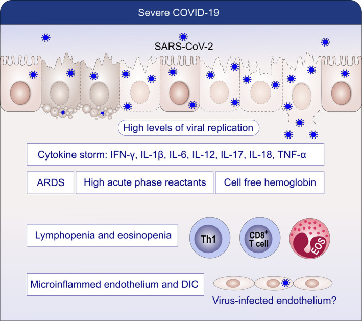
As a zoonotic disease that has already spread globally to several million human beings and possibly to domestic and wild animals, eradication of coronavirus disease 2019 (COVID-19) appears practically impossible. There is a pressing need to improve our understanding of the immunology of this disease to contain the pandemic by developing vaccines and medicines for the prevention and treatment of patients. In this review, we aim to improve our understanding on the immune response and immunopathological changes in patients linked to deteriorating clinical conditions such as cytokine storm, acute respiratory distress syndrome, autopsy findings and changes in acute-phase reactants, and serum biochemistry in COVID-19. Similar to many other viral infections, asymptomatic disease is present in a significant but currently unknown fraction of the affected individuals. In the majority of the patients, a 1-week, self-limiting viral respiratory disease typically occurs, which ends with the development of neutralizing antiviral T cell and antibody immunity. The IgM-, IgA-, and IgG-type virus-specific antibodies levels are important measurements to predict population immunity against this disease and whether cross-reactivity with other coronaviruses is taking place. High viral load during the first infection and repeated exposure to virus especially in healthcare workers can be an important factor for severity of disease. It should be noted that many aspects of severe patients are unique to COVID-19 and are rarely observed in other respiratory viral infections, such as severe lymphopenia and eosinopenia, extensive pneumonia and lung tissue damage, a cytokine storm leading to acute respiratory distress syndrome, and multiorgan failure. Lymphopenia causes a defect in antiviral and immune regulatory immunity. At the same time, a cytokine storm starts with extensive activation of cytokine-secreting cells with innate and adaptive immune mechanisms both of which contribute to a poor prognosis. Elevated levels of acute-phase reactants and lymphopenia are early predictors of high disease severity. Prevention of development to severe disease, cytokine storm, acute respiratory distress syndrome, and novel approaches to prevent their development will be main routes for future research areas. As we learn to live amidst the virus, understanding the immunology of the disease can assist in containing the pandemic and in developing vaccines and medicines to prevent and treat individual patients.

Keywords: COVID-19; cytokine storm; immune response; immunologic tests; immunopathology; infections; pandemic; virus.


Conflict of interest statement

LM reports personal fees from AHL and grants from GSK. KN reports grants and/or personal fees and/or other from NIAID, Novartis, Regeneron, FARE, EAT, Sanofi, Astellas, Nestle, BeforeBrands, Alladapt, ForTra, Genentech, AImmune Therapeutics, DBV Technologies, AstraZeneca, ImmuneWorks, Cour Pharmaceuticals, AllerGenis, Ukko Pharma, AnaptysBio, Adare Pharmaceuticals, Stallergenes‐Greer, NHLBI, NIEHS, EPA, WAO Center of Excellence, Iggenix, Probio, Vedanta, Centocor, Seed, Immune Tolerance Network, and NIH. In addition, KN has pending patents on Inhibition of Allergic Reaction to Peanut Allergen using an IL‐33 Inhibitor, Special Oral Formula for Decreasing Food Allergy Risk and Treatment for Food Allergy, Basophil Activation Based Diagnostic Allergy Test, Granulocyte‐based methods for detecting and monitoring immune system disorders, Methods and Assays for Detecting and Quantifying Pure Subpopulations of White Blood Cells in Immune System Disorders, Mixed Allergen Compositions and Methods for Using the Same, and Microfluidic Device and Diagnostic Methods for Allergy Testing Based on Detection of Basophil Activation. CA reports grants from Allergopharma, Idorsia, Swiss National Science Foundation, Christine Kühne‐Center for Allergy Research and Education, European Commission's Horizon's 2020 Framework Programme, Cure, Novartis Research Institutes, AstraZeneca, SciBase, and advisory role in Sanofi/Regeneron. All other authors have no conflict of interest within the scope of the submitted work.

Figures

Figure 1
Figure 1
Virus binding, internalization to epithelial cells, and replication. Schematic representation of the genomic and subgenomic organizations of SARS‐CoV and replication. SARS‐CoV‐2 uses receptor ACE2 and transmembrane protease, serine 2 (TMPRSS2) for host cell entry. Following entry to cell cytoplasm, the genomic RNA; the two large open reading frames (ORFs) 1ab are translated into a protein viral transcriptase complex (phosphatase activity and RNA‐dependent RNA polymerase (RdRp) and a helicase). Replication of the genome involves the synthesis of a full‐length negative‐strand RNA and serves as template for full‐length genomic RNA. After translation, structural proteins are localized to the Golgi intracellular membranes, the endoplasmic reticulum Golgi intermediate compartment (ERGIC) that is called site of budding. New virions that are assembled full genome RNA release from the cell
Figure 2
Figure 2
The iceberg of the COVID‐19 pandemic. 10%‐20% of currently diagnosed patients appear with severe cases and 60% with mild to moderate cases. False‐negative viral nucleic acid diagnosis with RT‐PCR should always be considered as 15%‐20% in best experienced hospital conditions, which can be higher in the field. Known asymptomatic cases are diagnosed by random screening of hospital staff and individuals with close contact to COVID‐19 cases in the household. However, there are also a high number of unproven asymptomatic individuals at the bottom of the iceberg with COVID‐like symptoms in the anamnesis without any diagnostic tests and hospital admission. Certain individuals have been reported, who never had symptoms although they had close contact to COVID‐19–positive family members. Overall death rate is 6% and is currently increasing worldwide. The reports on the number of recovered individuals are not currently convincing. Data are collected from https://www.worldometers.info/coronavirus/ and https://www.who.int/health-topics/coronavirus/
Figure 3
Figure 3
SARS‐CoV‐2 carriers and spreaders. There are different types of virus carriers in COVID‐19, an important factor for the containment of the pandemic. They are listed as asymptomatic virus carriers, current symptomatic patients, recently infected patients in window period before the onset of symptoms, clinically recovered patients who remain SARS‐CoV‐2‐positive, and susceptible domestic and wild animals. All of these have specific immunological mechanisms that are discussed in this review
Figure 4
Figure 4
Ideal immune response to SARS‐CoV‐2 infection or a vaccine. Acute infection and the vaccine are expected to develop same type of an immune response with T‐ and B‐cell immunity and development of virus‐specific neutralizing antibodies. The normal immune response is characterized with one‐week typical viral respiratory disease without the development of asymptomatic virus carriers. This type of immune response is observed in almost all mild cases
Figure 5
Figure 5
Immune response to coronaviruses and other respiratory viruses. After the epithelium is infected with SARS‐CoV‐2, the replicating virus can cause cell lysis and direct damage to the epithelium. The epithelium presents virus antigens to CD8+ T cells. With their perforin and granzymes, CD8+ T cells and natural killer (NK) cells can show cytotoxicity to virus‐infected epithelial cells and induce apoptosis. Subepithelial dendritic cells (DC) recognize virus antigens and present them to CD4 T cells and induce the differentiation of these T cells toward memory Th1, Th17, and memory T follicular helper (FH). TFH helps B cells to develop into plasma cells (PC) and promotes the production of IgM, IgA, and IgG isotype virus‐specific antibodies. Tissue macrophages (MΦ) and DCs also present viral antigens to CD4+ T cells
Figure 6
Figure 6
Specific antibody response to SARS‐CoV‐2. The incubation period of COVID‐19 is relatively long and has been reported to be 5‐10 d. A specific IgM response is the early antibody response that starts and peaks within 7 d. IgM continues as long as the acute phase of the disease continues. Specific IgA and IgG antibodies develop several days after IgM and do not decrease to undetectable levels and are assumed to continue lifelong as protective antibodies. This research requires an international consensus on the usage of correct methodology and antigens of SARS‐CoV‐2
Figure 7
Figure 7
Distinct response to high‐ and low‐dose virus exposure and infection. High‐dose exposure may occur in healthcare workers and individuals who are exposed to currently sick or asymptomatic virus spreading family members. Whereas low‐dose infection may lead to appropriate effector T‐ and B‐cell response, neutralizing antibodies, and rapid viral clearance, high‐dose exposure may cause severe disease and delayed viral clearance. This may be due to lymphopenia leading to inefficient T‐ and B‐cell immunity, subsequently the cytokine storm and destructive tissue inflammation
Figure 8
Figure 8
Different clinical phases of COVID‐19. COVID‐19 presents with three stages of diseases. It starts with upper respiratory disease symptoms followed by pneumonia and disseminated inflammation, and in the third‐stage cytokine storm, sepsis‐like syndrome, ARDS, DIC, and multiorgan failure take place. The disease that moves into second phage always carries the risk for secondary bacterial infections. Antiviral immune response and tissue and systemic inflammation are different in all stages
Figure 9
Figure 9
Pathogenesis of severe COVID‐19. Severe COVID‐19 presents with high viral load, cytokine storm, ARDS, cell‐free hemoglobin, high acute‐phase reactants, lymphopenia, eosinopenia, microinflammed endothelium, and DIC. Endothelial damage, cell‐free hemoglobin, cytokine storm, and lymphopenia show links to severe disease

References

    1. Coronaviridae Study Group of the International Committee on Taxonomy of V . The species Severe acute respiratory syndrome‐related coronavirus: classifying 2019‐nCoV and naming it SARS‐CoV‐2. Nat Microbiol. 2020;5(4):536‐544. - PMC - PubMed
    1. To KK, Hung IF, Chan JF, Yuen KY. From SARS coronavirus to novel animal and human coronaviruses. J Thorac Dis. 2013;5(Suppl 2):S103‐108. - PMC - PubMed
    1. Zaki AM, van Boheemen S, Bestebroer TM, Osterhaus AD, Fouchier RA. Isolation of a novel coronavirus from a man with pneumonia in Saudi Arabia. N Engl J Med. 2012;367(19):1814‐1820. - PubMed
    1. Zhu N, Zhang D, Wang W, et al. A Novel Coronavirus from Patients with Pneumonia in China, 2019. N Engl J Med. 2020;382(8):727‐733. - PMC - PubMed
    1. WHO . Coronavirus disease (COVID‐2019) situation reports. https://www.who.int/emergencies/diseases/novel-coronavirus-2019/situatio.... Accessed April 9

MeSH terms