. 2020 May 8;9(5):237.
doi: 10.3390/antibiotics9050237.

High Abundance of Proteobacteria in Ileo-Anal Pouch Anastomosis and Increased Abundance of Fusobacteria Associated with Increased Pouch Inflammation

Affiliations

High Abundance of Proteobacteria in Ileo-Anal Pouch Anastomosis and Increased Abundance of Fusobacteria Associated with Increased Pouch Inflammation

Andreas Munk Petersen et al. Antibiotics (Basel). .

Abstract

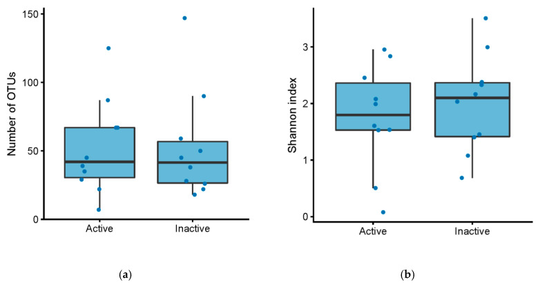
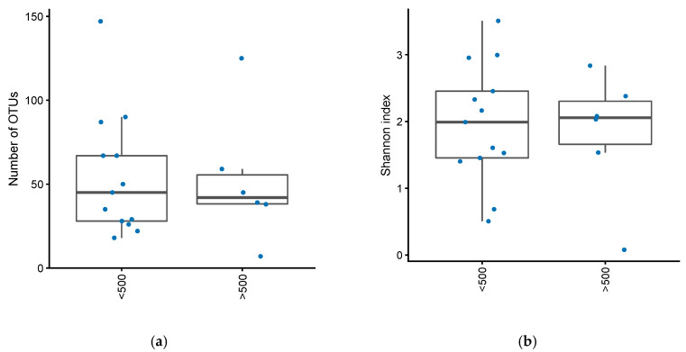
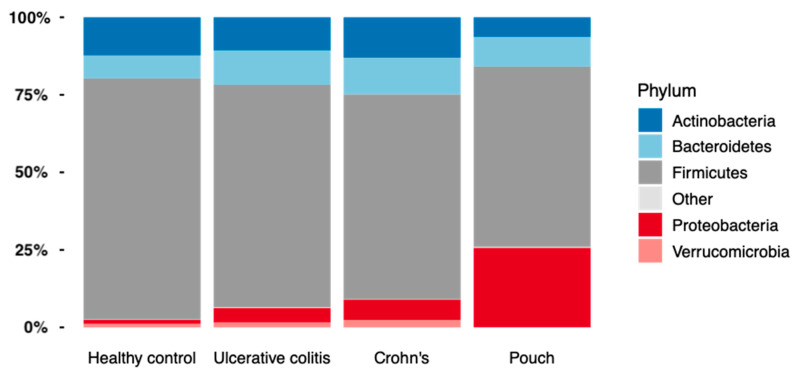
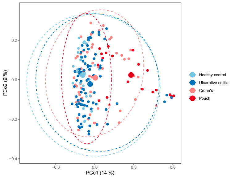
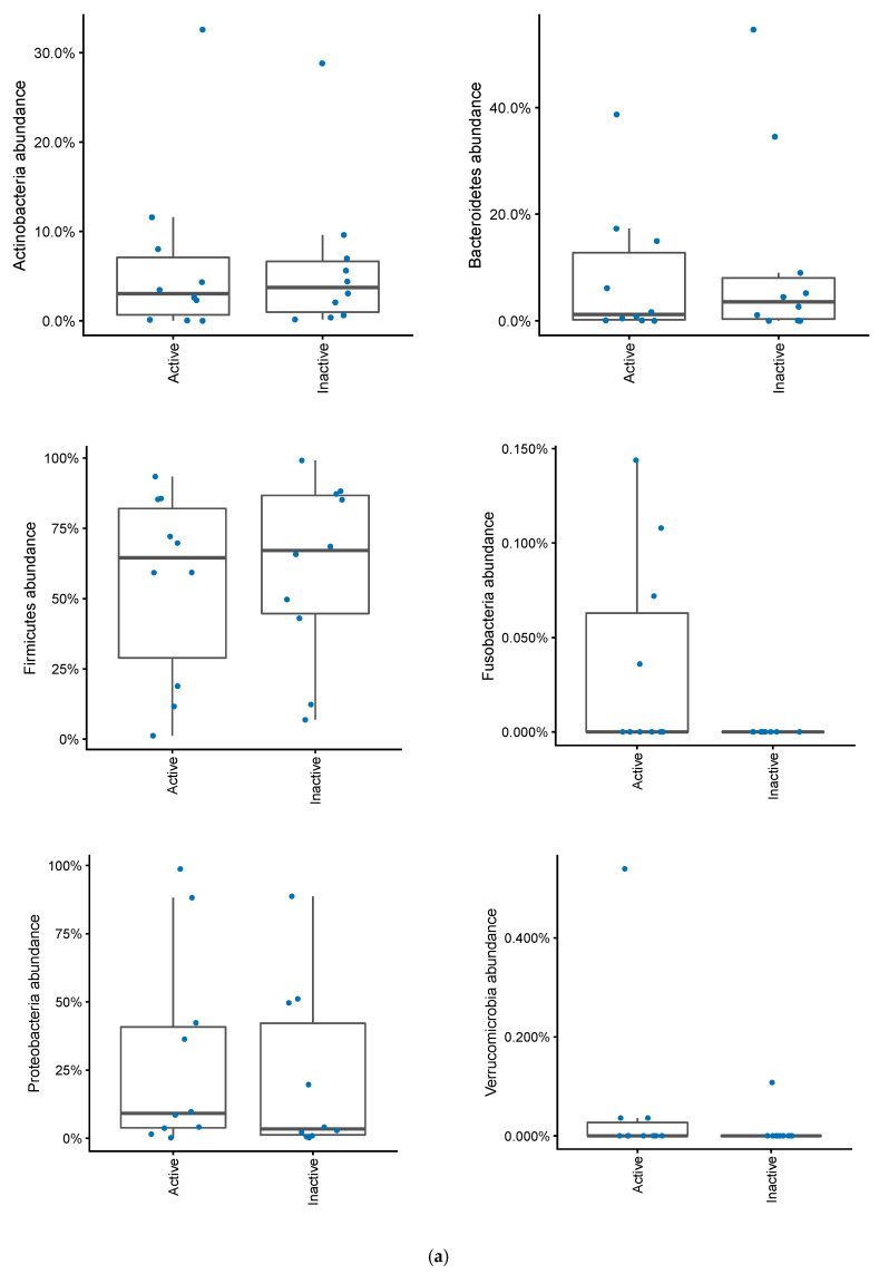
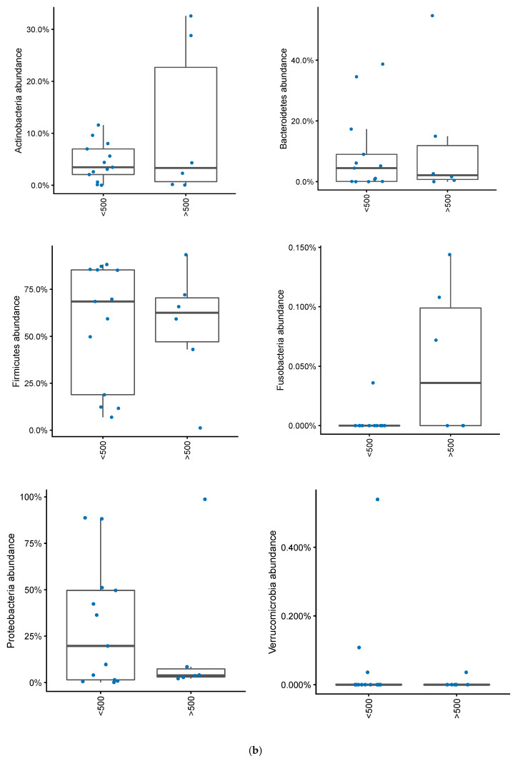
Low diversity intestinal dysbiosis has been associated with inflammatory bowel disease, including patients with ulcerative colitis with an ileo-anal pouch anastomosis. Furthermore, specific Escherichia coli phylogroups have been linked to inflammatory bowel disease. Our aim was to characterize the differences among microbiota and E. coli phylogroups in active and inactive pouchitis. Disease activity was assessed using the modified pouch disease activity index and by fecal calprotectin. Microbiota diversity was assessed by 16S rDNA MiSeq sequencing. E. coli phylogroup was determined after triplex PCR. Twenty patients with ulcerative colitis with an ileo-anal pouch anastomosis were included, 10 of whom had active pouchitis. Ileo-anal pouch anastomosis patients had an increased abundance of Proteobacteria colonization compared to patients with ulcerative colitis or Crohn's disease and healthy controls, p = 1.4·10-5. No differences in E. coli phylogroup colonization could be determined between cases of active and inactive disease. No significant link was found between α-diversity and pouch inflammation. However, higher levels of Fusobacteria colonization were found in patients with a pouch with a fecal calprotectin level above 500, p = 0.02. In conclusion, patients with a pouch had an increased Proteobacteria abundance, but only Fusobacteria abundance was linked to inflammation.

Keywords: Escherichia coli; Fusobacteria; Proteobacteria; calprotectin; inflammatory bowel disease; pouchitis.


Conflict of interest statement

The authors declare no conflict of interest.

Figures

Figure 1
Figure 1
(a) Number of OTUs in patients with an ileo-anal pouch anastomosis, with active vs. inactive pouchitis. Active pouchitis was defined as a modified pouchitis disease activity index (mPDAI) score of 3 or above. (b) Shannon diversity index in patients with an ileo-anal pouch anastomosis, with active vs. inactive pouchitis. Active pouchitis was defined as a mPDAI score of 3 or above.
Figure 2
Figure 2
(a) Number of OTUs in patients with an ileo-anal pouch anastomosis, with or without increased inflammation in their pouch. Increased inflammation defined as fecal calprotectin greater than 500. (b) Shannon diversity index in patients with an ileo-anal pouch anastomosis, with or without increased inflammation in their pouch. Increased inflammation was defined as a fecal calprotectin greater than 500.
Figure 3
Figure 3
Number of OTUs (a) and Shannon diversity index (b) in patients with Inflammatory Bowel Disease (Crohn’s disease, N = 58 and Ulcerative Colitis, N = 82), controls, N = 31, and in patients with an ileo-anal pouch anastomosis, N = 20.
Figure 4
Figure 4
The relative abundance of bacterial phyla found in Inflammatory Bowel Disease (Crohn’s disease, N = 58 and Ulcerative colitis, N = 82), controls, N = 30, and in patients with a pouch, N = 20.
Figure 5
Figure 5
PCoA plot of generalized UniFrac distances by patient group; Inflammatory Bowel Disease (Crohn’s disease, N = 58 and Ulcerative colitis, N = 82), controls, N = 30, and in patients with a pouch, N = 20. The small dots are samples, while the big dots are centroids of their respective groups.
Figure 6
Figure 6
(a) Changes in bacterial phyla in patients with an ileo-anal pouch anastomosis, with or without active pouchitis. Pouchits defined as as a mPDAI score of 3 or above. (b) Changes in bacterial phyla abundances in patients with an ileo-anal pouch anastomosis, with or without increased inflammation in their pouch. Increased inflammation was defined as a fecal calprotectin greater than 500.
Figure 6
Figure 6
(a) Changes in bacterial phyla in patients with an ileo-anal pouch anastomosis, with or without active pouchitis. Pouchits defined as as a mPDAI score of 3 or above. (b) Changes in bacterial phyla abundances in patients with an ileo-anal pouch anastomosis, with or without increased inflammation in their pouch. Increased inflammation was defined as a fecal calprotectin greater than 500.

References

    1. Feuerstein J.D., Cheifetz A.S. Ulcerative colitis: Epidemiology, diagnosis, and management. Mayo Clin. Proc. 2014;89:1553–1563. doi: 10.1016/j.mayocp.2014.07.002. - DOI - PubMed
    1. Hata K., Ishihara S., Nozawa H., Kawai K., Kiyomatsu T., Tanaka T., Kishikawa J., Anzai H., Watanabe T. Pouchitis after ileal pouch-anal anastomosis in ulcerative colitis: Diagnosis, management, risk factors, and incidence. Dig. Endosc. 2017;29:26–34. doi: 10.1111/den.12744. - DOI - PubMed
    1. Kartheuser A., Stangherlin P., Brandt D., Remue C., Sempoux C. Restorative proctocolectomy and ileal pouch-anal anastomosis for familial adenomatous polyposis revisited. Fam. Cancer. 2006;5:241–260. doi: 10.1007/s10689-005-5672-4. - DOI - PubMed
    1. Singh V.P., Proctor S.D., Willing B.P. Koch’s postulates, microbial dysbiosis and inflammatory bowel disease. Clin. Microbiol. Infect. 2016;22:594–599. doi: 10.1016/j.cmi.2016.04.018. - DOI - PubMed
    1. Khan K.J., Ullman T.A., Ford A.C., Abreu M.T., Abadir A., Marshall J.K., Talley N.J., Moayyedi P. Antibiotic therapy in inflammatory bowel disease: A systematic review and meta-analysis. Am. J. Gastroenterol. 2011;106:661–673. doi: 10.1038/ajg.2011.72. - DOI - PubMed

LinkOut - more resources