. 2020 May 8;8(2):212.
doi: 10.3390/vaccines8020212.

Investigating the Impact of Delivery System Design on the Efficacy of Self-Amplifying RNA Vaccines

Affiliations

Investigating the Impact of Delivery System Design on the Efficacy of Self-Amplifying RNA Vaccines

Giulia Anderluzzi et al. Vaccines (Basel). .

Abstract

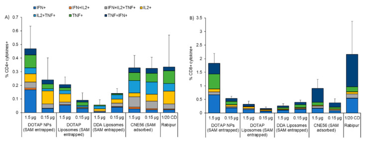
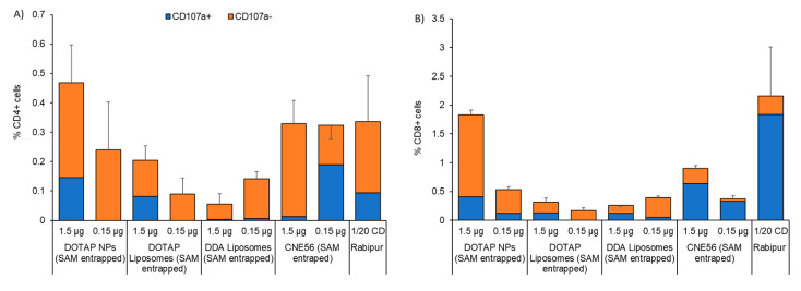
messenger RNA (mRNA)-based vaccines combine the positive attributes of both live-attenuated and subunit vaccines. In order for these to be applied for clinical use, they require to be formulated with delivery systems. However, there are limited in vivo studies which compare different delivery platforms. Therefore, we have compared four different cationic platforms: (1) liposomes, (2) solid lipid nanoparticles (SLNs), (3) polymeric nanoparticles (NPs) and (4) emulsions, to deliver a self-amplifying mRNA (SAM) vaccine. All formulations contained either the non-ionizable cationic lipid 1,2-dioleoyl-3-trimethylammonium-propane (DOTAP) or dimethyldioctadecylammonium bromide (DDA) and they were characterized in terms of physico-chemical attributes, in vitro transfection efficiency and in vivo vaccine potency. Our results showed that SAM encapsulating DOTAP polymeric nanoparticles, DOTAP liposomes and DDA liposomes induced the highest antigen expression in vitro and, from these, DOTAP polymeric nanoparticles were the most potent in triggering humoral and cellular immunity among candidates in vivo.

Keywords: antigen expression; emulsions; immunogenicity; liposomes; polymeric nanoparticles; self-amplifying RNA; solid lipid nanoparticles.


Conflict of interest statement

G.A. and G.L. participated in the European Marie Curie PHA-ST-TRAIN-VAC PhD project at the University of Strathclyde (Glasgow, UK) in collaboration with GSK (Siena, Italy). The project was co-sponsored between the University of Strathclyde and GlaxoSmithKline Biologicals SA. Y.P. declare no conflict of interest. S.G, M.B., R.J., D.T.O. and B.C.B are employees of the GSK group of companies. All the authors declare that they have no other relevant affiliations or financial interests in conflict with the subject matter or materials discussed in the manuscript.

Figures

Figure 1
Figure 1
Cell viability and cell association of delivery platforms with baby hamster kidney (BHK) cells. Cytotoxicity (A,B) and cell association (C,D) in BHK cell of DOTAP-based (A,C) and DDA-based (B,D) liposomes, SLNs, NPs and emulsions. Lipofectamine 2000 (LF2000) was used as positive control. For cell viability, cationic lipid concentrations of 3.7 to 100 µg/mL (N:P ratio of 8:1) were tested. For cell association, the results represent percentage of 1,1’-dioctadecyl-3,3,3’,3’-tetramethylindocarbocyanine perchlorate (DilC)-positive BHK cells after 16 hours of incubation with 11 µg/mL of DOTAP-based (C) and DDA (D) formulations in either 5% fetal calf serum (FCS) or FCS-free media. Results are represented as mean ± SD of 3 independent experiments.
Figure 2
Figure 2
Cell association of formulations in BHK cell line expressed as mean fluorescence intensity (MFI). Mean fluorescence intensity of BHK cells after 16 h incubation with DOTAP-based (A,C) and DDA-based (B,D) liposomes, SLNs, NPs and emulsions in either 5% FCS (A,B) or FCS-free media (C,D). Results are represented as mean ± SD of 3 independent experiments.
Figure 3
Figure 3
In vitro potency (IVP) in BHK cell line. IVP in BHK in either 5% FCS (A,B) or FCS-free (C,D) medium of DOTAP-based (A,C) and DDA-based (B,D) formulations prepared with SAM-RVG at different concentrations. Lipofectamine 2000 (LF2000) was used as a positive control. Results are represented as mean ± SD of 3 independent experiments.
Figure 4
Figure 4
Immunogenicity of SAM-RVG vaccine delivered by different cationic carriers. Groups of ten BALB/c mice were immunized i.m. on days 0 and 28 with either 1.5 or 0.15 μg of self-amplifying RNA encoding the rabies virus G protein formulated in DOTAP polymeric nanoparticles (NPs), DOTAP Liposomes or DDA Liposomes, and compared to the commercial vaccine Rabipur (1/20 of human dose). GSK trademark CNE56 was used as a positive control. Specific IgG titers were measured by enzyme-linked immunosorbent assay (ELISA). For each group, there were 5 samples, each representing data from pools of two mice (depicted as circles), and the geometric mean titers (GMTs) are solid lines. Sera were collected and analyzed (A) 4 weeks after the first immunization and (B) 2 weeks after the second immunization. Titers < 0.125 EU/mL (dotted blue line) were below the limit of detection, while titers > 0.5 EU/mL (dotted red line) were an indication of protection. Intergroup comparison was analyzed using the one-way analysis of variance (ANOVA) test (Dunnett’s multiple comparison test).
Figure 5
Figure 5
Percentages of antigen-specific CD4+ or CD8+ T-cells. Splenic (A) CD4+ T cells, and (B) CD8+ T-cells 2 weeks after two intramuscular immunizations spaced 4 weeks apart in BALB/c mice. Mice were immunized with either 1.5 or 0.15 μg/dose of self-amplifying RNA expressing rabies G glycoprotein adjuvanted with either polymeric nanoparticles (NPs), DOTAP liposomes or DDA liposomes, and spleens from 3 mice randomly selected out of the group were tested. Candidates were compared with the commercial vaccine Rabipur (1/20 of human dose) and GSK trademark CNE56 as positive controls. Splenocytes were stimulated with rabies G1/G2/G3 peptide, stained for intra-cellular cytokines, and analyzed by flow cytometry. The color code indicates the different combinations of cytokine produced by the respective cells. Unstimulated cells were used as control. Refer to Figure S3 in the Supplemental Material for the gating strategy.
Figure 6
Figure 6
Percentages of cytotoxic CD4+ or CD8+ T-cells. The induction of rabies-specific CD4+ or CD8+ T-cells by either 1.5 or 0.15 μg/dose of SAM encapsulating nanoparticles (NPs), DOTAP liposomes and DDA liposomes was characterized 2 weeks after the second immunization. Spleens from 3 mice were randomly selected from each group. Candidates were compared with the commercial vaccine Rabipur (1/20 of human dose) and GSK trademark CNE56 as positive controls. Surface expression of CD107a on splenocytes stimulated in vitro with rabies G1/G2/G3 peptide was assessed by flow cytometry. Data show the frequency of cytokine-secreting (A) CD4+ or (B) CD8+ T-cells that express (blue bars) or do not express (orange bars) CD107a. Unstimulated cells were used as control. Refer to Figure S3 in the Supplemental Material for the gating strategy.

References

    1. Wolff J.A., Malone R.W., Williams P., Chong W., Acsadi G., Jani A., Felgner P.L. Direct gene transfer into mouse muscle in vivo. Science. 1990;247:1465–1468. doi: 10.1126/science.1690918. - DOI - PubMed
    1. Wong J.P., Zabielski M.A., Schmaltz F.L., Brownlee G.G., Bussey L.A., Marshall K., Borralho T., Nagata L.P. DNA vaccination against respiratory influenza virus infection. Vaccine. 2001;19:2461–2467. doi: 10.1016/S0264-410X(00)00474-6. - DOI - PubMed
    1. Geall A.J., Mandl C.W., Ulmer J.B. RNA: The new revolution in nucleic acid vaccines. Semin. Immunol. 2013;25:152–159. doi: 10.1016/j.smim.2013.05.001. - DOI - PubMed
    1. Petsch B., Schnee M., Vogel A.B., Lange E., Hoffmann B., Voss D., Schlake T., Thess A., Kallen K.J., Stitz L., et al. Protective efficacy of in vitro synthesized, specific mRNA vaccines against influenza A virus infection. Nat. Biotechnol. 2012;30:1210–1216. doi: 10.1038/nbt.2436. - DOI - PubMed
    1. Akinc A., Maier M.A., Manoharan M., Fitzgerald K., Jayaraman M., Barros S., Ansell S., Du X., Hope M.J., Madden T.D., et al. The Onpattro story and the clinical translation of nanomedicines containing nucleic acid-based drugs. Nat. Nanotechnol. 2019;14:1084–1087. doi: 10.1038/s41565-019-0591-y. - DOI - PubMed