. 2020 May 8;21(9):3343.
doi: 10.3390/ijms21093343.

A Synergistic Effect of Reactive Oxygen and Reactive Nitrogen Species in Plasma Activated Liquid Media Triggers Astrocyte Wound Healing

Affiliations

A Synergistic Effect of Reactive Oxygen and Reactive Nitrogen Species in Plasma Activated Liquid Media Triggers Astrocyte Wound Healing

Eloisa Sardella et al. Int J Mol Sci. .

Abstract

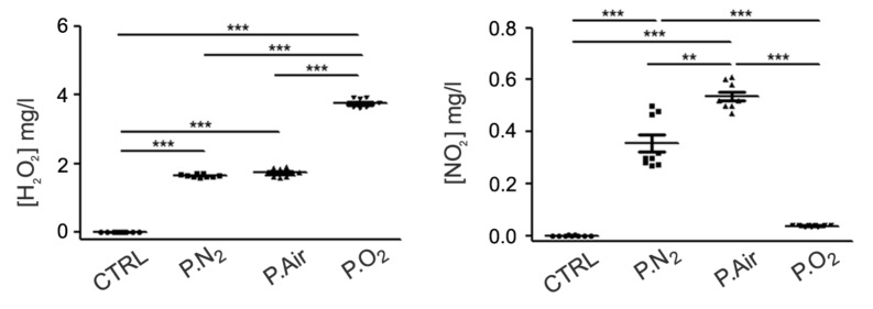
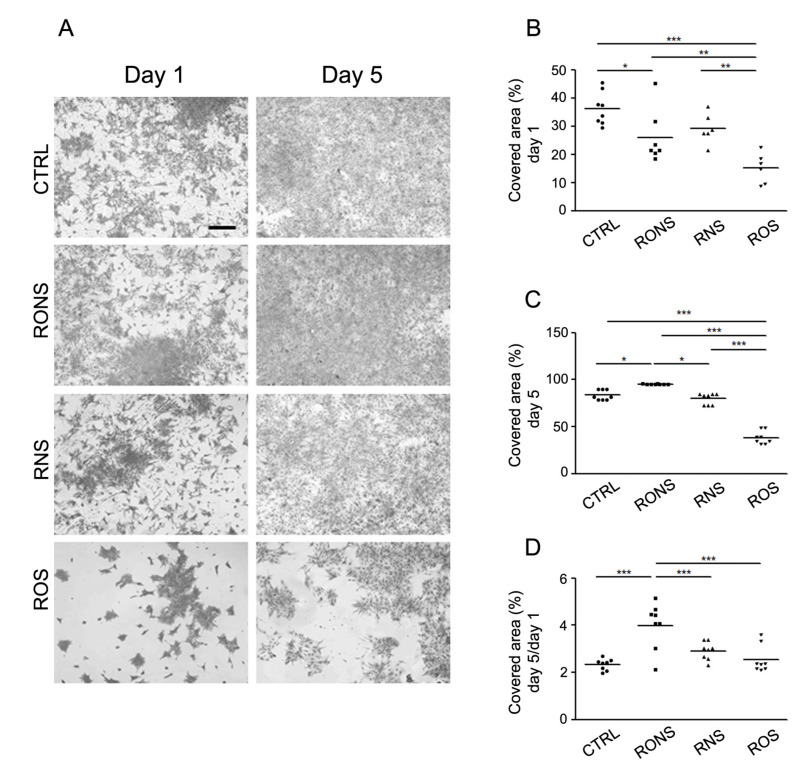
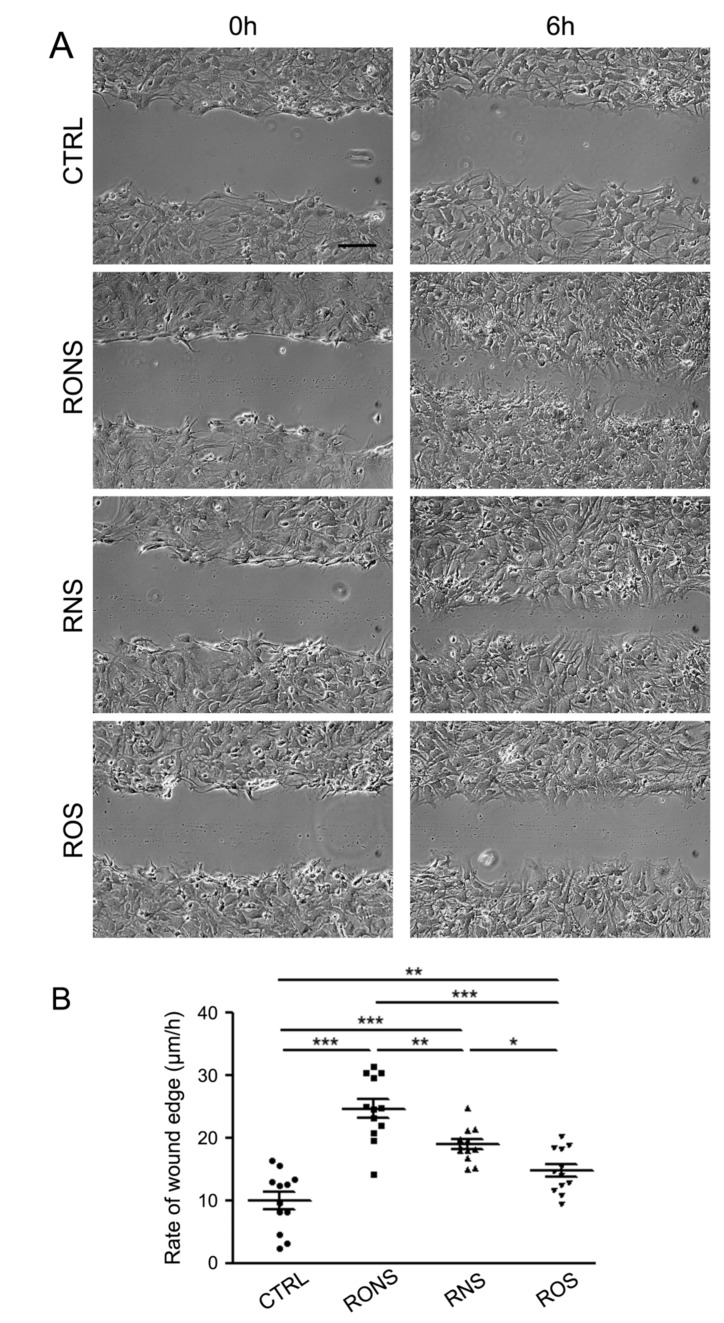
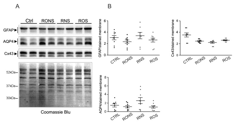
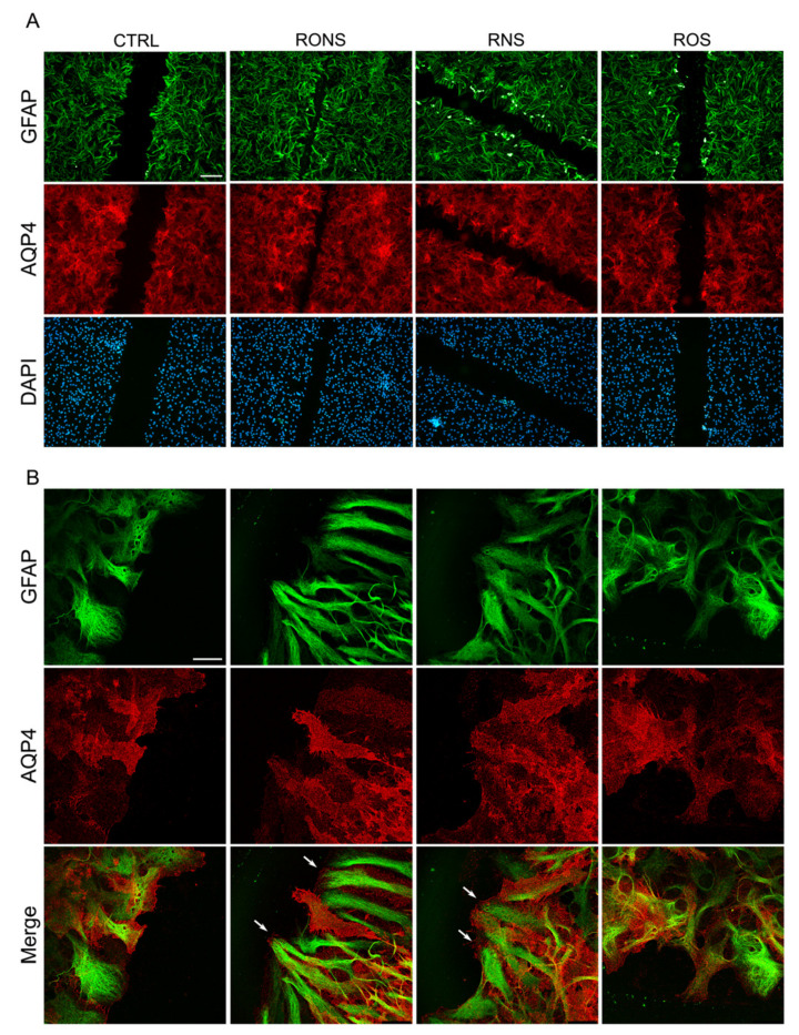
Astrocyte proliferation and migration toward injured Central Nervous System (CNS) areas are key features of astrogliosis and glial scar formation. Even though it is known that intracellular and environmental Reactive Oxygen and Nitrogen Species (RONS) affect astrocyte behaviour in physiological and pathophysiological conditions, their effects on the migration and growth of astrocytes are still unclear. Plasma-technologies are emerging in medicine as a tool to generate RONS for treating cells directly or through Plasma Activated Liquid Media (PALM). In this paper, we show for the first time how the use of PALM can modulate both astrocyte growth and migration as a function of active species produced by plasma in liquids. Our results show that PALM, generated by means of cold atmospheric pressure plasmas fed with N2, air or O2, can modulate astrocyte behaviour depending on the content of hydrogen peroxide and nitrite in the liquid. In particular, H2O2 enriched PALM induced a negative effect on cell growth associated with the mild wound healing improvement of primary astrocytes, in a scratch assay. Nitrite enriched PALM induced a selective effect on the wound healing without affecting cell growth. PALM containing a more balanced level of H2O2 and NO2- were able to affect cell growth, as well as significantly ameliorate wound healing. None of the PALM investigated induced upregulation of the gliotic inflammatory marker glial fibrillary acidic protein (GFAP), or of the astrocyte markers Aquaporin-4 (AQP4) and Connexin-43 (Cx-43) analysed by Western blot. Finally, immunofluorescence analysis revealed the presence of NO2- able to induce elongated protrusions at the front end of wounded astrocytes in the direction of cell migration. With our study we believe to have shown that PALM offer a novel tool to modulate astrocyte behaviour and that they are promising candidates for controlling astrogliosis in the case of CNS injuries.

Keywords: astrocytes; oxidative stress; plasma activated medium; wound healing.


Conflict of interest statement

The authors declare that there is no conflict of interest.

Figures

Figure 1
Figure 1
Chemical composition of the different Plasma Activated Liquid Media (PALM). Concentration of H2O2 and NO2 in untreated medium (Ctrl), in PALM N2 (P.N2), in PALM Air (P.Air) and in PALM O2 (P.O2). The following experimental conditions were kept constant: 6 KHz, 13 kV, 100 ms period, 25% D.C; 30 s treatment time, 0.5 slm gas feed. Significant differences of the means were calculated by One-way Anova followed by the Newman–Keuls Multiple Comparison Test. *: p < 0.05; **: p < 0.01; ***: p < 0.001.
Figure 2
Figure 2
Coomassie blue assay of astrocytes incubated with different PALM. (A) Coomassie blue staining of astrocytes, one and five days after a 2 h-PALM incubation. Images are compared with the untreated medium (Ctrl) used as control. Different PALM are indicated as follows: PALM N2 (RONS), PALM Air (RNS) and PALM O2 (ROS). Scale bar, 500 μm. (B) Scattered plots showing the percentage area covered by cells at day 1. (C) Scattered plots showing the percentage area covered by cells at day 5. (D) Scattered plot measuring the ratio of the area covered by astrocytes after five days over that covered after one day. A One-way Anova and Newman–Keuls Multiple Comparison Test were performed. *: p < 0.05; **: p < 0.01; ***: p < 0.001.
Figure 3
Figure 3
Scratch-induced migration assay in PALM-incubated astrocyte monolayers. (A) Phase-contrast micrographs showing astrocytes exposed to untreated medium (Ctrl) and to P.N2 (RONS), P.Air (RNS) and P.O2 (ROS) PALM immediately after the scratch (0 h), and 6 h post-wounding. Scale bar, 100 μm. (B) Scattered plots showing the mean ± SE values of the migration rate of astrocytes incubated as indicated. A One-way Anova and Newman-Keuls Multiple Comparison Test were performed: *: p < 0.05; **: p < 0.01; ***: p < 0.001.
Figure 4
Figure 4
GFAP, AQP4 and Cx43 immunoblotting and quantification in PALM-incubated astrocytes. (A) Representative Western blot analysis of GFAP, AQP4 and Cx43 expression in astroglial cultures exposed to untreated medium (Ctrl), to P.Air (RNS), to P.O2 (ROS) and to P.N2 (RONS). Protein samples were collected 24 h after 2 h-PALM incubation. (B) Summary of the densitometric analysis of GFAP, AQP4 and Cx43 corresponding signals normalised to the Coomassie blue-stained membrane. No significant differences by One-way Anova test were found between PALM-treated and Ctrl astrocytes.
Figure 5
Figure 5
GFAP and AQP4 protein localisation in astrocyte cultures after PALM incubation. Immunostaining at 6 h after scratch for AQP4 (red) and GFAP (green) in PALM-exposed and control astrocytes. Untreated medium (Ctrl), P.N2 (RONS), P.Air (RNS) and P.O2 (ROS) PALM. (A) Fluorescence signal for AQP4 and GFAP in migrating cells at the wound. Nuclear staining with DAPI is also shown. (Scale bar: 100 μm). (B) Confocal images at the leading edge of wounded astrocytes as in (A). Merged images (Merge) of the two proteins’ localisation are reported (Scale bar: 25 μm). White arrows in RONS and RNS indicate enhanced foot processes at the front end of the scratch.

References

    1. Allen N.J., Barres B.A. Neuroscience: Glia—more than just brain glue. Nature. 2009;457:675–677. doi: 10.1038/457675a. - DOI - PubMed
    1. Sofroniew M.V., Vinters H.V. Astrocytes: Biology and pathology. Acta Neuropathol. 2010;119:7–35. doi: 10.1007/s00401-009-0619-8. - DOI - PMC - PubMed
    1. Iadecola C. The neurovascular unit coming of age: A journey through neurovascular coupling in health and disease. Neuron. 2017;96:17–42. doi: 10.1016/j.neuron.2017.07.030. - DOI - PMC - PubMed
    1. Sofroniew M.V. Molecular dissection of reactive astrogliosis and glial scar formation. Trends Neurosci. 2009;32:638–647. doi: 10.1016/j.tins.2009.08.002. - DOI - PMC - PubMed
    1. Nicchia G.P., Pisani F., Simone L., Cibelli A., Mola M.G., Dal Monte M., Frigeri A., Bagnoli P., Svelto M. Glio-vascular modifications caused by aquaporin-4 deletion in the mouse retina. Exp. Eye Res. 2016;146:259–268. doi: 10.1016/j.exer.2016.03.019. - DOI - PubMed

LinkOut - more resources