. 2020 May 9;10(5):918.
doi: 10.3390/nano10050918.

Toxicological Profile of Nanostructured Bone Substitute Based on Hydroxyapatite and Poly(lactide-co-glycolide) after Subchronic Oral Exposure of Rats

Affiliations

Toxicological Profile of Nanostructured Bone Substitute Based on Hydroxyapatite and Poly(lactide-co-glycolide) after Subchronic Oral Exposure of Rats

Smiljana Paraš et al. Nanomaterials (Basel). .

Abstract

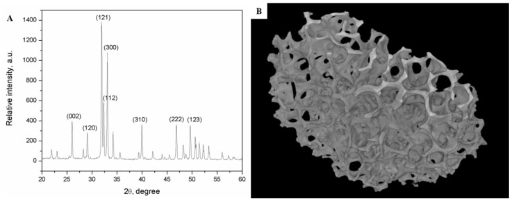
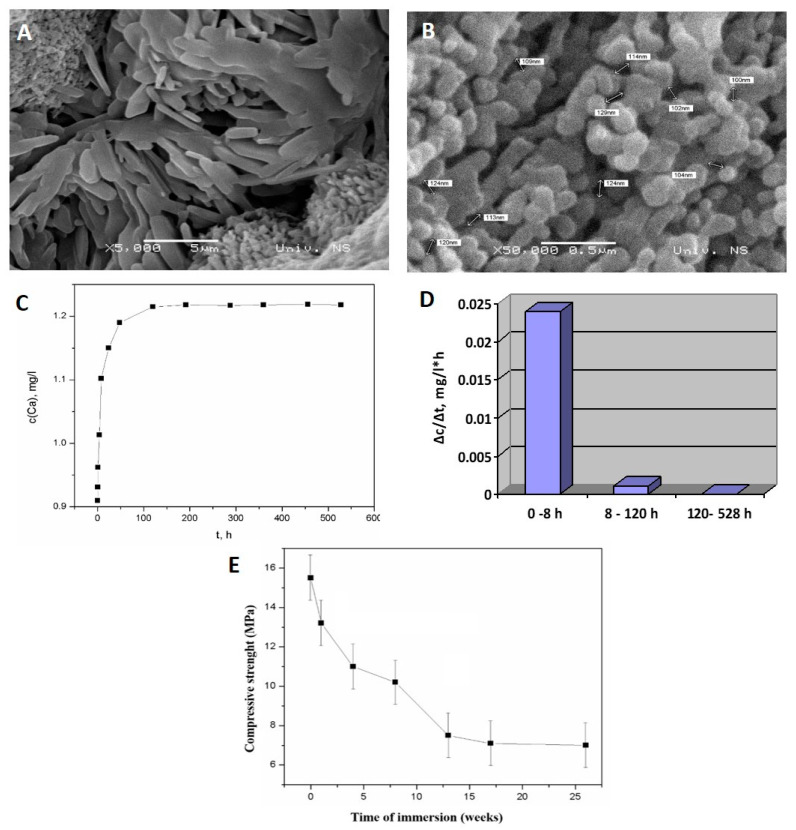
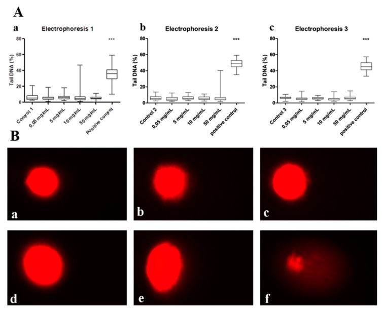
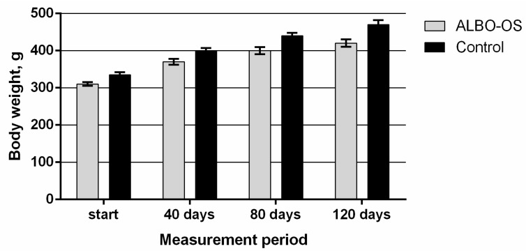
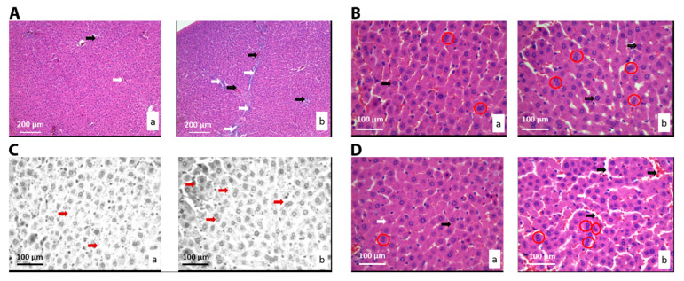
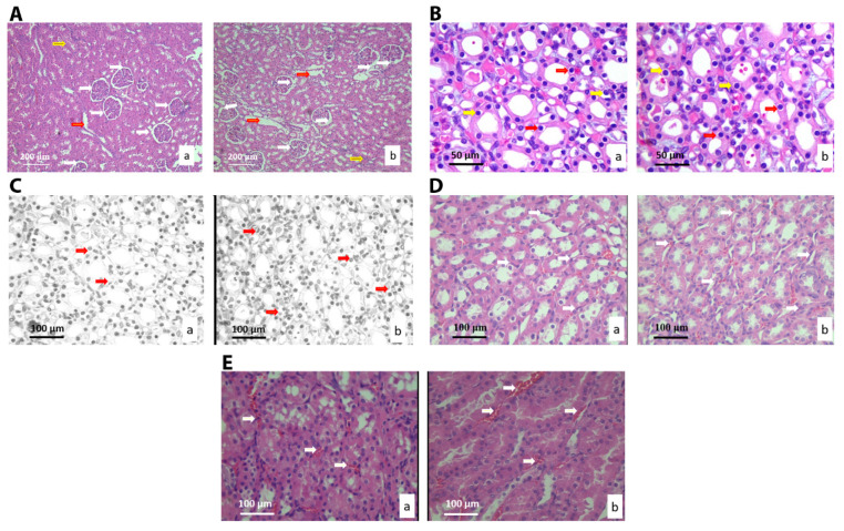
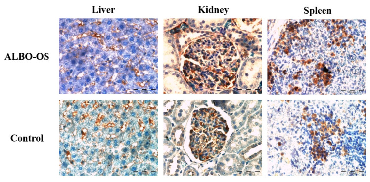
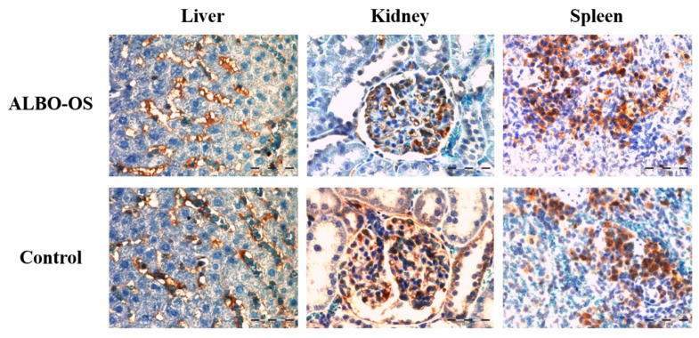
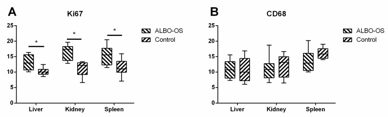
Novel three-dimensional (3D) nanohydroxyapatite-PLGA scaffolds with high porosity was developed to better mimic mineral component and microstructure of natural bone. To perform a final assessment of this nanomaterial as a potential bone substitute, its toxicological profile was particularly investigated. Therefore, we performed a comet assay on human monocytes for in vitro genotoxicity investigation, and the systemic subchronic toxicity investigation on rats being per oral feed with exactly administrated extract quantities of the nano calcium hydroxyapatite covered with tiny layers of PLGA (ALBO-OS) for 120 days. Histological and stereological parameters of the liver, kidney, and spleen tissue were analyzed. Comet assay revealed low genotoxic potential, while histological analysis and stereological investigation revealed no significant changes in exposed animals when compared to controls, although the volume density of blood sinusoids and connective tissue, as well as numerical density and number of mitosis were slightly increased. Additionally, despite the significantly increased average number of the Ki67 and slightly increased number of CD68 positive cells in the presence of ALBO-OS, immunoreactive cells proliferation was almost neglected. Blood analyses showed that all of the blood parameters in rats fed with extract nanomaterial are comparable with corresponding parameters of no feed rats, taken as blind probe. This study contributes to the toxicological profiling of ALBO-OS scaffold for potential future application in bone tissue engineering.

Keywords: biocompatibility; bone substitute; genotoxicity; hydroxyapatite; subchronic toxicity.


Conflict of interest statement

The authors declare no conflict of interest. The funders had no role in the design of the study; in the collection, analyses, or interpretation of data; in the writing of the manuscript, or in the decision to publish the results.

Figures

Figure A1
Figure A1
(A) XRD pattern of HA; (B) Micro-CT scan of nanoHAP-PLGA.
Figure A2
Figure A2
The SEM micrographs of nanoHAP-PLGA (A) 5000×, (B) 50,000×; (C) Change of the Ca2+ concentration in solution containing nanoHAP granules with time; (D) The rate of nanoHAP degradation during prolonged time period (0–428 h); and, (E) Change of compressive strength during nanoHAP degradation.
Figure 1
Figure 1
(A) Comet assay results: (a) first repeat, (b) second repeat, (c) third repeat. Asterisks denote the significant differences with respect to the untreated control cells (*** p < 0.001; one-way ANOVA, Dunnett’s test). (B) Images of comets for: (a) negative control, different concentration of material’s extract: (b) 0.05 mg/mL, (c) 5 mg/mL, (d) 10 mg/mL, (e) 50 mg/mL and (f) positive control.
Figure 2
Figure 2
Average body weights of experimental and control animals during the study of chronic systemic toxicity of ALBO-OS.
Figure 3
Figure 3
Micrographs of the histological cross-section of the liver of the control group (a) and treated group (b), (hematoxylin-eosin (H&E)). (A) White arrows show connective tissue and black show blood vessels. Magnification 20×; (B) Black arrows show hepatocytes’ nuclei, and red circles show hepatocytes with two nuclei. Magnification 50×; (C) Red scalpers show hepatocytes’ nuclei. Magnification 50×, digitally processed RGB technique; (D) Black arrows show capillary sinusoids, and red circles show hepatocytes with two nuclei. Magnification 50×.
Figure 4
Figure 4
Micrographs of histological cross-sections of the kidney of the control (a) and treated group (b), (H&E). (A) White arrows show glomeruli, red arrows show blood sinusoids and yellow arrows show connective tissue. Magnification 20×; (B) Yellow arrows show nuclei, and red arrows show epithelial cells of collecting ducts. Magnification 50×; (C) Red arrows show the epithelial cells of collecting ducts. Magnification 50×; digitally processed RGB technique; (D) White arrows show the connective tissue of collecting ducts. Magnification 50×; and, (E) White arrows show the blood sinusoids of the collection ducts. Magnification 100×.
Figure 5
Figure 5
Micrographs of the histological cross-section of the rat spleen of the control (a) and treated group (b), (H&E). (A) White arrows show connective tissue and red arrows show lymphatic tissue. Magnification 200×; (B) White arrows show epithelial cells. Magnification 50×; (C) White arrows show lymphocytes and red arrows show blood vessel. Magnification 50× and, (D) Red arrows show lymphocytes. Magnification 50×; digitally processed RGB technique.
Figure 6
Figure 6
Representative images of Ki67 protein expression in rats’ liver, kidney and spleen tissue sections of experimental and control group, 40× magnification. Significant expression of Ki67+ cells was observed in all groups.
Figure 7
Figure 7
Representative images of CD68 protein expression in rats’ liver, kidney and spleen tissue sections of experimental and control group, 40× magnification. Mild expression of CD68+ immunoreactive cells was observed in all groups.
Figure 8
Figure 8
Percentage area of Ki67 (A) and immunoreactive CD68+ (B) stained cells was calculated with ImageJ using 3 images per organ. The results were analyzed with a two-way ANOVA, a p value < 0.05 was considered significant; * p < 0.05.

Similar articles

Cited by

References

    1. Boga J.C., Miguel S.P., de Melo-Diogo D., Mendonça A.G., Louro R.O., Correia I.J. In Vitro characterization of 3D printed scaffolds aimed at bone tissue regeneration. Colloids Surf. B Biointerfaces. 2018;165:207–218. doi: 10.1016/j.colsurfb.2018.02.038. - DOI - PubMed
    1. Erasmus E.P., Sule R., Johnson O.T., Massera J., Sigalas I. In Vitro evaluation of porous borosilicate, borophosphate and phosphate bioactive glasses scaffolds fabricated using foaming agent for bone regeneration. Sci. Rep. 2018;8:3699. doi: 10.1038/s41598-018-22032-2. - DOI - PMC - PubMed
    1. Jeon O.H., Elisseeff J. Orthopedic tissue regeneration: Cells, scaffolds, and small molecules. Drug Deliv. Transl. Res. 2016;6:105–120. doi: 10.1007/s13346-015-0266-7. - DOI - PubMed
    1. Tohamy K.M., Mabrouk M., Soliman I.E., Beherei H.H., Aboelnasr M.A. Novel alginate/hydroxyethyl cellulose/hydroxyapatite composite scaffold for bone regeneration: In Vitro cell viability and proliferation of human mesenchymal stem cells. Int. J. Biol. Macromol. 2018;112:448–460. doi: 10.1016/j.ijbiomac.2018.01.181. - DOI - PubMed
    1. Liu X., Zhao K., Gong T., Song J., Bao C., Luo E., Weng J., Zhou S. Delivery of growth factors using a smart porous nanocomposite scaffold to repair a mandibular bone defect. Biomacromolecules. 2014;15:1019–1030. doi: 10.1021/bm401911p. - DOI - PubMed

LinkOut - more resources