. 2020 May 9;10(5):740.
doi: 10.3390/biom10050740.

Ticagrelor Prevents Endothelial Cell Apoptosis through the Adenosine Signalling Pathway in the Early Stages of Hypoxia

Affiliations

Ticagrelor Prevents Endothelial Cell Apoptosis through the Adenosine Signalling Pathway in the Early Stages of Hypoxia

Catherine Feliu et al. Biomolecules. .

Abstract

Background: Several studies have reported the beneficial effects of anti-platelet drugs in cardioprotection against ischaemia-reperfusion injuries. To date, no studies have focused on the indirect cytoprotective effects of ticagrelor via adenosine receptor on the endothelium.

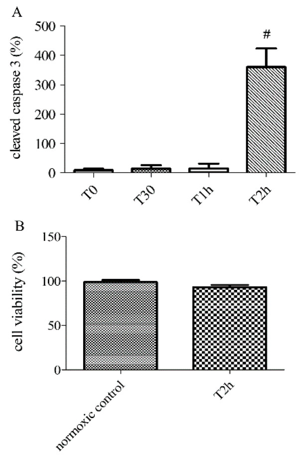
Method: By evaluating cell viability and cleaved caspase 3 expression, we validated a model of endothelial cell apoptosis induced by hypoxia. In hypoxic endothelial cells treated with ticagrelor, we quantified the extracellular concentration of adenosine, and then we studied the involvement of adenosine pathways in the cytoprotective effect of ticagrelor.

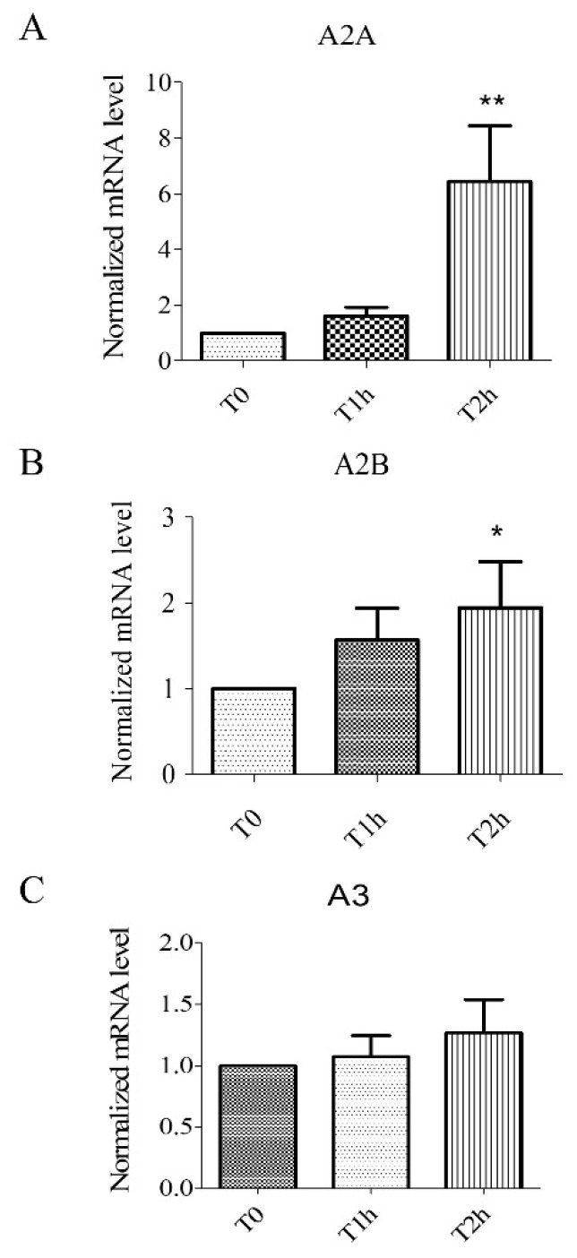
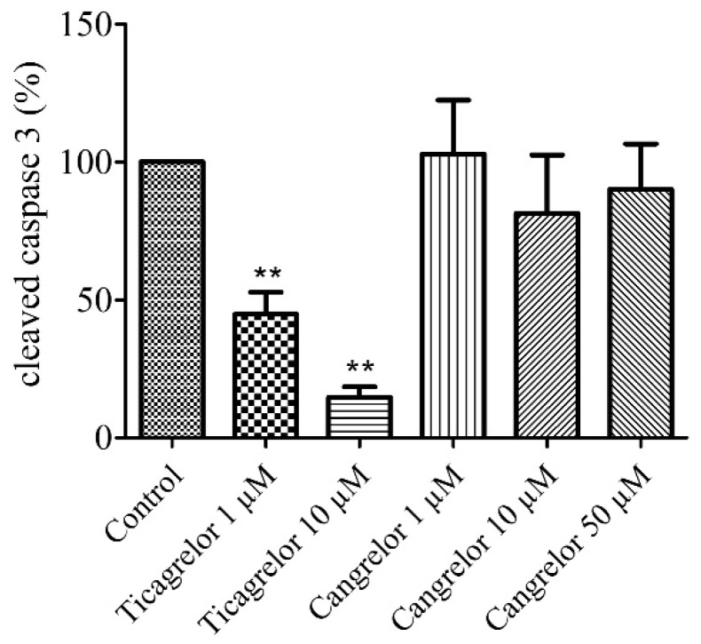
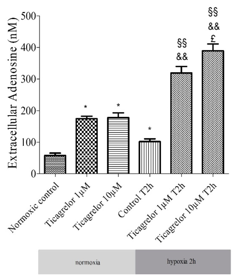
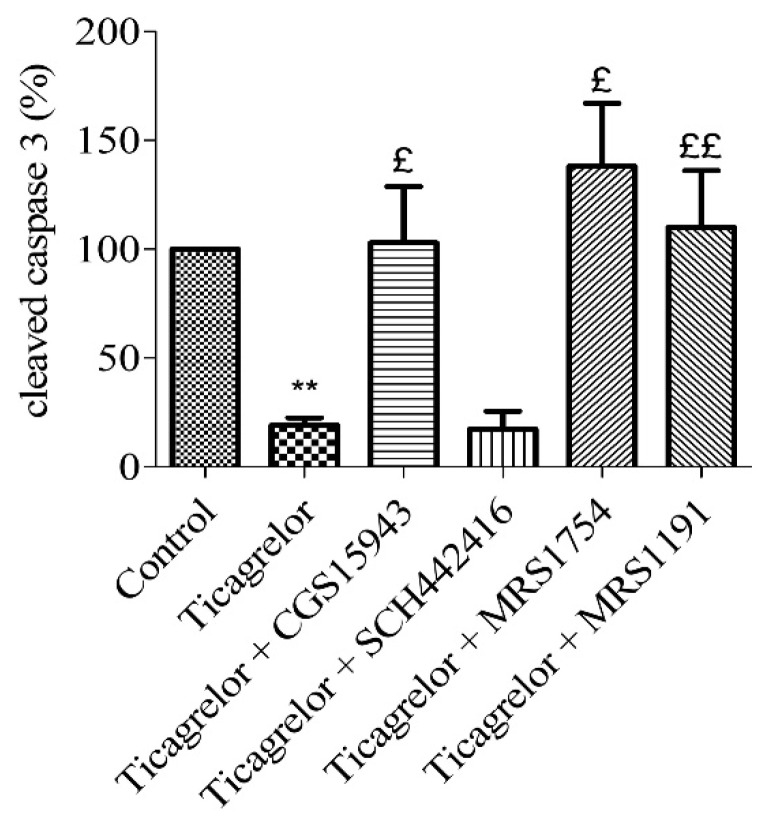
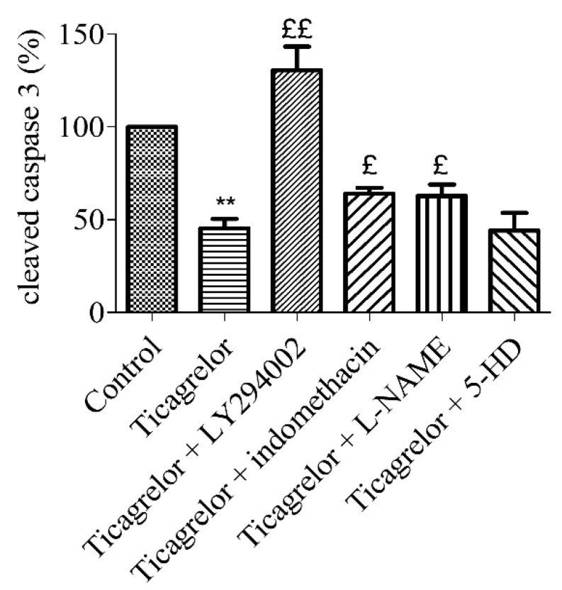
Results: Our results showed that 10 µM ticagrelor induced an anti-apoptotic effect in our model associated with an increase of extracellular adenosine concentration. Similar experiments were conducted with cangrelor but did not demonstrate an anti-apoptotic effect. We also found that A2B and A3 adenosine receptors were involved in the anti-apoptotic effect of ticagrelor in endothelial cells exposed to 2 h of hypoxia stress.

Conclusion: we described an endothelial cytoprotective mechanism of ticagrelor against hypoxia stress, independent of blood elements. We highlighted a mechanism triggered mainly by the increased extracellular bioavailability of adenosine, which activates A2B and A3 receptors on the endothelium.

Keywords: adenosine receptors; cytoprotective effect; endothelium; extracellular adenosine; ticagrelor.


Conflict of interest statement

C.F., H.P., G.P., S.B.-P., F.O., P.N., H.M., and Z.D. declare no financial relationships with any organizations that might have an interest in the submitted work. No other relationships or activities that could appear to have influenced the submitted work.

Figures

Figure 1
Figure 1
Expression of mRNA for adenosine receptors A2A (A), A2B (B), and A3 (C). Results are expressed with normalized mRNA levels using the following formula: 2−ΔΔCT as a function of time. Overexpression of mRNA for A2A and A2B receptors was observed. Results are expressed as means ± sem (n = 6/group). *: p < 0.05, **: p < 0.01, compared to a normoxic control group.
Figure 2
Figure 2
Apoptosis at different time-points of hypoxia (0, 30 min, 1 and 2 h) and cell viability after 0 (normoxic control) or 2 h of hypoxia (T2h), determined by the relative expression of cleaved caspase-3 by immunoblotting (A) and PrestoBlue assays (B). Results are expressed as means ± sem (n = 6/group). #: p < 0.05, compared to normoxic control.
Figure 3
Figure 3
Ticagrelor but not cangrelor induced an anti-apoptotic effect. Cells were treated with ticagrelor 1 µM, 10 µM or with cangrelor 1 µM, 10 µM and 50 µM. Results are expressed as means ± sem (n = 6/group) of relative cleaved caspase 3 expression (%) in the human umbilical vein endothelial cells (HUVECs) after 2 h of hypoxia. **: p < 0.01 compared to control without any treatment.
Figure 4
Figure 4
Extracellular adenosine concentration: effect of 1 µM and 10 µM ticagrelor after 2 h of hypoxia stress in HUVECs. Extracellular adenosine concentrations are expressed in nM. Results are expressed as means ± sem (n = 6/group). *: p < 0.05, compared to the normoxic control group; §§: p < 0.01 compared to control T2h, &&: p < 0.01 compared to ticagrelor normoxia for each corresponding concentration, £: p < 0.05, compared to ticagrelor 1 µM T2h group.
Figure 5
Figure 5
Anti-apoptotic effect of ticagrelor mediated by adenosine receptors. Five minutes prior to 10 µM ticagrelor treatment, HUVECs were treated with different adenosine antagonists. Thirty minutes after ticagrelor treatment, cells were exposed to simulated hypoxia for 2 h. The involvement of adenosine receptors was studied using a non-selective adenosine receptor antagonist (1 µM CGS15943) and selective receptors antagonists of A2A (10 µM SCH442416), A2B (0.1 µM MRS1754) and A3 (10 µM MRS1191) receptors. Data are means ± sem (n = 6/group) of relative cleaved caspase 3 expressions (%) in HUVECs after 2 h of hypoxia. £: p < 0.05, ££: p < 0.01, compared to 10 µM ticagrelor group. **: p < 0.01, compared to control without treatment.
Figure 6
Figure 6
Anti-apoptotic effect of ticagrelor involved PI3K, NOS, and COX. Five minutes prior to 10 µM ticagrelor treatment, HUVECs were pretreated with different selective inhibitors of signalling pathways. Thirty minutes after ticagrelor treatment, cells were exposed to simulated hypoxia for 2 h. Signalling pathways were studied using selective inhibitors pathways of PI3K (10 µM LY294002), mitoKATP (100 µM 5-HD), NOS (10 µM L-NAME) and COX (5 µM, indomethacin). Data are means ± sem (n = 6/group) of relative cleaved caspase 3 expression (%) in HUVECs after 2 h of hypoxia. £: p < 0.05, ££: p < 0.01, compared to 10 µM ticagrelor group. **: p < 0.01, compared to control without treatment.

Similar articles

Cited by

References

    1. Favero G., Paganelli C., Buffoli B., Rodella L.F., Rezzani R. Endothelium and Its Alterations in Cardiovascular Diseases: Life Style Intervention. BioMed Res. Int. 2014;2014 doi: 10.1155/2014/801896. - DOI - PMC - PubMed
    1. Fisher M. Injuries to the vascular endothelium: Vascular wall and endothelial dysfunction. Rev. Neurol. Dis. 2008;5(Suppl. S1):S4–S11. - PubMed
    1. Mason J.C. Cytoprotective pathways in the vascular endothelium. Do they represent a viable therapeutic target? Vascul. Pharmacol. 2016;86:41–52. doi: 10.1016/j.vph.2016.08.002. - DOI - PubMed
    1. Singhal A.K., Symons J.D., Boudina S., Jaishy B., Shiu Y.-T. Role of Endothelial Cells in Myocardial Ischemia-Reperfusion Injury. Vasc. Dis. Prev. 2010;7:1–14. doi: 10.2174/1874120701007010001. - DOI - PMC - PubMed
    1. Scarabelli T., Stephanou A., Rayment N., Pasini E., Comini L., Curello S., Ferrari R., Knight R., Latchman D. Apoptosis of endothelial cells precedes myocyte cell apoptosis in ischemia/reperfusion injury. Circulation. 2001;104:253–256. doi: 10.1161/01.CIR.104.3.253. - DOI - PubMed

Publication types

MeSH terms