. 2020 May 9;21(9):3367.
doi: 10.3390/ijms21093367.

Systemic Dermatitis Model Mice Exhibit Atrophy of Visceral Adipose Tissue and Increase Stromal Cells via Skin-Derived Inflammatory Cytokines

Affiliations

Systemic Dermatitis Model Mice Exhibit Atrophy of Visceral Adipose Tissue and Increase Stromal Cells via Skin-Derived Inflammatory Cytokines

Kento Mizutani et al. Int J Mol Sci. .

Abstract

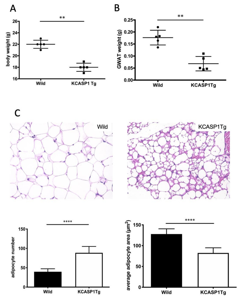
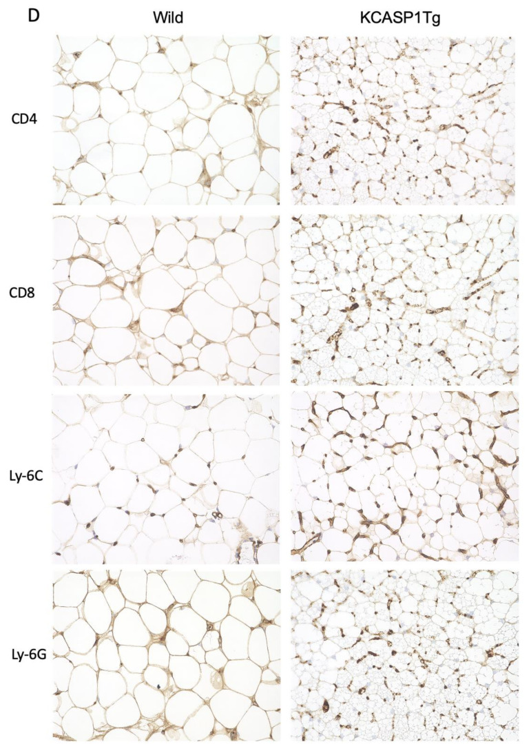
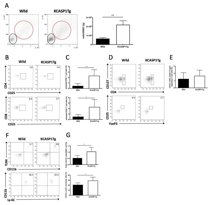
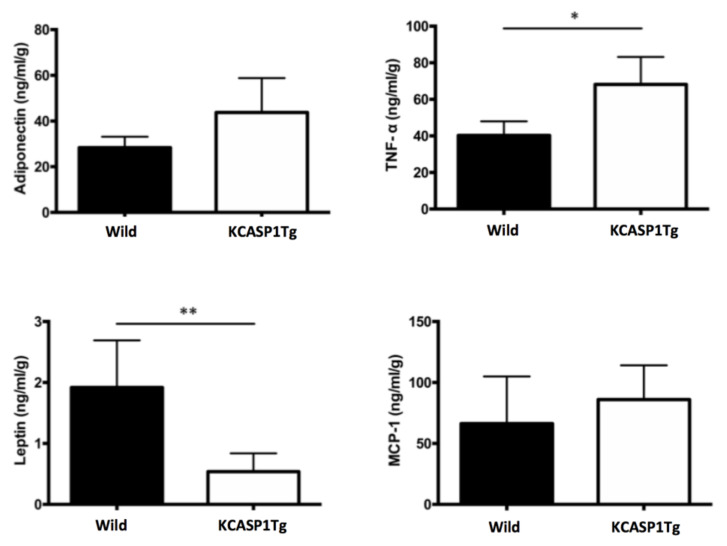
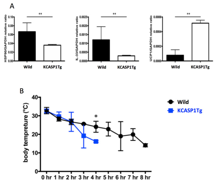
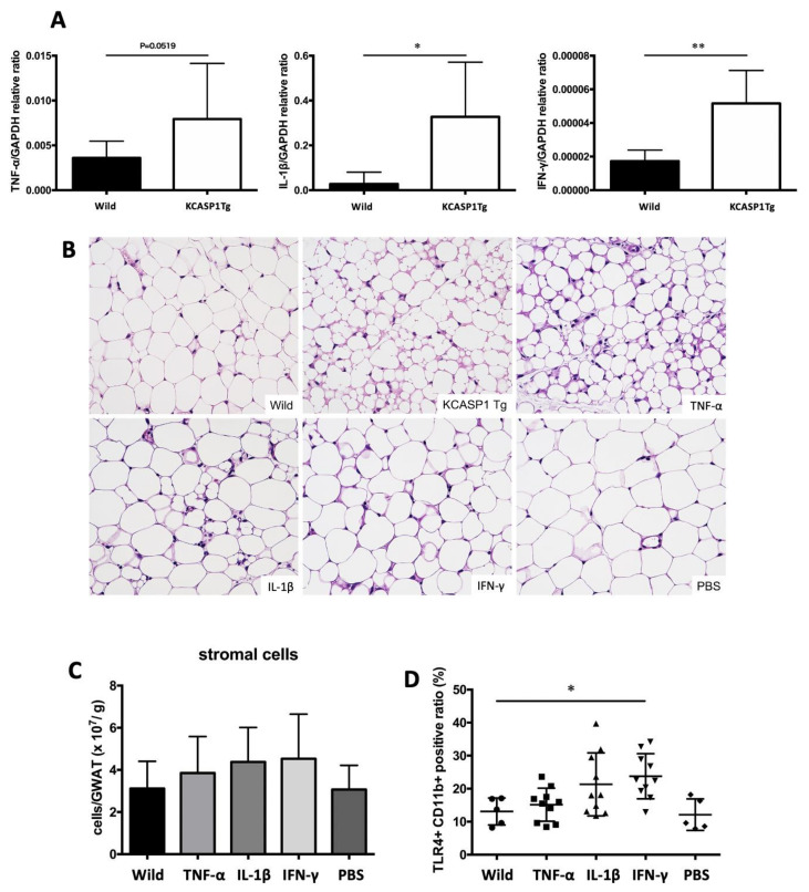
: Adipose tissue (AT) is the largest endocrine organ, producing bioactive products called adipocytokines, which regulate several metabolic pathways, especially in inflammatory conditions. On the other hand, there is evidence that chronic inflammatory skin disease is closely associated with vascular sclerotic changes, cardiomegaly, and severe systemic amyloidosis in multiple organs. In psoriasis, a common chronic intractable inflammatory skin disease, several studies have shown that adipokine levels are associated with disease severity. Chronic skin disease is also associated with metabolic syndrome, including abnormal tissue remodeling; however, the mechanism is still unclear. We addressed this problem using keratin 14-specific caspase-1 overexpressing transgenic (KCASP1Tg) mice with severe erosive dermatitis from 8 weeks of age, followed by re-epithelization. The whole body and gonadal white AT (GWAT) weights were decreased. Each adipocyte was large in number, small in size and irregularly shaped; abundant inflammatory cells, including activated CD4+ or CD8+ T cells and toll-like receptor 4/CD11b-positive activated monocytes, infiltrated into the GWAT. We assumed that inflammatory cytokine production in skin lesions was the key factor for this lymphocyte/monocyte activation and AT dysregulation. We tested our hypothesis that the AT in a mouse dermatitis model shows an impaired thermogenesis ability due to systemic inflammation. After exposure to 4 °C, the mRNA expression of the thermogenic gene uncoupling protein 1 in adipocytes was elevated; however, the body temperature of the KCASP1Tg mice decreased rapidly, revealing an impaired thermogenesis ability of the AT due to atrophy. Tumor necrosis factor (TNF)-α, IL-1β and interferon (INF)-γ levels were significantly increased in KCASP1Tg mouse ear skin lesions. To investigate the direct effects of these cytokines, BL/6 wild mice were administered intraperitoneal TNF-α, IL-1β and INF-γ injections, which resulted in small adipocytes with abundant stromal cell infiltration, suggesting those cytokines have a synergistic effect on adipocytes. The systemic dermatitis model mice showed atrophy of AT and increased stromal cells. These findings were reproducible by the intraperitoneal administration of inflammatory cytokines whose production was increased in inflamed skin lesions.

Keywords: adiponectin; adipose tissue; cytokine; leptin; skin inflammation; thermogenesis.


Conflict of interest statement

The authors declare no conflict of interest.

Figures

Figure 1
Figure 1
KCASP1Tg mice showed low gonadal white adipose tissue (GWAT) weights and degenerated adipose tissue (AT). (A) The whole body and (B) GWAT weights were decreased in KCASP1Tg compared to wild littermate mice at 10 weeks of age. (C) Representative photomicrographs of the hematoxylin and eosin (HE) (×400) staining of the GWAT of wild littermate and KCASP1Tg mice at 10 weeks of age. Histological sections were 3.5 µm thick. Adipocyte number and adipocyte area were measured by using ImageJ and Adiposoft (three parts of each slide, n = 3 in each group). Adipocytes were significantly smaller and larger in number in KCASP1Tg. (D) Immunohistochemical staining (×400) was performed to characterize stromal cells. Lymphocytes, monocytes and neutrophils were slightly higher in KCASP1Tg but not significantly so. ** p < 0.01, **** p < 0.0001 versus control.
Figure 1
Figure 1
KCASP1Tg mice showed low gonadal white adipose tissue (GWAT) weights and degenerated adipose tissue (AT). (A) The whole body and (B) GWAT weights were decreased in KCASP1Tg compared to wild littermate mice at 10 weeks of age. (C) Representative photomicrographs of the hematoxylin and eosin (HE) (×400) staining of the GWAT of wild littermate and KCASP1Tg mice at 10 weeks of age. Histological sections were 3.5 µm thick. Adipocyte number and adipocyte area were measured by using ImageJ and Adiposoft (three parts of each slide, n = 3 in each group). Adipocytes were significantly smaller and larger in number in KCASP1Tg. (D) Immunohistochemical staining (×400) was performed to characterize stromal cells. Lymphocytes, monocytes and neutrophils were slightly higher in KCASP1Tg but not significantly so. ** p < 0.01, **** p < 0.0001 versus control.
Figure 2
Figure 2
AT cell infiltration in KCASP1Tg mice. (A) Flow cytometric analysis of infiltrated stromal cells in GWAT. The black and red circles correspond to the lymphocyte and monocyte populations, respectively, in the left figure. Stromal cell infiltration, including lymphocytes and monocytes, was significantly increased in KCASP1Tg mice. (B,C) The population of CD25+ activated lymphocytes was measured in CD4+ or CD8+ lymphocytes. Activated T cells were significantly increased in KCASP1Tg. (D,E) The proportion of regulatory T cells in AT was measured. There was no significant difference between KCASP1Tg and wild mice. (F,G) The proportion of Ly-6C+ CD11b+ and number of TLR4+ CD11b+ activated monocytes were evaluated and increased in the AT of KCASP1Tg mice. n = 5–6 in each group. * p < 0.05, ** p < 0.01 versus control.
Figure 3
Figure 3
Adipokine expression in AT. We quantified adipocytokines such as adiponectin, TNF-α, leptin and MCP-1 from the adipocytes of GWAT by using a specific ELISA kit. The TNF-α level was elevated in KCASP1Tg mice compared to in controls; conversely, the leptin level was decreased in KCASP1Tg (n = 5 in each group). * p < 0.05 and ** p < 0.01 versus control.
Figure 4
Figure 4
mRNA expression of heat-related proteins in GWAT after cold exposure. (A) The mRNA expression of HSP90 and IL-33 was decreased, while that of UCP1 was increased in the adipocytes of KCASP1Tg mice after exposure to 4 °C (n = 5, each group). (B) The body temperature of mice exposed to 4 °C was measured every 1 hour using thermal imaging camera. The body temperature of the KCASP1Tg mice decreased more rapidly in a cold environment (n = 5, each group). * p < 0.05, ** p < 0.01 versus control.
Figure 5
Figure 5
Production of inflammatory cytokines in the skin and the direct effect on AT. (A) The TNFα, IL-1β and INF-γ levels were significantly increased in KCASP1Tg mouse ear skin lesions. (B) Eight-week-old BL/6 wild mice were treated by intraperitoneal injections of TNF-α, IL-1β, IFN-γ (250 µg/kg body weight/each time) or PBS three times per week for two weeks. The AT of TNF-α-treated mice showed a similar appearance to that of KCASP1Tg mice; IL-1β- and IFN-γ-administrated mice also showed a similar trend; adipocytes were large in number, small and irregularly shaped (HE, ×400). (C,D) The number of infiltrating cells in GWAT was counted, and the infiltrating cells were analyzed by flowcytometry. The number of infiltrating cells in GWAT was increased in cytokine-administrated mice, while TLR4+ CD11b+ activated monocytes were significantly increased in IFN-γ-treated mice. n = 5 per group. * p < 0.05, ** p < 0.01 versus control.

Similar articles

Cited by

References

    1. Scherer P.E. Adipose tissue: From lipid storage compartment to endocrine organ. Diabetes. 2006;55:1537–1545. doi: 10.2337/db06-0263. - DOI - PubMed
    1. Gerdes S., Rostami-Yazdi M., Mrowietz U. Adipokines and psoriasis. Exp. Dermatol. 2011;20:81–87. doi: 10.1111/j.1600-0625.2010.01210.x. - DOI - PubMed
    1. Blaton V. How is the metabolic syndrome related to the dyslipidemia? EJIFCC. 2007;18:15–22. doi: 10.11613/BM.2008.003. - DOI - PMC - PubMed
    1. Suganami T., Ogawa Y. Adipose tissue macrophages: Their role in adipose tissue remodeling. J. Leukoc. Biol. 2010;88:33–39. doi: 10.1189/jlb.0210072. - DOI - PubMed
    1. Caton P.W., Evans E.A., Philpott M.P., Hannen R.F. Can the skin make you fat? A role for the skin in regulating adipose tissue function and whole-body glucose and lipid homeostasis. Curr. Opin. Pharmacol. 2017;37:59–64. doi: 10.1016/j.coph.2017.08.011. - DOI - PubMed

MeSH terms