. 2020 May 10;13(9):2189.
doi: 10.3390/ma13092189.

Comparative Surface Morphology, Chemical Composition, and Cytocompatibility of Bio-C Repair, Biodentine, and ProRoot MTA on hDPCs

Affiliations

Comparative Surface Morphology, Chemical Composition, and Cytocompatibility of Bio-C Repair, Biodentine, and ProRoot MTA on hDPCs

James Ghilotti et al. Materials (Basel). .

Abstract

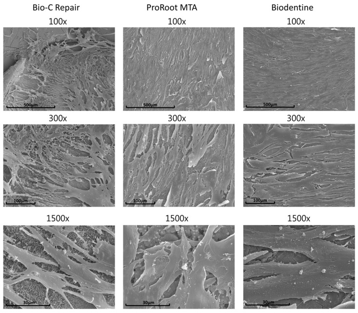
Biocompatibility is an essential property for any vital pulp material that may interact with the dental pulp tissues. Accordingly, this study aimed to compare the chemical composition and ultrastructural morphology of Biodentine (Septodont, Saint Maur-des-Fosses, France), ProRoot MTA (Dentsply Tulsa Dental Specialties, Johnson City, TN, USA), and Bio-C Repair (Angelus, Londrina, PR, Brazil), as well as their biological effects on human dental pulp cells. Chemical element characterization of the materials was undertaken using scanning electron microscopy and energy dispersive X-ray analysis (SEM-EDX). The cytotoxicity was assessed by analyzing the cell viability (MTT assay), cell morphology (immunofluorescence assay), and cell attachment (flow cytometry assay). The results were statistically analyzed using ANOVA and Tukey's test (p < 0.05). EDX revealed that ProRoot MTA and Biodentine were mostly composed of calcium, carbon, and oxygen (among others), whereas Bio-C Repair evidenced a low concentration of calcium and the highest concentration of zirconium. SEM showed adequate attachment of human dental pulp cells (hDPCS) to vital pulp materials and cytoskeletal alterations were not observed in the presence of material eluates. Remarkably, the undiluted Biodentine group showed higher viability than the control group cells (without eluates) at 24 h, 48 h, and 72 h (p < 0.001). Based on the evidence derived from an in vitro cellular study, it was concluded that Bio-C Repair showed excellent cytocompatibility that was similar to Biodentine and ProRoot MTA.

Keywords: calcium silicate materials; cytocompatibility; dental pulp cells; endodontic; vital pulp materials.


Conflict of interest statement

The authors declare no conflict of interest.

Figures

Figure 1
Figure 1
Sample morphology under scanning electron microscopy (SEM) after 1 week of setting at 37 °C and 95% humidity: (A) ProRoot MTA sample, 500×; (B) ProRoot MTA sample, 10,000×; (C) Biodentine sample, 500×; (D) Biodentine sample, 10,000×; (E) Bio-C Repair sample, 500×; and (F) Bio-C Repair sample, 10,000×. The arrows indicate exophytic areas.
Figure 2
Figure 2
Energy dispersive X-ray (EDX) analysis results for the different samples analyzed. (a) Biodentine, (b) ProRoot MTA, (c) Bio-C Repair.
Figure 3
Figure 3
SEM images of human dental pulp cells (hDPCs) cultivated on vital pulp material (Bio-C Repair, ProRoot MTA, and Biodentine) surfaces after 72 h of culture. Magnifications of 100×, 300×, and 1500×. Scale bars: 500 μm, 100 μm, and 30 μm.
Figure 4
Figure 4
Relative formazan formation of hDPCs that were exposed to Biodentine, ProRoot MTA, and Bio-C Repair eluates, as well as the culture medium (control) for 24, 48, and 72 h. (a): Non–diluted, (b) Diluted 1:2, (c) Diluted 1:4. The bar heights represent the mean values and the line extensions represent the standard deviations. Significant differences compared to the control are marked with an *, where * means p < 0.05, and *** means p < 0.001.
Figure 5
Figure 5
Confocal analysis was used to analyze changes in the actin cytoskeleton of hDPCs exposed to material eluates. The confocal microscopy images show F-actin labeling with CruzFluor 594-conjugated phalloidin (red) and nuclei with DAPI (blue). Scale bar: 100 μm.

References

    1. Shah D., Lynd T., Ho D., Chen J., Vines J., Jung H.-D., Kim J.-H., Zhang P., Wu H., Jun H.-W., et al. Pulp–Dentin Tissue Healing Response: A Discussion of Current Biomedical Approaches. J. Clin. Med. 2020;9:434. doi: 10.3390/jcm9020434. - DOI - PMC - PubMed
    1. Hashemi-Beni B., Khoroushi M., Foroughi M.R., Karbasi S., Khademi A. Tissue engineering: Dentin – pulp complex regeneration approaches (A review) Tissue Cell. 2017;49:552–564. doi: 10.1016/j.tice.2017.07.002. - DOI - PubMed
    1. Shi X., Mao J., Liu Y. Pulp stem cells derived from human permanent and deciduous teeth: Biological characteristics and therapeutic applications. Stem Cells Transl. Med. 2020;9:445–464. doi: 10.1002/sctm.19-0398. - DOI - PMC - PubMed
    1. DClinDent B.K., Ricucci D., Saoud T.M., Sigurdsson A., Kahler B. Vital pulp therapy of mature permanent teeth with irreversible pulpitis from the perspective of pulp biology. Aust. Endod. J. 2019;46:154–166. doi: 10.1111/aej.12392. - DOI - PubMed
    1. Spagnuolo G., Codispoti B., Marrelli M., Rengo C., Rengo S., Tatullo M. Commitment of Oral-Derived Stem Cells in Dental and Maxillofacial Applications. Dent. J. 2018;6:72. doi: 10.3390/dj6040072. - DOI - PMC - PubMed