Review
. 2020 May 10;21(9):3369.
doi: 10.3390/ijms21093369.

Autophagy in Neurodegenerative Diseases: A Hunter for Aggregates

Affiliations
Review

Autophagy in Neurodegenerative Diseases: A Hunter for Aggregates

Hyungsun Park et al. Int J Mol Sci. .

Abstract

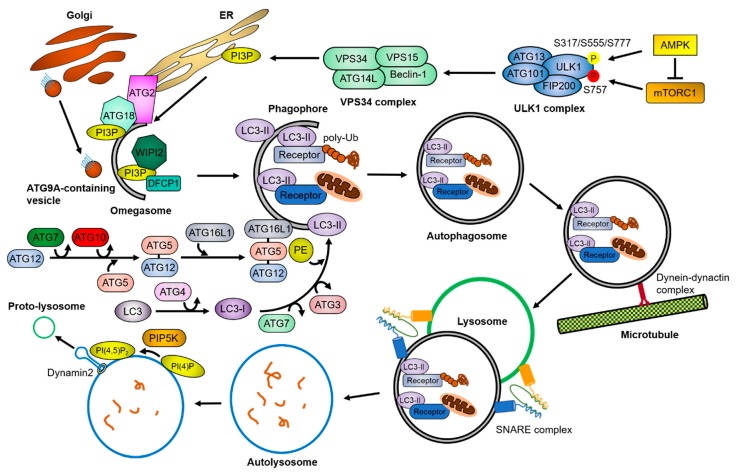
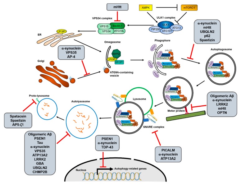
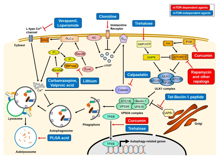
Cells have developed elaborate quality-control mechanisms for proteins and organelles to maintain cellular homeostasis. Such quality-control mechanisms are maintained by conformational folding via molecular chaperones and by degradation through the ubiquitin-proteasome or autophagy-lysosome system. Accumulating evidence suggests that impaired autophagy contributes to the accumulation of intracellular inclusion bodies consisting of misfolded proteins, which is a hallmark of most neurodegenerative diseases. In addition, genetic mutations in core autophagy-related genes have been reported to be linked to neurodegenerative diseases, such as Alzheimer's disease, Parkinson's disease, and Huntington's disease. Conversely, the pathogenic proteins, such as amyloid β and α-synuclein, are detrimental to the autophagy pathway. Here, we review the recent advances in understanding the relationship between autophagic defects and the pathogenesis of neurodegenerative diseases and suggest autophagy induction as a promising strategy for the treatment of these conditions.

Keywords: autophagy; neurodegenerative disease; protein aggregates.


Conflict of interest statement

The authors declare that they have no conflict of interest.

Figures

Figure 1
Figure 1
Overview of the autophagy pathway. Upon cellular stresses, mTORC1 and AMPK phosphorylates the ULK1 complex and the VPS34 complex sequentially (P in yellow circle, activation; P in red circle, inhibition). The VPS34 complex generates PI3P in lipid-enriched membranes, which recruit PI3P-binding proteins. This lipid–protein complex facilitates the assembly of autophagy-related proteins, resulting in the formation of omegasomes. During the growth of the omegasomes to the autophagosomes, the ubiquitin-like conjugation systems of ATG proteins convert LC3 to LC3-II by conjugating PE, and the autophagic receptors and adaptor proteins transport cargoes to the autophagosomes. The matured autophagosomes are transported near the lysosomes by a dynein-dynactin complex, where they form the autolysosomes through the SNARE complex. After the fusion, lysosomal proteases degrade the cargoes. Meanwhile, PI(4,5)P2 builds lysosomal tubule formation sites, generating the proto-lysosomes required for maintaining the free-lysosome pool in the cytosol. The detailed mechanisms are described in Section 1.
Figure 2
Figure 2
Interactions between the autophagy process and neurodegenerative disease-associated proteins. Increasing evidence for interactions between autophagy and neurodegenerative diseases suggests that not only the decrease of autophagic activity is one of causes of the diseases, but mutations in disease-associated genes also inhibit autophagy in various stages. The possible interruptions in each autophagic process from initiation to reformation of free lysosomes by the different neurodegenerative disease-associated mutant proteins are shown. The detailed relationships between the mutant proteins and autophagy pathway are described in Section 2.
Figure 3
Figure 3
Mechanisms of therapeutics of autophagy-inducing agents. Treatment of various autophagy-inducing agents enhances the clearance of aggregate-prone proteins in an mTOR-dependent or -independent manner. The mTOR-dependent autophagy inducers (red), rapamycin and curcumin, directly inhibit mTORC1 activity, resulting in activation of the ULK1 complex. Conversely, mTOR-independent agents (blue) upregulate autophagy activity through various intracellular signaling cascade or lysosome biogenesis. The simplified actions of the agents are drawn, and detailed mechanisms are described in Section 3. AC = adenylyl cyclase, PLC-ε = phospholipase C-epsilon, DAG = diacylglycerol.

Similar articles

Cited by

References

    1. Di Nardo A., Wertz M.H., Kwiatkowski E., Tsai P.T., Leech J.D., Greene-Colozzi E., Goto J., Dilsiz P., Talos D.M., Clish C.B., et al. Neuronal Tsc1/2 complex controls autophagy through AMPK-dependent regulation of ULK1. Hum. Mol. Genet. 2014;23:3865–3874. doi: 10.1093/hmg/ddu101. - DOI - PMC - PubMed
    1. Jang M., Park R., Kim H., Namkoong S., Jo D., Huh Y.H., Jang I.S., Lee J.I., Park J. AMPK contributes to autophagosome maturation and lysosomal fusion. Sci. Rep. 2018;8:12637. doi: 10.1038/s41598-018-30977-7. - DOI - PMC - PubMed
    1. Kim J., Kundu M., Viollet B., Guan K.L. AMPK and mTOR regulate autophagy through direct phosphorylation of Ulk1. Nat. Cell Biol. 2011;13:132–141. doi: 10.1038/ncb2152. - DOI - PMC - PubMed
    1. Lin M.G., Hurley J.H. Structure and function of the ULK1 complex in autophagy. Curr. Opin. Cell Biol. 2016;39:61–68. doi: 10.1016/j.ceb.2016.02.010. - DOI - PMC - PubMed
    1. Turco E., Fracchiolla D., Martens S. Recruitment and Activation of the ULK1/Atg1 Kinase Complex in Selective Autophagy. J. Mol. Biol. 2020;432:123–134. doi: 10.1016/j.jmb.2019.07.027. - DOI - PMC - PubMed

MeSH terms