. 2020 May 10;8(5):701.
doi: 10.3390/microorganisms8050701.

Development and Evaluation of a Single Dye Duplex Droplet Digital PCR Assay for the Rapid Detection and Quantification of Mycobacterium tuberculosis

Affiliations

Development and Evaluation of a Single Dye Duplex Droplet Digital PCR Assay for the Rapid Detection and Quantification of Mycobacterium tuberculosis

Raphael Nyaruaba et al. Microorganisms. .

Abstract

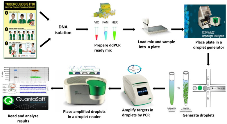
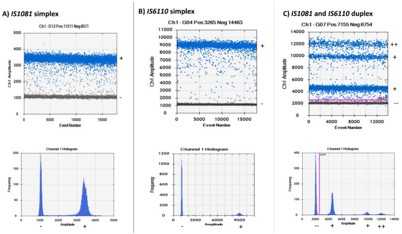
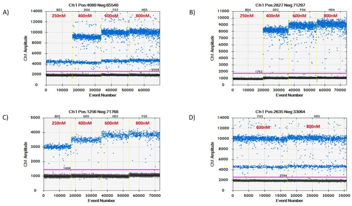
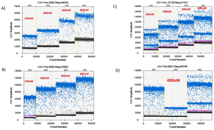
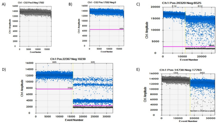
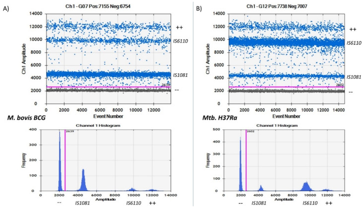
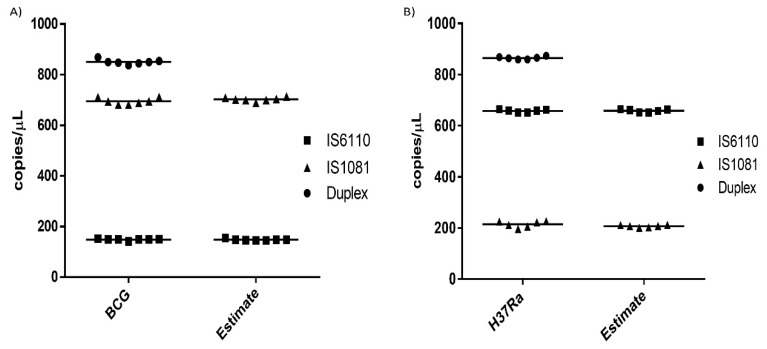
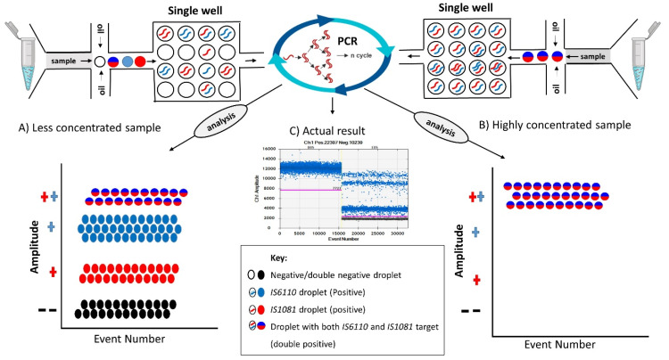
Droplet digital PCR (ddPCR) is a third generation of PCR that was recently developed to overcome the challenges of real-time fluorescence-based quantitative PCR (qPCR) in absolute quantification of pathogens. Few studies have been done on tuberculosis (TB) detection and quantification using ddPCR despite its many advantages over qPCR. From the few studies, none explores a single dye duplex assay for the detection and quantification of TB. In this study, steps toward developing and evaluating a duplex single dye (FAM) assay for detecting two targets (IS6110 and IS1081) are clearly described using simplex and duplex experiments. To achieve this, various parameters are investigated, including annealing temperature, primer and probe concentration, sensitivity and specificity, sample concentration, and inter/intra-assay variability. From the results, primer and probe concentration, annealing temperature, and sample concentration have an effect on the position and separation of droplets in both simplex and duplex assays. The copies of target genes in a duplex assay can be estimated accurately using the threshold tool with little inter-assay (CV <1%) and intra-assay (CV <6%) variability when compared to simplex assays. The ddPCR assay specificity and sensitivity are both 100% when compared to qPCR. This work shows steps toward the detection and quantification of two targets in a single channel, enabling higher multiplexing to include more targets in future works.

Keywords: Detection; Duplex assay; IS1081; IS6110; Mycobacterium tuberculosis; Quantification; Simplex assay; ddPCR; qPCR.


Conflict of interest statement

The authors declare no conflict of interest.

Figures

Figure 1
Figure 1
Overview of the droplet digital PCR (ddPCR) assay workflow for tuberculosis (TB) detection.
Figure 2
Figure 2
Channel 1 and histogram results for droplet separation based on simplex and duplex assays. (A) Droplet separation based on the IS1081 simplex assay results. Positive droplets were observed at an amplitude of about 3500 while negative droplets were observed at an amplitude of about 1000. (B) Droplet separation based on the IS6110 simplex assay results. Positive droplets were observed at an amplitude of about 9000 while negative droplets were observed at an amplitude of about 1000. (C) Droplet separation based on a duplex assay containing primers for both IS6110 and IS1081 genes. Four droplet partitions observed with double negative droplets at an amplitude of about 2000, IS1081 droplets at an amplitude of about 4500, IS6110 droplets at an amplitude of about 10,000, and double positive droplets at an amplitude of about 12,000. (−) negative droplets, (+) positive droplets, (++) double positive droplets, (− −) double negative droplets.
Figure 3
Figure 3
Primer concentration test of both simplex and duplex assays. (B,C) represent simplex assay results of both IS6110 and IS1081 genes, respectively, using various primer concentrations ranging from 250 nM to 800 nM. (A) is a duplex assay result containing both genes in a single reaction well. (D) is the optimal concentration result to be used in subsequent experiments.
Figure 4
Figure 4
Simplex and duplex assay probe concentration (250 nM, 400 nM, 600 nM, and 800 nM) test results. (A,B) represent simplex assays results of IS1081 and IS6110 genes, respectively, at different probe concentrations ranging from 250 nM to 800 nM. (C) represents duplex assay results. (D) shows the result of the optimal concentration chosen for further experiments.
Figure 5
Figure 5
Droplet separation based on sample concentration. (A,B) represent the results of a highly concentrated sample based on automatic results (A) or manually generated results by thresholding (B) in the QuantaSoft™ software. (C,E) are the results of a simplex assay for high and low concentrated samples. (D) represents the duplex assay result of high (well D05) and low (well E05) concentrated samples.
Figure 6
Figure 6
Histogram and 1D droplet concentrations of IS6110 and IS1081 genes based on M. bovis BCG and Mtb. H37Ra strains. (A) BCG result showing a high droplet concentration of IS1081 gene than IS6110 gene. (B) Mtb. H37Ra result showing a high concentration of IS6110 gene droplets rather than IS1081 gene droplets. In A, a higher concentration of droplets can be clearly observed in the IS1081 gene amplitude in both the histogram and 1D plot when compared to double positive droplets and IS6110 droplet concentrations. For B, the reverse is true. Less droplet concentrations can be observed in the IS1081 and double positive droplets with high concentrations of IS6110 droplets.
Figure 7
Figure 7
Reproducibility of replicate wells. (A) M. bovis BCG reproducibility results with BCG showing the actual duplex and simplex assay results while “Estimate” shows the approximated result calculated using the thresholding tool. (B) Mtb. H37Ra reproducibility results with H37Ra showing the actual duplex and simplex assay results while “Estimate” shows the approximated result calculated using the thresholding tool.
Figure 8
Figure 8
Droplet separation in a single dye duplex ddPCR assay based on sample concentration. (A) Less concentrated samples; droplets are generated and distributed randomly with some droplets containing only a single target (positive droplets), double targets (double positive droplets), and no targets (double negative droplets). After PCR amplification, the distribution of droplets gives four clear partitions where targets can be clearly identified. (B) Highly concentrated samples; each droplet generated here has both targets in a single droplet, meaning all the droplets generated are double positive droplets. PCR amplification will consequently result in highly concentrated double positive droplets that during analysis and reading will exhibit double fluorescence, which automatically results in a single double positive droplet partition.

Similar articles

Cited by

References

    1. WHO Global Tuberculosis Report. [(accessed on 22 March 2019)];2018 Available online: https://www.who.int/tb/publications/global_report/en/
    1. Nyaruaba R., Mwaliko C., Kering K.K., Wei H. Droplet digital PCR applications in the tuberculosis world. Tuberculosis. 2019;117:85–92. doi: 10.1016/j.tube.2019.07.001. - DOI - PubMed
    1. Luo J., Luo M., Li J., Yu J., Yang H., Yi X., Chen Y., Wei H. Rapid direct drug susceptibility testing of Mycobacterium tuberculosis based on culture droplet digital polymerase chain reaction. Int. J. Tuberc. Lung Dis. 2019;23:219–225. doi: 10.5588/ijtld.18.0182. - DOI - PubMed
    1. Li H., Bai R., Zhao Z., Tao L., Ma M., Ji Z., Jian M., Ding Z., Dai X., Bao F., et al. Application of droplet digital PCR to detect the pathogens of infectious diseases. Biosci. Rep. 2018;38:BSR20181170. doi: 10.1042/BSR20181170. - DOI - PMC - PubMed
    1. Kuypers J., Jerome K.R. Applications of Digital PCR for Clinical Microbiology. J. Clin. Microbiol. 2017;55:1621–1628. doi: 10.1128/JCM.00211-17. - DOI - PMC - PubMed

LinkOut - more resources