. 2020 May 10;25(9):2247.
doi: 10.3390/molecules25092247.

Conserved Dynamic Mechanism of Allosteric Response to L-arg in Divergent Bacterial Arginine Repressors

Affiliations

Conserved Dynamic Mechanism of Allosteric Response to L-arg in Divergent Bacterial Arginine Repressors

Saurabh Kumar Pandey et al. Molecules. .

Abstract

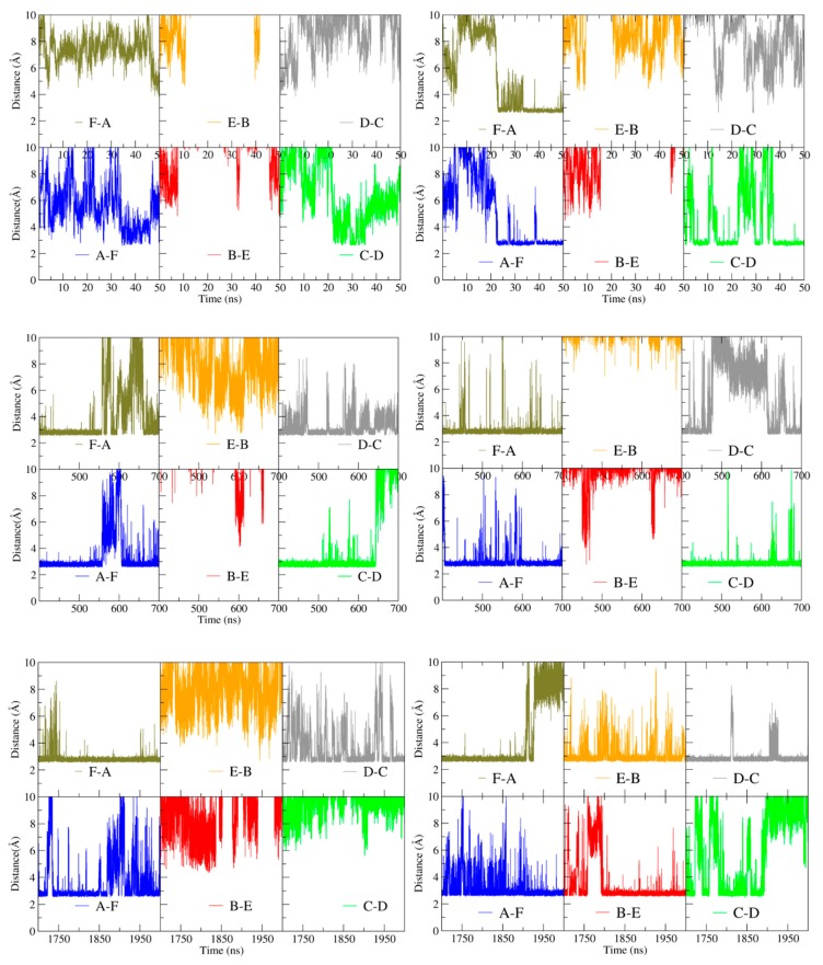
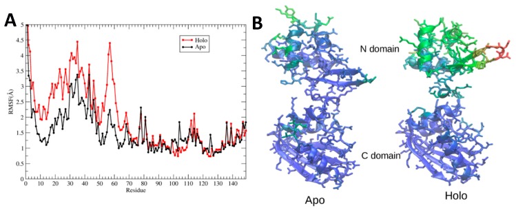
Hexameric arginine repressor, ArgR, is the feedback regulator of bacterial L-arginine regulons, and sensor of L-arg that controls transcription of genes for its synthesis and catabolism. Although ArgR function, as well as its secondary, tertiary, and quaternary structures, is essentially the same in E. coli and B. subtilis, the two proteins differ significantly in sequence, including residues implicated in the response to L-arg. Molecular dynamics simulations are used here to evaluate the behavior of intact B. subtilis ArgR with and without L-arg, and are compared with prior MD results for a domain fragment of E. coli ArgR. Relative to its crystal structure, B. subtilis ArgR in absence of L-arg undergoes a large-scale rotational shift of its trimeric subassemblies that is very similar to that observed in the E. coli protein, but the residues driving rotation have distinct secondary and tertiary structural locations, and a key residue that drives rotation in E. coli is missing in B. subtilis. The similarity of trimer rotation despite different driving residues suggests that a rotational shift between trimers is integral to ArgR function. This conclusion is supported by phylogenetic analysis of distant ArgR homologs reported here that indicates at least three major groups characterized by distinct sequence motifs but predicted to undergo a common rotational transition. The dynamic consequences of L-arg binding for transcriptional activation of intact ArgR are evaluated here for the first time in two-microsecond simulations of B. subtilis ArgR. L-arg binding to intact B. subtilis ArgR causes a significant further shift in the angle of rotation between trimers that causes the N-terminal DNA-binding domains lose their interactions with the C-terminal domains, and is likely the first step toward adopting DNA-binding-competent conformations. The results aid interpretation of crystal structures of ArgR and ArgR-DNA complexes.

Keywords: entropy; global motion; ligand binding; molecular evolution; salt bridges.


Conflict of interest statement

The authors declare no conflict of interest. The funders had no role in the design of the study; in the collection, analyses, or interpretation of data; in the writing of the manuscript; or in the decision to publish the results.

Figures

Figure 1
Figure 1
Structure-based sequence alignment. α-helices (cyan) and β-strands (red) are based on each respective PDB structure (BsArgR, PDB ID: 1F9N; MtArgR, PDB ID: 3FHZ; VvArgR, PDB ID: 3V4G; EcArgR: EcArgRN, PDB ID: 1AOY; EcArgRC, PDB ID: 1XXA). Although EcArgR and VvArgR structures may lack α4, numbering of their secondary structural elements counts the linker region as if α4 is present.
Figure 2
Figure 2
Phylogenetic tree of selected ArgR sequences. The three major branches are colored: green is defined by MtArgR and is closest in evolutionary distance to the common ancestor (scale bar, bottom, average number of residue substitutions per position); orange is defined by BsArgR; and yellow is defined by EcArgR. Entries shown in grey represent sequences that belong to the assigned phylogenetic group but lack the corresponding sequence motif.
Figure 3
Figure 3
Model of holoBsArgR. The model was prepared as described in the text by docking six molecules of L-arg in the empty binding sites of the apoBsArgR crystal structure (PDB ID: 1F9N). Monomers are shown in cartoon representation in unique colors and labeled A–F. Atoms of L-arg are shown as spheres with atomic colors and cyan carbons. (Left) Top view down the 32 axis. The central C-terminal domains form two trimers stacked directly atop one another to form a hexameric core, and pairs of peripheral N-terminal DNA-binding domains (CE, green-orange; BF, red-tan; and AD, blue-grey) can be identified. (Right) Side view, rotated from the left panel by 90° about the z-axis and enlarged. This view shows that L-arg ligands are located at the trimer-trimer interface, and N-terminal domain pairs form across the trimer interface, illustrated for the green and orange CE subunits; all remaining subunits are in grey for clarity.
Figure 4
Figure 4
Global rotational shift of BsArgR. (A) Each initial structure (0 time) undergoes a very fast initial rotational shift of ~10 degrees, then evolves by further rotation, reaching a mean value of rotation during the last 500 ns of the equilibrated part of the trajectories of 14 ± 1.4 (mean ± std. deviation) degrees for apoBsArgR and 22 ± 1.3 degrees for holoBsArgR. Rotation angle is calculated as depicted in Figure S6. As described in the text, the variance observed in each time course is uncoordinated motions of each subunit, not rotational oscillation of trimers. (B) Sim 1 represents the equilibrated holoArgR simulation from 1–2 us from panel A but with averaging over a 1-ns window. Sims 2–5 are replicas of that simulation starting from the equilibrated holoBsAgR conformation, each averaged over a 1-ns window.
Figure 5
Figure 5
Inter-trimer interactions. Residues Lys75, Arg78, and Asp82 (in one-letter code) on helix α4 (blue), and their symmetry-equivalent residues on antiparallel helix α4′ from the closest subunit on the other trimer (tan), are shown as stick models in atomic colors with cyan carbons. Dashed lines show all hydrogen-bonded salt bridges that form during the simulation, although not all are within hydrogen-bonding distance simultaneously (as shown in Figure 6); e.g., in this snapshot taken from 1903 ns, the distance between the left-most Lys75–Asp82 pair is longer than hydrogen-bonding distance.
Figure 6
Figure 6
Interactions driving rotation. Left, apoBsArgR; right, holoBsArgR. Colors correspond to subunits in Figure 3. Distance between guanidino nitrogen atoms of Arg78 and carboxylate oxygen atoms of Asp82 is plotted for subunit pairs during three time windows of the rotational time course shown in Figure 4; these distances over the full time course are presented in Figure S2. Letters in lower right of each panel indicate the subunit pairs whose inter-residue distances are measured as identified in Figure 3. The first 50 ns of the simulations is shown in the top panels; 400–700 ns is shown in the middle panels; the final 300 ns is shown in the bottom panels.
Figure 7
Figure 7
L-arginine binding-site interactions. The three subunits that form representative binding site A, shown, are colored according to Figure 3. Site A is defined as the binding site adjacent to helix α5 of chain A (blue) and is formed by chains A and C (green) from one trimer and F (tan) from the other trimer. Residues whose sidechain functional groups are within hydrogen-bonding distance of the ligand are shown as sticks with atomic colors and black carbons. Residue numbers (one-letter code) are colored according to their subunit of origin. L-arg is shown in CPK spheres with atomic colors and cyan carbons. The position of the Gly107 alpha carbon is indicated by an orange sphere.
Figure 8
Figure 8
Domain movements upon L-arg binding. (A) RMSF was calculated for all non-hydrogen atoms during the final 500 ns of each simulation and averaged over the six monomers of the hexamer. (B) RMSF calculated for representative monomer F was converted to crystallographic B-factors (see Methods) and colored from lowest (blue) to highest (red).
Figure 9
Figure 9
Distance between N-terminal domain pairs. The distance between Cα atoms of residue Arg43 in neighboring N-terminal domains was calculated during the time course of the simulations. The background color of each panel corresponds to one monomer of one trimer (A, B, or C; blue, red, or green) as in Figure 3. The colored lines on each panel indicate the N-terminal domain from the other trimer (D, E, or F; grey, orange, or tan) as in Figure 3. Left six panels, apoBsArgR; right six panels, holoBsArgR.

References

    1. Maas W.K., Clark A.J. Studies on the mechanism of repression of arginine biosynthesis in Escherichia coli. II. Dominance of repressibility in diploids. J. Mol. Biol. 1964;8:365–370. doi: 10.1016/S0022-2836(64)80200-X. - DOI - PubMed
    1. Maas W.K. The arginine repressor of Escherichia coli. Microbiol. Rev. 1994;58:631–640. doi: 10.1128/MMBR.58.4.631-640.1994. - DOI - PMC - PubMed
    1. Van Duyne G.D., Ghosh G., Maas W.K., Sigler P.B. Structure of the oligomerization and L-arginine binding domain of the arginine repressor of Escherichia coli. J. Mol. Biol. 1996;256:377–391. doi: 10.1006/jmbi.1996.0093. - DOI - PubMed
    1. Ni J., Sakanyan V., Charlier D., Glansdorff N., Van Duyne G.D. Structure of the arginine repressor from Bacillus stearothermophilus. Nat. Struct. Biol. 1999;6:427–432. - PubMed
    1. Dennis C.A., Glykos N.M., Parsons M.R., Phillips S.E.V. The structure of AhrC, the arginine repressor/activator protein from Bacillus subtilis. Acta. Cryst. D. 2002;58:421–430. doi: 10.1107/S0907444901021692. - DOI - PubMed

MeSH terms

LinkOut - more resources