Case Reports
. 2020 May 12;13(1):76.
doi: 10.1186/s13041-020-00612-6.

Endoplasmic reticulum retention and degradation of a mutation in SLC6A1 associated with epilepsy and autism

Affiliations
Case Reports

Endoplasmic reticulum retention and degradation of a mutation in SLC6A1 associated with epilepsy and autism

Jie Wang et al. Mol Brain. .

Abstract

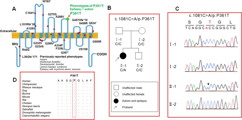
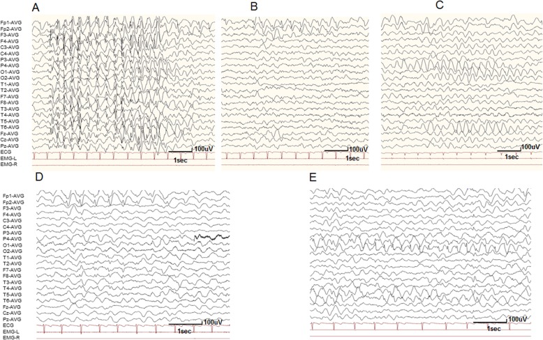
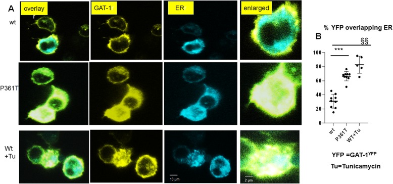
Mutations in SLC6A1, encoding γ-aminobutyric acid (GABA) transporter 1 (GAT-1), have been recently associated with a spectrum of epilepsy syndromes, intellectual disability and autism in clinic. However, the pathophysiology of the gene mutations is far from clear. Here we report a novel SLC6A1 missense mutation in a patient with epilepsy and autism spectrum disorder and characterized the molecular defects of the mutant GAT-1, from transporter protein trafficking to GABA uptake function in heterologous cells and neurons. The heterozygous missense mutation (c1081C to A (P361T)) in SLC6A1 was identified by exome sequencing. We have thoroughly characterized the molecular pathophysiology underlying the clinical phenotypes. We performed EEG recordings and autism diagnostic interview. The patient had neurodevelopmental delay, absence epilepsy, generalized epilepsy, and 2.5-3 Hz generalized spike and slow waves on EEG recordings. The impact of the mutation on GAT-1 function and trafficking was evaluated by 3H GABA uptake, structural simulation with machine learning tools, live cell confocal microscopy and protein expression in mouse neurons and nonneuronal cells. We demonstrated that the GAT-1(P361T) mutation destabilizes the global protein conformation and reduces total protein expression. The mutant transporter protein was localized intracellularly inside the endoplasmic reticulum (ER) with a pattern of expression very similar to the cells treated with tunicamycin, an ER stress inducer. Radioactive 3H-labeled GABA uptake assay indicated the mutation reduced the function of the mutant GAT-1(P361T), to a level that is similar to the cells treated with GAT-1 inhibitors. In summary, this mutation destabilizes the mutant transporter protein, which results in retention of the mutant protein inside cells and reduction of total transporter expression, likely via excessive endoplasmic reticulum associated degradation. This thus likely causes reduced functional transporter number on the cell surface, which then could cause the observed reduced GABA uptake function. Consequently, malfunctioning GABA signaling may cause altered neurodevelopment and neurotransmission, such as enhanced tonic inhibition and altered cell proliferation in vivo. The pathophysiology due to severely impaired GAT-1 function may give rise to a wide spectrum of neurodevelopmental phenotypes including autism and epilepsy.

Keywords: 3H GABA uptake; Autism; Degradation; Endoplasmic reticulum; Epilepsy; GABA transporter 1; Mutation; Protein stability.


Conflict of interest statement

The authors declare that they are no competing interests.

Figures

Fig. 1
Fig. 1
GABA transporter 1 (GAT-1) protein topology, mutations and identification of a novel SLC6A1 missense mutation GAT1(P361T). a. Schematic representation of GAT-1 protein topology and locations of GAT-1 variants previously identified in patients associated with a spectrum of epilepsy syndromes. It is predicted that GAT-1 contains 12 transmembrane domains. P361 is located at the extracellular loop between the 7th and 8th transmembrane helices of the GAT-1 protein. The positions of variants are based on the published LeuT crystal structure. b Pedigree and the genotype. A missense mutation was only found in the proband but not in the rest of the family members. c Chromatogram of PCR-Sanger sequencing. DNA sequences of the proband and the immediate family members were shown. Arrow indicated a C-to-A transversion. d Amino acid sequence homology shows that proline (P) at residue 361 is highly conserved in SCL6A1 in humans (Accession NO.NP_003033.3) and across species as shown in boxed region
Fig. 2
Fig. 2
Modeling of the mutant GAT-1 protein with machine learning tools. a-b. Tertiary structures of both the wildtype (a) and P361T mutant (b) GAT-1 protein are predicted by I-TASSER and DynaMut. The proline at residue 361 is mutated to threonine, both highlighted in light green, alongside with the surrounding residues. The interatomic interactions were predicted by DynaMut, where halogen bonds are depicted in blue and hydrogen bonds are colored in red. The P361T mutation results in the loss of two hydrogen bonds, those between residues 361 and 365 (yellow arrow with red border) and between 361 and 364 (yellow arrow with blue border). This supports the result in Table 1 that this mutation destabilized the global conformation of the GAT-1 protein. c. Machine learning tools predicted ΔΔG (Kcal/mol) of the mutant GAT-1 protein. Bars in the positive direction are predicted as stabilizing while bars in the negative direction are predicted as destabilizing
Fig. 3
Fig. 3
Electroencephalogram (EEG) of a 6-year-old girl carrying GAT-1(P361T) mutation. Interictal video EEG recordings showed 2.5–3.0 Hz generalized spike and slow waves (a), 2.0–3.0 Hz spike and slow waves in the bilateral prefrontal lobes (b) and 2.0–3.0 Hz slow waves predominantly in the bilateral occipital area (c) during both wakefulness and sleep when the patient was 3.5 years old. Interictal video EEG recordings demonstrated 2.0–3.0 Hz spike and slow waves in the bilateral prefrontal lobes (d), and 2.0–3.0 Hz spike and slow waves predominantly in the bilateral occipital and posterior-temporal area (e) during both wakefulness and sleep when the patient was 6 years old
Fig. 4
Fig. 4
The expression of the mutant of GAT-1(P361T) protein was reduced in non-neuronal cells and neurons. a-b. Mouse cortical neurons were transfected with the wildtype or the mutant GAT-1(P361T) cDNAs at day 7 in culture. The total lysates were harvested from mouse cortical neurons expressing the wildtype GAT-1YFP (wt) or mutant GAT-1(P361T)YFP transporters after 8 days of transfection (a). HeLa cells were transfected with the wildtype GAT-1YFP (wt) or mutant GAT-1(P361T)YFP transporters for 48 h (b). The total lysates were then analyzed by SDS-PAGE. Membranes were immunoblotted with rabbit anti-GAT-1 for both neuronal and HeLa cell lysates (1:200). In neurons, the protein band of endogenous GAT-1, at 67 KDa, was intense. The main protein bands run at 108 KDa in both the wildtype and the mutant conditions, representing the YFP-tagged GAT-1. c. The total protein integrated density values (IDVs) were measured. The abundance of the mutant GAT-1(P361T) transporter was normalized to the wildtype condition. In c, the total protein abundance was measured by adding up all the bands between 90 and 110 KDa. The total protein IDVs of either the wildtype or the mutant was normalized to its loading control. The abundance of the mutant transporter was then normalized to the wildtype. (*p < 0.05 vs wt in HeLa; **p < 0.01 vs. wt in Neuron, n = 4–5 different transfections)
Fig. 5
Fig. 5
There was reduced YFP fluorescence in cells expressing the mutant GAT-1(P361T) transporters, which were retained inside the endoplasmic reticulum. a HEK293T cells were transfected with wildtype GAT-1YFP or the mutant GAT-1(P361T)YFP with the pECFP-ER marker (ERCFP) at 2:1 ratio (2 μg:1 μg cDNAs) for 48 h. Live cells were examined under a confocal microscopy with excitation at 458 nm for CFP, 514 nm for YFP. All images were single confocal sections averaged from 8 times to reduce noise, except when otherwise specified. b The GAT-1YFP fluorescence overlapping with ERCFP fluorescence was quantified by Metamorph with colocalization percentage. (***p < 0.001 P361T vs. wt, §§ p < 0.01 wt + Tunicamycin vs wt untreated, n = 5–9 representative fields from different transfections)
Fig. 6
Fig. 6
Impaired GABA uptake of the mutant GAT-1(P361T) transporters. (A) HEK293T cells were transfected with wildtype GAT-1YFP (wt), or the mutant GAT-1(P361T)YFP cDNAs (1 μg/35mm2) for 48 h. The GABA uptake assay was carried out with 3H radioactive labeling in HEK 293 T cells. GABA flux was measured after 30 min transport at room temperature. The influx of GABA, expressed in pmol/μg protein/min, was averaged from duplicates for each condition and for each transfection. The average counting was taken as n = 1. The untransfected condition was taken as baseline flux, which was subtracted from both the wild-type and the mutant conditions. The pmol/μg protein/min in the mutant was then normalized to the wildtype from each experiment, which was arbitrarily set as 100%. (**p < 0.01 vs. wt, n = 4–5 different transfections)

References

    1. Abrahams BS, Geschwind DH. Advances in autism genetics: on the threshold of a new neurobiology. Nat Rev Genet. 2008;9:341–355. doi: 10.1038/nrg2346. - DOI - PMC - PubMed
    1. Allen AS, Berkovic SF, Cossette P, Delanty N, Dlugos D, Eichler EE, Epstein MP, Glauser T, Goldstein DB, Han Y, Heinzen EL, Hitomi Y, Howell KB, Johnson MR, Kuzniecky R, Lowenstein DH, Lu YF, Madou MR, Marson AG, Mefford HC, Esmaeeli NS, O'Brien TJ, Ottman R, Petrovski S, Poduri A, Ruzzo EK, Scheffer IE, Sherr EH, Yuskaitis CJ, Abou-Khalil B, Alldredge BK, Bautista JF, Berkovic SF, Boro A, Cascino GD, Consalvo D, Crumrine P, Devinsky O, Dlugos D, Epstein MP, Fiol M, Fountain NB, French J, Friedman D, Geller EB, Glauser T, Glynn S, Haut SR, Hayward J, Helmers SL, Joshi S, Kanner A, Kirsch HE, Knowlton RC, Kossoff EH, Kuperman R, Kuzniecky R, Lowenstein DH, McGuire SM, Motika PV, Novotny EJ, Ottman R, Paolicchi JM, Parent JM, Park K, Poduri A, Scheffer IE, Shellhaas RA, Sherr EH, Shih JJ, Singh R, Sirven J, Smith MC, Sullivan J, Lin TL, Venkat A, Vining EP, Von Allmen GK, Weisenberg JL, Widdess-Walsh P, Winawer MR. De novo mutations in epileptic encephalopathies. Nature. 2013;501:217–221. doi: 10.1038/nature12439. - DOI - PMC - PubMed
    1. Andang M, Hjerling-Leffler J, Moliner A, Lundgren TK, Castelo-Branco G, Nanou E, Pozas E, Bryja V, Halliez S, Nishimaru H, Wilbertz J, Arenas E, Koltzenburg M, Charnay P, El MA, Ibanez CF, Ernfors P. Histone H2AX-dependent GABA(a) receptor regulation of stem cell proliferation. Nature. 2008;451:460–464. doi: 10.1038/nature06488. - DOI - PubMed
    1. Ben-Ari Y. Excitatory actions of gaba during development: the nature of the nurture. Nat Rev Neurosci. 2002;3:728–739. doi: 10.1038/nrn920. - DOI - PubMed
    1. Bennett ER, Kanner BI. The membrane topology of GAT-1, a (Na+ + cl-)-coupled gamma-aminobutyric acid transporter from rat brain. J Biol Chem. 1997;272:1203–1210. doi: 10.1074/jbc.272.2.1203. - DOI - PubMed

Publication types

MeSH terms