. 2020 May 12;20(1):112.
doi: 10.1186/s12866-020-01807-6.

Morin inhibits Listeria monocytogenes virulence in vivo and in vitro by targeting listeriolysin O and inflammation

Affiliations

Morin inhibits Listeria monocytogenes virulence in vivo and in vitro by targeting listeriolysin O and inflammation

Gen Li et al. BMC Microbiol. .

Abstract

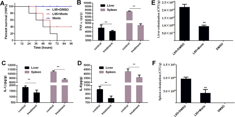
Background: Listeria monocytogenes (L. monocytogenes) is a global opportunistic intracellular pathogen that can cause many infections, including meningitis and abortion in humans and animals; thus, L. monocytogenes poses a great threat to public safety and the development of the aquaculture industry. The isolation rate of Listeria monocytogenes in fishery products has always been high. And the pore-forming toxin listeriolysin O (LLO) is one of the most important virulence factors of L. monocytogenes. LLO can promote cytosolic bacterial proliferation and help the pathogen evade attacks from the host immune system. In addition, L. monocytogenes infection can trigger a series of severe inflammatory reactions.

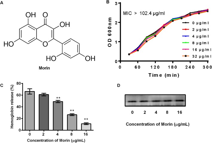
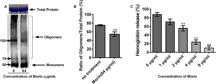
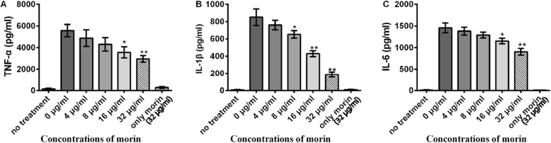
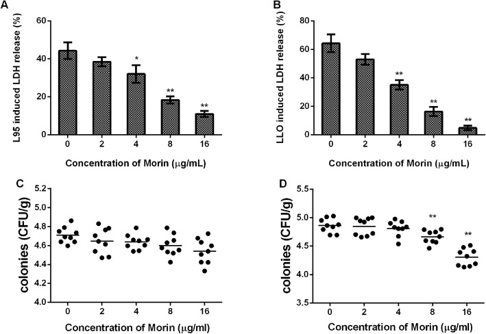
Results: Here, we further confirmed that morin lacking anti-Listeria activity could inhibit LLO oligomerization. We also found that morin can effectively alleviate the inflammation induced by Listeria in vivo and in vitro and exerted an obvious protective effect on infected cells and mice.

Conclusions: Morin does not possess anti-Listeria activity, neither does it interfere with secretion of LLO. However, morin inhibits oligomerisation of LLO and morin does reduce the inflammation caused during Listeria infection.

Keywords: Anti-infection; Inflammation; Listeria monocytogenes; Listeriolysin O,morin.


Conflict of interest statement

The authors declare that they have no conflicts of interest.

Figures

Fig. 1
Fig. 1
Inhibition of L. monocytogenes culture by morin. a Chemical structure of morin. b Growth kinetics curve of L95 cells treated with different concentrations of morin. L95 cells were incubated in TSB in the presence of various dosages of morin, and the growth of each sample was monitored every 30 min by measuring the value of OD600 nm at each indicated time point. c Suppression of the hemolytic activity of L95 culture supernatants by morin. L95 cells were co-cultured with morin, and the co-cultured supernatants were harvested by centrifugation. The hemolytic activity was assessed by a hemolysis assay. The hemolysis percent of each co-cultured supernatant was compared with that of the positive control group (treatment with Triton X-100). d The expression of LLO in L. monocytogenes cultures treated with different concentrations of morin was assessed by Western blotting using a specific antibody against LLO. Three independent trials were performed for each index. * indicates P < 0.05, and ** indicates P < 0.01. (Student’s t-test)
Fig. 2
Fig. 2
Inhibitory effects of morin on oligomer formation and the hemolytic activity of LLO. a and b Purified LLO proteins were treated with morin, and the inhibition of LLO oligomerization by morin was assessed by Western blotting. The total protein level in the groups without oligomerization induction was determined by coomassie blue staining. c Purified LLO protein was co-incubated with morin in PBS, and the co-cultured systems were harvested by centrifugation. The hemolytic activity was assessed by a hemolysis assay. Three independent trials were performed for each index.* indicates P < 0.05, and ** indicates P < 0.01. (Student’s t-test)
Fig. 3
Fig. 3
Inhibition of the production of inflammatory cytokines by morin in cells infected with L. monocytogenes. J774 macrophage-like cells were co-cultured with L. monocytogenes in the presence or absence of morin (0, 8, 16, and 32 μg/mL) at an MOI of 10 for 5 h. The co-culture supernatants were harvested by centrifugation, and the levels of TNF-α (a), IL-1β (b) and IL-6 (c) were detected by ELISA. Cells treated with morin in the absence of L. monocytogenes were used as a control. Three independent trials were performed for each index.* indicates P < 0.05, and ** indicates P < 0.01. (Student’s t-test)
Fig. 4
Fig. 4
Morin-mediated attenuation of cell damage after infection with L95 cells or treatment with LLO. The level of LDH released into the supernatants of cells infected with L95 cells (a) or treated with LLO (b) in the presence or absence of various concentrations of morin was detected using a cytotoxicity detection kit. The extracellular (c) and intracellular (d) colonies were calculated by colonization. Three independent trials were performed for each index. * indicates P < 0.05, and ** indicates P < 0.01. (Student’s t-test)
Fig. 5
Fig. 5
Protective effect of morin against L. monocytogenes infection in vivo. a Mice were intraperitoneally injected with 1 × 107 CFUs of L95 cells and treated with 100 mg/kg morin or DMSO 2 h after infection and at 8-h intervals. The survival rate of the infected mice was observed for 96 h. The mice were intraperitoneally injected with 2 × 106 CFUs of L95 cells and treated with 100 mg/kg morin or DMSO 2 h after infection and again at 8-h intervals. Forty-eight hours after infection, all of the mice were euthanized, the tissues of the liver and spleen were homogenized, and the number of colonies in ground tissue was calculated viaserial dilution (e-f). The levels of IL-6, IL-1β and TNF-α were detected by ELISA (b-d). Ten mice were arranged to each group for survival assays and three independent trials were performed (30 in total). For the inflammation and burden of the bacteria assays three mice were arranged to each group and three independent trials were performed (9 in total).* indicates P < 0.05, and ** indicates P < 0.01. (Student’s t-test)

Similar articles

Cited by

References

    1. Pensinger DA, Aliota MT, Schaenzer AJ, Boldon KM, Ansari IU, Vincent WJB, Knight B, Reniere ML, Striker R, Sauer JD. Selective pharmacologic inhibition of a PASTA kinase increases Listeria monocytogenes susceptibility to beta-lactam antibiotics. Antimicrob Agents Chemother. 2014;58(8):4486–4494. - PMC - PubMed
    1. Cossart P. Illuminating the landscape of host-pathogen interactions with the bacterium Listeria monocytogenes. Proc Natl Acad Sci U S A. 2011;108(49):19484–19491. - PMC - PubMed
    1. Jordan K, McAuliffe O. Listeria monocytogenes in foods. Adv Food Nutr Res. 2018;86:181–213. - PubMed
    1. Scallan E, Hoekstra RM, Angulo FJ, Tauxe RV, Widdowson MA, Roy SL, Jones JL, Griffin PM. Foodborne illness acquired in the United States-major pathogens. Emerg Infect Dis. 2011;17(1):7–15. - PMC - PubMed
    1. Williams MA, Schmidt RL, Lenz LL. Early events regulating immunity and pathogenesis during Listeria monocytogenes infection. Trends Immunol. 2012;33(10):488–495. - PMC - PubMed

Publication types

MeSH terms