. 2020 May 12;8(1):67.
doi: 10.1186/s40478-020-00936-3.

Behavioral and electrophysiological evidence for a neuroprotective role of aquaporin-4 in the 5xFAD transgenic mice model

Affiliations

Behavioral and electrophysiological evidence for a neuroprotective role of aquaporin-4 in the 5xFAD transgenic mice model

Yoichiro Abe et al. Acta Neuropathol Commun. .

Abstract

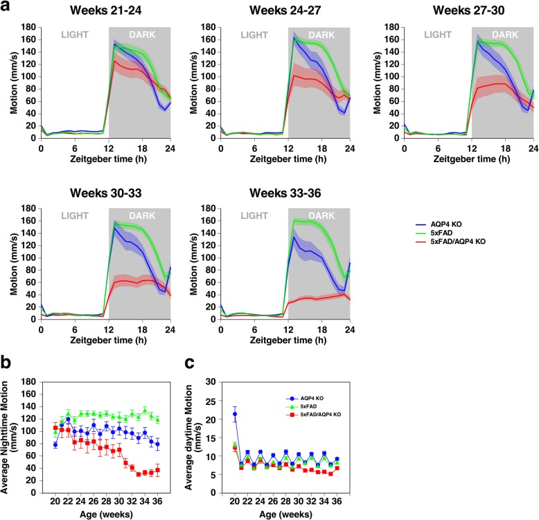
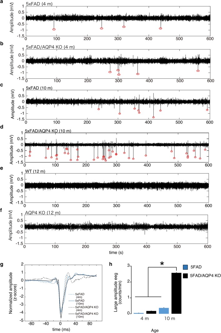
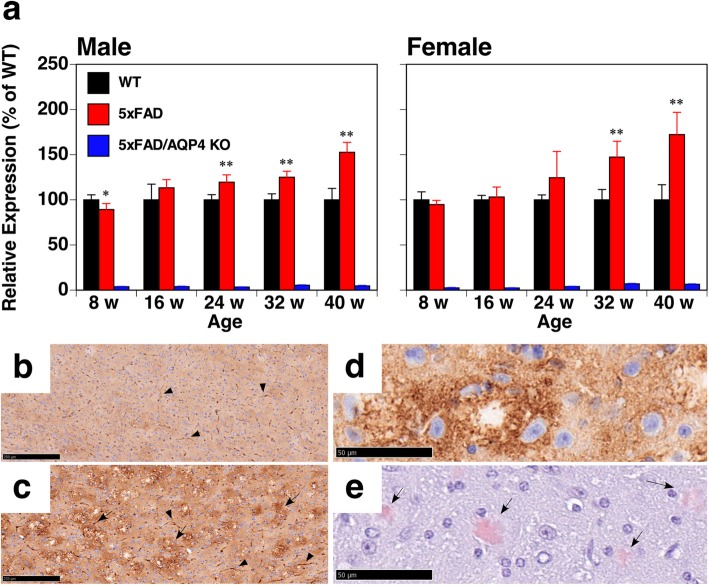
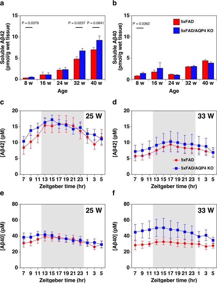
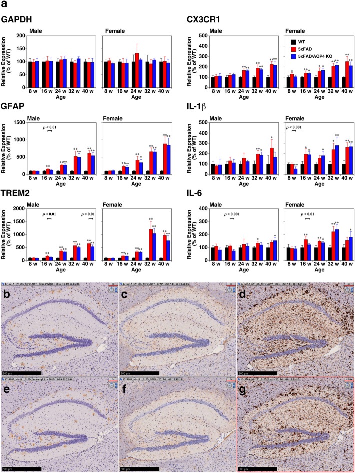
Aquaporin-4 (AQP4) has been suggested to be involved in the pathogenesis of neurodegenerative diseases including Alzheimer's disease (AD), which may be due to the modulation of neuroinflammation or the impairment of interstitial fluid bulk flow system in the central nervous system. Here, we show an age-dependent impairment of several behavioral outcomes in 5xFAD AQP4 null mice. Twenty-four-hour video recordings and computational analyses of their movement revealed that the nighttime motion of AQP4-deficient 5xFAD mice was progressively reduced between 20 and 36 weeks of age, with a sharp deterioration occurring between 30 and 32 weeks. This reduction in nighttime motion was accompanied by motor dysfunction and epileptiform neuronal activities, demonstrated by increased abnormal spikes by electroencephalography. In addition, all AQP4-deficient 5xFAD mice exhibited convulsions at least once during the period of the analysis. Interestingly, despite such obvious phenotypes, parenchymal amyloid β (Aβ) deposition, reactive astrocytosis, and activated microgliosis surrounding amyloid plaques were unchanged in the AQP4-deficient 5xFAD mice relative to 5xFAD mice. Taken together, our data indicate that AQP4 deficiency greatly accelerates an age-dependent deterioration of neuronal function in 5xFAD mice associated with epileptiform neuronal activity without significantly altering Aβ deposition or neuroinflammation in this mouse model. We therefore propose that there exists another pathophysiological phase in AD which follows amyloid plaque deposition and neuroinflammation and is sensitive to AQP4 deficiency.

Keywords: 5xFAD; Alzheimer’s disease; Amyloid β; Aquaporin-4; Epilepsy.


Conflict of interest statement

Masato Yasui has received funding for the research from Suntory Global Innovation Center Ltd.

The other authors declare that they have no competing interests.

Figures

Fig. 1
Fig. 1
Effect of AQP4 deficiency on the activity of 5xFAD female mice. a Average circadian motion of 5xFAD (green), AQP4 KO (blue), and 5xFAD/AQP4 KO (red) mice. b and c Average nighttime (b) and daytime (c) motions of 5xFAD (green triangles), AQP4 KO (blue circles), and 5xFAD/AQP4 KO (red squares) mice. Mice became active during the daytime after cage change occurred every 2 weeks. Values are mean ± S.E.M of 11 individuals
Fig. 2
Fig. 2
Electroencephalography of 5xFAD female mice. a-f Representative electroencephalographic (EEG) traces from 4-month-old 5xFAD (a), 4-month-old 5xFAD/AQP4 KO (b), 10-month-old 5xFAD (c), 10-month-old 5xFAD/AQP4 KO (d), 12-month-old wild-type (e), and 12-month-old AQP4 KO (f) mice. Red circles indicate epileptiform activity. g Mean traces of epileptiform spike waveforms from 5xFAD (4 months), 5xFAD/AQP4 KO (4 months), 5xFAD (10 months), and 5xFAD/AQP4 KO (10 months) mice. h Mean epileptiform spike counts per minute were obtained from 5xFAD (4 months, n = 5), 5xFAD/AQP4 KO (4 months, n = 5), 5xFAD (10 months, n = 6), and 5xFAD/AQP4 KO (10 months, n = 10) mice. Values are mean ± S.E.M. * (P < 0.01) represents significant differences of 10-month-old 5xFAD/AQP4 KO versus each of the other groups
Fig. 3
Fig. 3
Expression of AQP4 in 5xFAD mice. a qPCR analysis of brain hemispheres from wild-type (black columns), 5xFAD (red columns), and 5xFAD/AQP4 KO (blue columns) mice. Values are mean ± S.E.M of 5–6 individuals. *(P < 0.01) and **(P < 0.001) represent significant differences versus wild-type mice. b and c Comparison of AQP4 expression in the cortices of wild-type (b) and 5xFAD (c) mice. Perivascular staining of AQP4 is denoted by arrow heads. The expression of AQP4 around unstained spots is denoted by arrows. d and e Aberrant expression of AQP4 (d) was observed around amyloid plaques stained with Congo red (e). Scale bars = 250 μm (b and c) and 50 μm (d and e)
Fig. 4
Fig. 4
Effect of AQP4 deficiency on the deposition of Aβ in parenchyma of 5xFAD female mice. a-h Representative immunohistochemical images of sagittal brain sections of 40-week-old 5xFAD/AQP4 KO (a, b, e, and f) and 5xFAD (c, d, g, and h) mice stained with an anti-Aβ42 antibody. b, d, f, and h are magnified images of a, c, e, and g, respectively, indicated by boxes. Scale bars = 1 mm (a, c, e, and g) and 100 μm (b, d, f, and h). i and j Quantification of the number of amyloid plaques and plaque size in the cortical region. The average number of plaques (i) and plaque size (j) in ten fields in two sections of cortical regions from each animal (n = 3), including (a) and (c), were counted using ImageJ. Values are mean ± S.E.M of 3 individuals. k and l Quantification of the number of amyloid plaques of approximately 25 and 100 μm in diameter calculated by Imaris in the 3D imaging of cleared hemispheres from 45-week-old 5xFAD (red columns, Supplemental Movie S5) and 5xFAD/AQP4 KO (blue columns, Supplemental Movie S6) mice stained with Alexa Fluor 488-labeled 6E10 to identify total Aβ (k, green in movies) and Amylo-Glo to identify Aβ fibrils (l, magenta in movies). Values are mean ± S.E.M of 3 individuals. m and n The amounts of insoluble Aβ42 (m) and Aβ40 (n) extracted from the cerebral hemispheres of 5xFAD (red columns) and 5xFAD/AQP4 KO (blue columns) mice were determined by ELISA. Values are mean ± S.E.M of 6–7 individuals
Fig. 5
Fig. 5
Effect of AQP4 deficiency on soluble Aβ levels in 5xFAD female mice. a and b The amounts of soluble Aβ42 (a) and Aβ40 (b) extracted from the cerebral hemispheres of 5xFAD (red columns) and 5xFAD/AQP4 KO (blue columns) mice were determined by ELISA. One individual 32-week-old 5xFAD female mouse showed more than 4.0 and 7.5 times higher soluble Aβ42 and Aβ40 concentrations, respectively, compared with the average of the other seven individuals of the same group. Therefore, this mouse was excluded from all analyses, including insoluble Aβ level shown in Fig. 4m and n. Values are mean ± S.E.M of 6–7 individuals. c-f Time course of ISF-soluble Aβ42 (c and d) and Aβ40 (e and f) concentrations in hippocampal region of 5xFAD (red circles) and 5xFAD/AQP4 KO (blue squares) females at 25 weeks (c and e) and 33 weeks (d and f) of ages measured by ELISA of samples obtained by microdialysis. Values are mean ± S.E.M of 6–9 individuals
Fig. 6
Fig. 6
Effect of AQP4 deficiency on neuroinflammatory responses in 5xFAD mice. a qPCR analysis of brain hemispheres from wild-type (black columns), 5xFAD (red columns), and 5xFAD/AQP4 KO (blue columns) mice. The level of the transcript of each gene in 5xFAD and 5xFAD/AQP4 KO mice was determined as the % of that of age-matched wild-type mice. Values are mean ± S.E.M of 5–6 individuals. *(P < 0.01) and **(P < 0.001) represent significant differences versus wild-type mice. The primers used are listed in Supplemental Table S2. b-g Immunostaining of Aβ (b and e), GFAP (c and f), and Iba1 (d and g) in the hippocampal region of 5xFAD/AQP4 KO (b-d) and 5xFAD (e-g) mice. Scale bars = 500 μm

References

    1. Bezzina C, Verret L, Juan C, Remaud J, Halley H, Rampon C, et al. Early onset of hypersynchronous network activity and expression of a marker of chronic seizures in the Tg2576 mouse model of Alzheimer's disease. PLoS One. 2015;10:e0119910. - PMC - PubMed
    1. Born HA. Seizures in Alzheimer's disease. Neuroscience. 2015;286:251–263. - PubMed
    1. Born HA, Kim JY, Savjani RR, Das P, Dabaghian YA, Guo Q, et al. Genetic suppression of transgenic APP rescues hypersynchronous network activity in a mouse model of Alzeimer's disease. J Neurosci. 2014;34:3826–3840. - PMC - PubMed
    1. Brown R, Lam AD, Gonzalez-Sulser A, Ying A, Jones M, Chou RC, et al. Circadian and brain state modulation of network hyperexcitability in Alzheimer's disease. eNeuro. 2018;5:e0426–e0417. - PMC - PubMed
    1. Carare RO, Bernardes-Silva M, Newman TA, Page AM, Nicoll JA, Perry VH, et al. Solutes, but not cells, drain from the brain parenchyma along basement membranes of capillaries and arteries: significance for cerebral amyloid angiopathy and neuroimmunology. Neuropathol Appl Neurobiol. 2008;34:131–144. - PubMed

Publication types