. 2020 Jul;12(4):608-625.
doi: 10.4168/aair.2020.12.4.608.

Toll-like Receptor 4 Deficiency Aggravates Airway Hyperresponsiveness and Inflammation by Impairing Neutrophil Apoptosis in a Toluene Diisocyanate-Induced Murine Asthma Model

Affiliations

Toll-like Receptor 4 Deficiency Aggravates Airway Hyperresponsiveness and Inflammation by Impairing Neutrophil Apoptosis in a Toluene Diisocyanate-Induced Murine Asthma Model

Shuyu Chen et al. Allergy Asthma Immunol Res. 2020 Jul.

Abstract

Purpose: Accumulating evidence has suggested that toll-like receptor 4 (TLR4) is critically involved in the pathogenesis of asthma. The aim of this study was to investigate the role of TLR4 in toluene diisocyanate (TDI)-induced allergic airway inflammation.

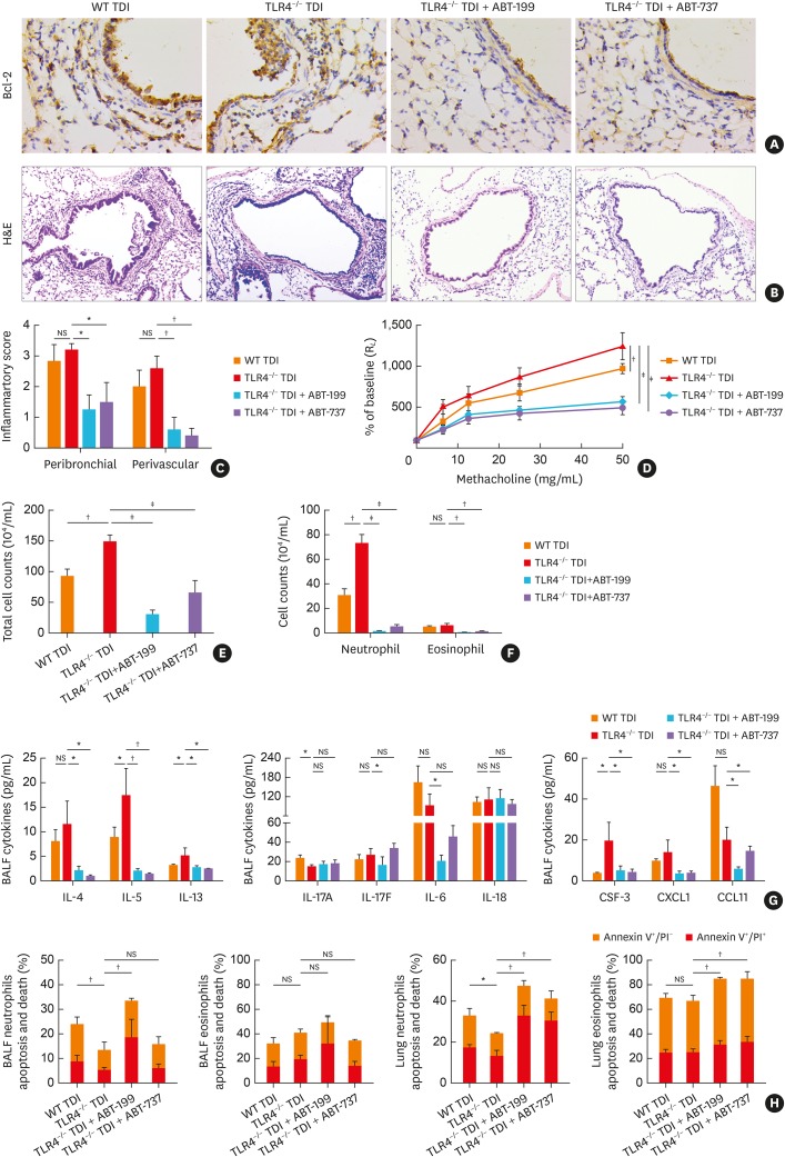
Methods: TLR4-/- and wild-type (WT) C57BL/10J mice were sensitized and challenged with TDI to generate a TDI-induced asthma model. B-cell lymphoma 2 (Bcl-2) inhibitors, ABT-199 (4 mg/kg) and ABT-737 (4 mg/kg), were intranasally given to TDI-exposed TLR4-/- mice after each challenge.

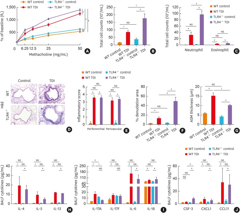
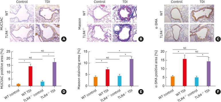
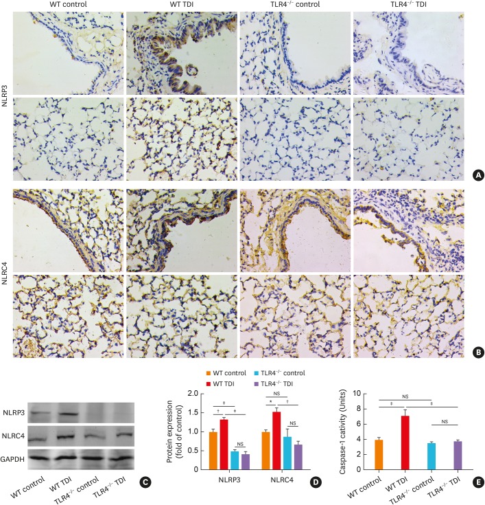
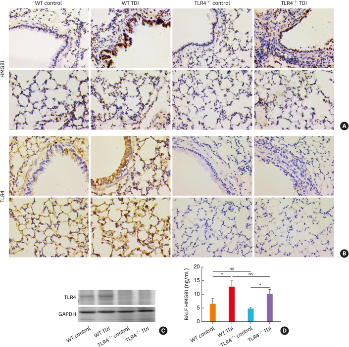
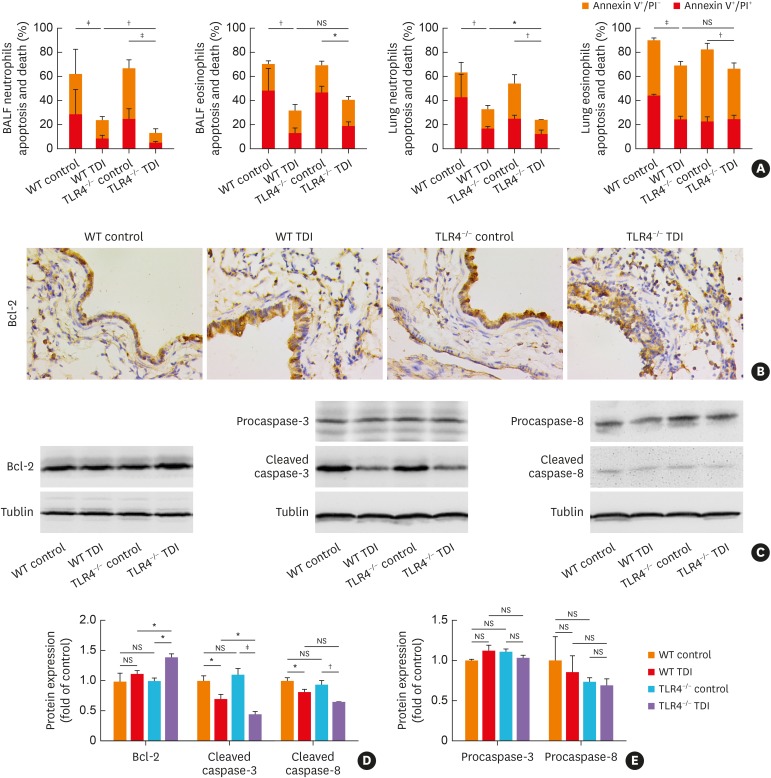
Results: TDI exposure led to increased airway hyperresponsiveness (AHR), granulocyte flux, bronchial epithelial shedding and extensive submucosal collagen deposition, which were unexpectedly aggravated by TLR4 deficiency. Following TDI challenge, TLR4-/- mice exhibited down-regulated interleukin-17A and increased colony-stimulating factor 3 in bronchoalveolar lavage fluid (BALF), while WT mice did not. In addition, TLR4 deficiency robustly suppressed the expression of NOD-like receptor family pyrin domain containing 3 and NLR family CARD domain containing 4, decreased caspase-1 activity in TDI-exposed mice, but had no effect on the level of high mobility group box 1 in BALF. Flow cytometry revealed that TDI hampered both neutrophil and eosinophil apoptosis, of which neutrophil apoptosis was further inhibited in TDI-exposed TLR4-/- mice, with marked up-regulation of Bcl-2. Moreover, inhibition of Bcl-2 with either ABT-199 or ABT-737 significantly alleviated neutrophil recruitment by promoting apoptosis.

Conclusions: These data indicated that TLR4 deficiency promoted neutrophil infiltration by impairing its apoptosis via up-regulation of Bcl-2, thereby resulting in deteriorated AHR and airway inflammation, which suggests that TLR4 could be a negative regulator of TDI-induced neutrophilic inflammation.

Keywords: Toll-like receptor 4; apoptosis; asthma; neutrophlic inflammation; toluene diisocyanate.


Conflict of interest statement

There are no financial or other issues that might lead to conflict of interest.

Figures

Fig. 1
Fig. 1. TLR4 deficiency aggravated TDI-induced airway hyperreactivity and inflammation. (A) Airway hyperresponsiveness was measured by RL. Results are shown as percentage of baseline (n = 4). (B) Numbers of total inflammatory cell in BALF. (C) Numbers of neutrophils and eosinophils in BALF (n = 6). (D) Representative H&E-stained lung sections of different groups. Original magnification was 200×. (E) Semi-quantification of airway inflammation was performed (n = 4–6). (F) Semi-quantification of epithelial denudation was performed (n = 4–6). (G) Analysis of ASM thickness was performed (n = 4–6). (H) Levels of Th2-related cytokines IL-4, IL-5 and IL-13 in BALF (n = 4–6). (I) Levels of Th17-related cytokines IL-17A, IL-17F, IL-6, IL-18 and in BALF (n = 4–6). (J) Levels of neutrophil chemoattractant CSF-3, CXCL1 and eosinophil chemoattractant CCL11 in BALF (n = 4–6).
WT, wild-type; TLR4, toll-like receptor 4; TDI, toluene diisocyanate; RL, lung resistance; H&E, hematoxylin and eosin; BALF, bronchoalveolar lavage fluid; NS, not significant; IL, interleukin; Th, T helper; CCL11, C-C motif chemokine 11; CXCL1, chemokine (C-X-C motif) ligand 1; CSF-3, colony-stimulating factor 3; ASM, airway smooth muscle. *P < 0.05; P < 0.01; P < 0.001.
Fig. 2
Fig. 2. TLR4−/− mice exhibited exacerbated airway remodeling after TDI exposure. (A) Representative immunohistochemical staining of MUC5AC in the lung sections of different groups. Original magnification was 200×. (B) Representative Masson trichrome-stained lung sections showing the collagen deposition of different groups. Original magnification was 200×. (C) Representative immunohistochemical staining of α-SMA indicates smooth muscle density. Original magnification was 200×. (D) Semi-quantification of MUC5AC staining was performed by ImageJ software (n = 4–6). (E) Semi-quantification of Masson staining was performed by ImageJ software (n = 4–6). (F) Semi-quantification of α-SMA staining was performed. The percentage of α-SMA positive staining area was measured by ImageJ software (n = 4–6).
WT, wild-type; TLR4, toll-like receptor 4; TDI, toluene diisocyanate; NS, not significant; MUC5AC, mucin-5AC; α-SMA, α-smooth muscle actin. *P < 0.01; P < 0.001.
Fig. 3
Fig. 3. TLR4-deficiency remarkably inhibited the activation of NLRP3, but slightly blunted NLRC4 in TDI-exposed mice. (A) Representative immunohistochemistry of NLRP3 in the bronchial regions (upper panel) and alveolar regions (lower panel). TLR4 deficiency significantly inhibited expression of NLRP3. Original magnification was 400×. (B) Representative immunohistochemistry of NLRC4 in the bronchial regions (upper panel) and alveolar regions (lower panel). TLR4 deficiency in C57/BL10 mice slightly blunted pulmonary expression of NLRC4. Original magnification was 400×. (C, D) Protein expressions of NLRP3, NLRC4 in lung homogenates were detected by Western blotting and densitometric analysis was performed (n = 6). (E) Levels of caspase-1 activity in lung homogenates of different groups (n = 4–6).
WT, wild-type; TLR4, toll-like receptor 4; TDI, toluene diisocyanate; NS, not significant; GAPDH, glyceraldehyde 3-phosphate dehydrogenase; NLRP3, NOD-like receptor family, pyrin domain containing 3; NLRC4, NLR family CARD domain containing 4. *P < 0.05; P < 0.01; P < 0.001.
Fig. 4
Fig. 4. TLR4 deficiency did not affect HMGB1 production in TDI-treated mice. (A) Representative immunohistochemistry of HMGB1 in the bronchial regions (upper panel) and alveolar regions (lower panel). Original magnification was 400×. (B) Representative immunohistochemical staining of TLR4 in the bronchial regions (upper panel) and alveolar regions (lower panel). (C) Protein expression of TLR4, HMGB1 in lung homogenates were detected by Western blotting (n = 6). (D) Levels of HMGB1 in BALF of different groups were detected by ELISA (n = 4–6).
WT, wild-type; TLR4, toll-like receptor 4; TDI, toluene diisocyanate; NS, not significant; HMGB1, high mobility group box 1; BALF, bronchoalveolar lavage fluid; GAPDH, glyceraldehyde 3-phosphate dehydrogenase; ELISA, enzyme-linked immunosorbent assay. *P < 0.05.
Fig. 5
Fig. 5. TLR4 deficiency impaired apoptosis of neutrophils, with pulmonary up-regulation of Bcl-2. (A) Numbers of apoptotic ex vivo eosinophils and neutrophils were determined by flow cytometry. (B) Representative immunohistochemistry of Bcl-2 in the bronchial regions. Original magnification was 400×. (C-E) Protein expressions of Bcl-2, procaspase-3, procaspase-8, cleaved caspase-3 and cleaved caspase-8 in lung homogenates were detected by Western blotting and densitometric analysis was performed (n = 6).
WT, wild-type; TLR4, toll-like receptor 4; TDI, toluene diisocyanate; NS, not significant; Bcl-2, B-cell lymphoma 2; BALF, bronchoalveolar lavage fluid; PI, propidium iodide. *P < 0.05; P < 0.01; P < 0.001.
Fig. 6
Fig. 6. Bcl-2 inhibitors alleviated the aggravated airway inflammation in TDI-exposed TLR4–/– mice. (A) Representative immunohistochemistry of Bcl-2 in the bronchial regions. Original magnification was 400×. (B) Representative H&E-stained lung sections of different groups. Original magnification was 200×. (C) Semi-quantification of peribronchial and perivascular inflammation was performed (n = 5-6). (D) Airway hyperresponsiveness was measured by RL. Results were shown as percentage of baseline (n = 4). (E) Numbers of total inflammatory cell in BALF (n = 5–6). (F) Numbers of neutrophils, and eosinophils in BALF (n = 5–6). (G) Levels of Th2-related cytokines (IL-4, IL-5 and IL-13), Th17-related cytokines (IL-17A, IL-17F, IL-6 and IL-18), eosinophil chemoattractant CCL11, and neutrophil chemoattractant CSF-3, CXCL1 in BALF (n = 4–6). (H) Numbers of apoptotic ex vivo eosinophils and neutrophils were determined.
WT, wild-type; TLR4, toll-like receptor 4; TDI, toluene diisocyanate; NS, not significant; RL, lung resistance; Bcl-2, B-cell lymphoma 2; IL, interleukin; CCL11, C-C motif chemokine 11; CXCL1, chemokine (C-X-C motif) ligand 1; CSF-3, colony-stimulating factor 3; BALF, bronchoalveolar lavage fluid; Th, T helper; H&E, hematoxylin and eosin; PI, propidium iodide. *P < 0.05; P < 0.01; P < 0.001.

References

    1. Masoli M, Fabian D, Holt S, Beasley R Global Initiative for Asthma (GINA) Program. The global burden of asthma: executive summary of the GINA Dissemination Committee report. Allergy. 2004;59:469–478. - PubMed
    1. Kim RY, Pinkerton JW, Essilfie AT, Robertson AA, Baines KJ, Brown AC, et al. Role for NLRP3 inflammasome-mediated, IL-1β-dependent responses in severe, steroid-resistant asthma. Am J Respir Crit Care Med. 2017;196:283–297. - PubMed
    1. Zakeri A, Russo M. Dual role of toll-like receptors in human and experimental asthma models. Front Immunol. 2018;9:1027. - PMC - PubMed
    1. Hammad H, Chieppa M, Perros F, Willart MA, Germain RN, Lambrecht BN. House dust mite allergen induces asthma via toll-like receptor 4 triggering of airway structural cells. Nat Med. 2009;15:410–416. - PMC - PubMed
    1. Tang H, Li T, Han X, Sun J. TLR4 antagonist ameliorates combined allergic rhinitis and asthma syndrome (CARAS) by reducing inflammatory monocytes infiltration in mice model. Int Immunopharmacol. 2019;73:254–260. - PubMed