. 2020 Jun 1;123(6):2326-2342.
doi: 10.1152/jn.00179.2020. Epub 2020 May 13.

The serotonin reuptake blocker citalopram destabilizes fictive locomotor activity in salamander axial circuits through 5-HT1A receptors

Affiliations

The serotonin reuptake blocker citalopram destabilizes fictive locomotor activity in salamander axial circuits through 5-HT1A receptors

Aurélie Flaive et al. J Neurophysiol. .

Abstract

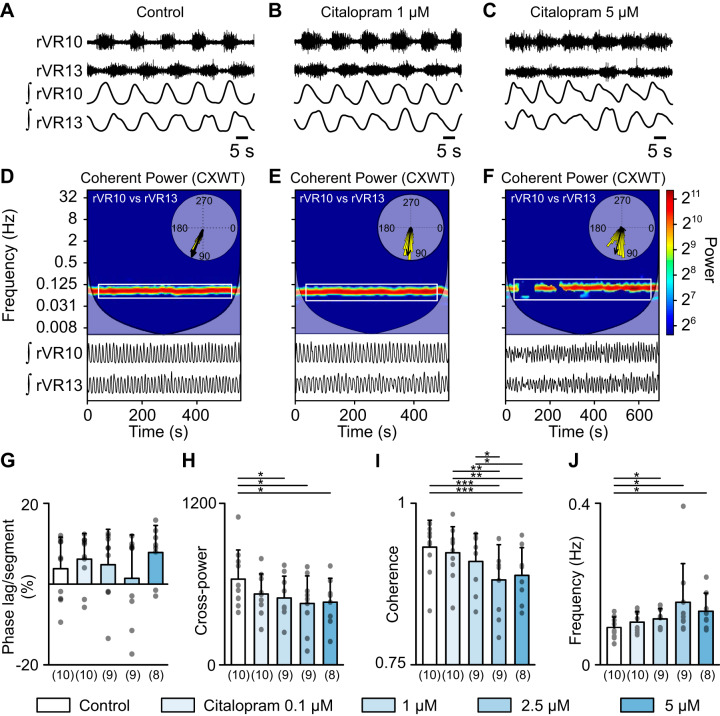
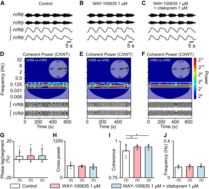
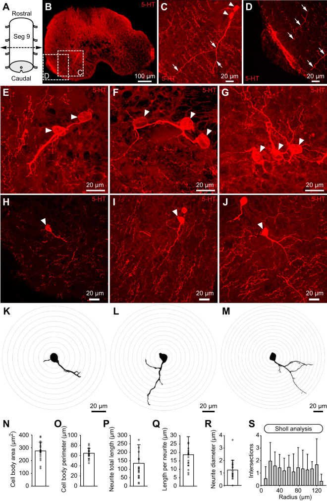
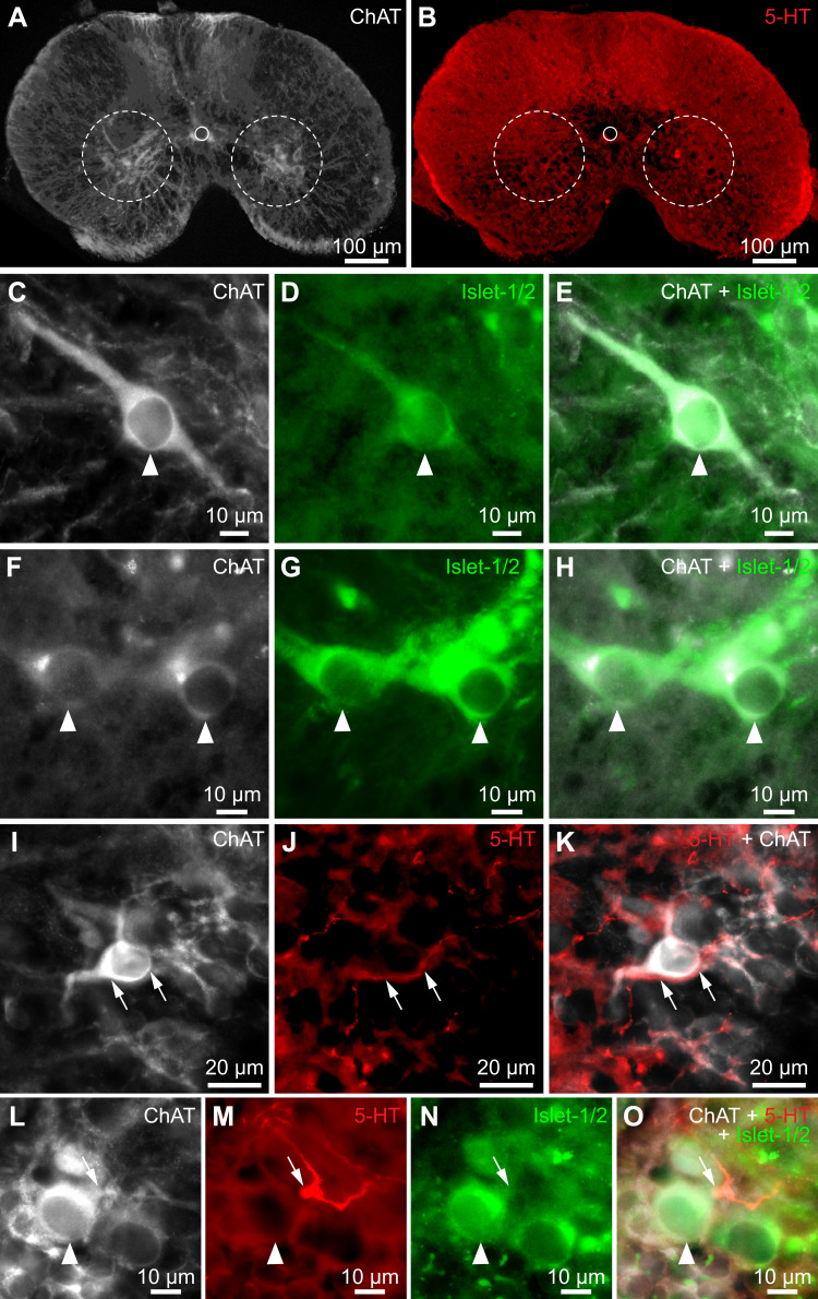
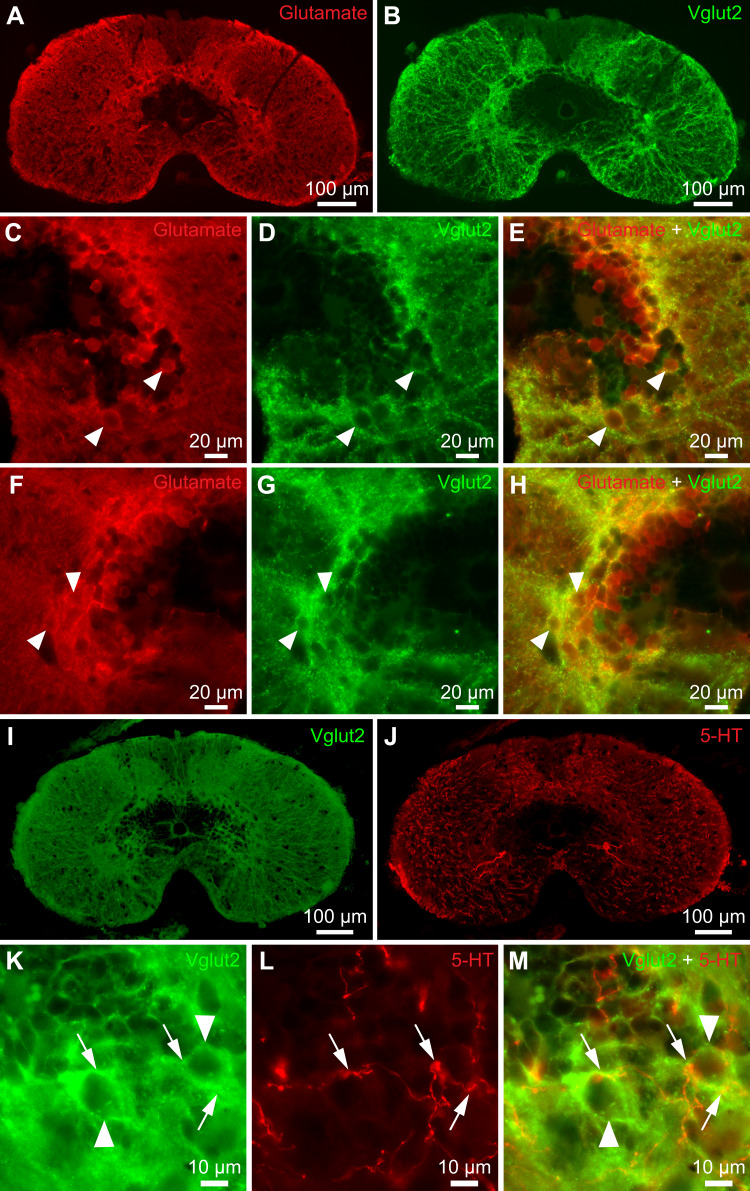
Serotoninergic (5-HT) neurons are powerful modulators of spinal locomotor circuits. Most studies on 5-HT modulation focused on the effect of exogenous 5-HT and these studies provided key information about the cellular mechanisms involved. Less is known about the effects of increased release of endogenous 5-HT with selective serotonin reuptake inhibitors. In mammals, such molecules were shown to destabilize the fictive locomotor output of spinal limb networks through 5-HT1A receptors. However, in tetrapods little is known about the effects of increased 5-HT release on the locomotor output of axial networks, which are coordinated with limb circuits during locomotion from basal vertebrates to mammals. Here, we examined the effect of citalopram on fictive locomotion generated in axial segments of isolated spinal cords in salamanders, a tetrapod where raphe 5-HT reticulospinal neurons and intraspinal 5-HT neurons are present as in other vertebrates. Using electrophysiological recordings of ventral roots, we show that fictive locomotion generated by bath-applied glutamatergic agonists is destabilized by citalopram. Citalopram-induced destabilization was prevented by a 5-HT1A receptor antagonist, whereas a 5-HT1A receptor agonist destabilized fictive locomotion. Using immunofluorescence experiments, we found 5-HT-positive fibers and varicosities in proximity with motoneurons and glutamatergic interneurons that are likely involved in rhythmogenesis. Our results show that increasing 5-HT release has a deleterious effect on axial locomotor activity through 5-HT1A receptors. This is consistent with studies in limb networks of turtle and mouse, suggesting that this part of the complex 5-HT modulation of spinal locomotor circuits is common to limb and axial networks in limbed vertebrates.NEW & NOTEWORTHY Little is known about the modulation exerted by endogenous serotonin on axial locomotor circuits in tetrapods. Using axial ventral root recordings in salamanders, we found that a serotonin reuptake blocker destabilized fictive locomotor activity through 5-HT1A receptors. Our anatomical results suggest that serotonin is released on motoneurons and glutamatergic interneurons possibly involved in rhythmogenesis. Our study suggests that common serotoninergic mechanisms modulate axial motor circuits in amphibians and limb motor circuits in reptiles and mammals.

Keywords: 5-HT1A; citalopram; locomotion; salamander; serotonin.


Conflict of interest statement

No conflicts of interest, financial or otherwise, are declared by the authors.

Figures

Fig. 1.
Fig. 1.
Effects of the selective serotonin reuptake inhibitor citalopram on axial fictive locomotor activity. AC: fictive locomotor activity evoked by bath application of the glutamatergic agonists N-methyl-d-aspartate (NMDA; 20 µM) and d-serine (10 µM) on isolated spinal cord preparations. Typical extracellular recordings obtained from ventral roots (VR) located on the right (r) side of spinal segments 10 and 13 (i.e., innervating trunk muscles) using suction electrodes. The traces were rectified and smoothed (ʃ traces; see materials and methods). After the control period, increasing concentrations of citalopram were applied each 30 min. D and E: coherent cross-power representation (CXWT; see Mor and Lev-Tov 2007) illustrating in time-frequency domains the coordination of the motor activities recorded from the two VRs. The analysis was performed on longer bouts of activities from the preparation illustrated in AC. The frequency domain showing high cross-power (color coded) in close neighborhood of the locomotor rhythm was delineated (solid rectangle) for analysis in GJ. In the insets, the phase relationships (degrees) between the rhythmic activities of the two VRs are illustrated with vectors on circular plots. The mean vector was used to determine the corresponding intersegmental phase lag. GJ: effects of increasing concentrations of citalopram on the intersegmental phase lag (G), cross-power (H), rhythm coherence (I), and rhythm frequency (J). The number of animals is indicated in parentheses. Note that in some animals some concentrations of citalopram were not applied. Each gray dot represents one animal. Data from AF were obtained from the same animal. *P < 0.05, **P < 0.01, ***P < 0.001, Student–Newman–Keuls post hoc test after a significant one-way ANOVA for repeated measures (P < 0.05 for H, P < 0.01 for I, P < 0.001 for J).
Fig. 2.
Fig. 2.
Effects of the 5-HT1A antagonist WAY-100635 on axial fictive locomotor activity. AC: fictive locomotor activity evoked by bath application of N-methyl-d-aspartate (NMDA; 20 µM) and d-serine (10 µM) on isolated spinal cord preparations. Typical extracellular recordings obtained from ventral roots (VR) located on the right (r) side of spinal segments 8 and 9 (i.e., innervating trunk muscles) using suction electrodes. The traces were rectified and smoothed (ʃ traces; see materials and methods). After the control period, the 5-HT1A antagonist WAY-100635 (1 µM) was applied during 30 min, then the selective serotonin reuptake inhibitor citalopram (1 µM) was added during 30 min. DF: coherent cross-power representation (CXWT; see Mor and Lev-Tov 2007) illustrating in time-frequency domains the coordination of the motor activities recorded from the two VRs. The analysis was performed on longer bouts of activities from the preparation illustrated in AC. The frequency domain showing high cross-power (color coded) in close neighborhood of the locomotor rhythm was delineated (solid rectangle) for analysis in GJ. In the insets, the phase relationships (degrees) between the rhythmic activities of the two VRs are illustrated with vectors on circular plots. The mean vector was used to determine the corresponding intersegmental phase lag. GJ: effects of drug applications on the intersegmental phase lag (G), cross-power (H), rhythm coherence (I), and rhythm frequency (J). The number of animals is indicated in parentheses. Each gray dot represents one animal. Data from AF were obtained from the same animal. *P < 0.05, Fisher LSD post hoc test after a one-way ANOVA for repeated measures (P < 0.05 in I).
Fig. 3.
Fig. 3.
Effects of the 5-HT1A/7 agonist 8-OH-DPAT on axial fictive locomotor activity. A and B: fictive locomotor activity evoked by bath application of N-methyl-d-aspartate (NMDA; 20 µM) and d-serine (10 µM) on isolated spinal cord preparations in control conditions and in presence of the 5-HT1A/7 agonist 8-OH-DPAT. Typical extracellular recordings obtained from ventral roots (VR) located on the right (r) side of spinal segments 6 and 8 (i.e., innervating trunk muscles) using suction electrodes. The traces were rectified and smoothed (ʃ traces; see materials and methods). After the control period, the 5-HT1A/7 agonist 8-OH-DPAT was applied during 30 min at 0.1 µM, and then during 30 min at 1 µM. C and D: coherent cross-power representation (CXWT; see Mor and Lev-Tov 2007) illustrating in time-frequency domains the coordination of the motor activities recorded from the two VRs. The analysis was performed on longer bouts of activities from the preparation illustrated in A and B. The frequency domain showing high cross-power (color coded) in close neighborhood of the locomotor rhythm was delineated (solid rectangle) for analysis in EH. In the insets, the phase relationships (degrees) between the rhythmic activities of the two VRs are illustrated with vectors on circular plots. The mean vector was used to determine the corresponding intersegmental phase lag. EH: effects of increasing concentrations of the 5-HT1A/7 agonist 8-OH-DPAT on the intersegmental phase lag (E), cross-power (F), rhythm coherence (G), and rhythm frequency (H). The number of animals is indicated in parentheses. Each gray dot represents one animal. Data from AD were obtained from the same animal. *P < 0.05, Student–Newman–Keuls post hoc test after a significant one-way ANOVA for repeated measures (P < 0.05 both in E and G).
Fig. 4.
Fig. 4.
Serotoninergic (5-HT) neurons in the axial spinal cord. A: schematic representation of some axial salamander spinal segments. B: transverse section of axial spinal segment 9 (Seg 9) showing fibers and cell bodies positive for 5-HT (red). C: 5-HT-positive cells bodies (white arrowheads) were found on the ventral side of the spinal cord, with their fibers (white arrows) extending ventrolaterally, where the ventrolateral 5-HT-immunoreactive plexus (D) is located. Note the dense presence of 5-HT-positive fibers in the ventral part of the spinal cord in C. D: a dense plexus of 5-HT-positive fibers was consistently found at the border between the nervous system and the dura. Some 5-HT-positive fibers, likely originating from the 5-HT cell bodies, were pointing at the plexus (white arrows). Many other 5-HT fibers, likely from a different origin, were not pointing toward the plexus. EG: clusters of 5-HT cell bodies (white arrowheads) located in the ventromedial region, likely sending projections to each other. HM: three examples of 5-HT-positive neurons (HJ) and the corresponding 3D reconstructions that here appear projected in 2D (KM). NS: morphometric measurements obtained from 22 reconstructed cells obtained from three animals (3, 8, and 11 cells per animal). In S, the Sholl analysis gives an index of cell morphological complexity by quantifying the number of intersections between their neurites and spheres with increasing diameters centered on the cell body. Data from BM were obtained from three animals.
Fig. 5.
Fig. 5.
Serotoninergic (5-HT) fibers in close apposition with motoneurons in the axial spinal cord. A and B: transverse section of axial spinal segment 9 showing fibers and cell bodies positive for choline acetyltransferase (ChAT, white, A) or 5-HT (red, B). The white dashed circles delineate the approximate position of the motoneurons. The white solid circles delineate the position of the central canal. CH: two series of photomicrographs (CE, FH) showing examples of motoneurons (arrowheads) in the ventral spinal cord showing immunoreactivity for ChAT (white, C and F) and for the motoneuron marker Islet-1/2 (green, D and G), and the double labeling (E and H). IK: photomicrographs showing a putative motoneuron (ChAT-positive, white, I) closely surrounded by a 5-HT-positive fiber (red, J) and the double labeling (K). LO: triple-labeling experiment (O) showing a 5-HT-positive fiber and varicosity (red, M) in close apposition with a motoneuron cell body positive for ChAT (white, L) and Islet-1/2 (green, N). Data from AO were obtained from three animals.
Fig. 6.
Fig. 6.
Serotoninergic (5-HT) fibers in close apposition with glutamatergic neurons in the axial spinal cord. A and B: transverse section of axial spinal segment 11 showing cell bodies positive for glutamate (red, A) or the vesicular glutamatergic transporter (Vglut2, green, B). CH: two series of photomicrographs (CE, FH) showing examples of neurons (arrowheads) immunoreactive for glutamate (red, C and F) and Vglut2 (green, D and G), and the double labeling (E and H). I and J: transverse section of axial spinal segment 9 showing fibers and cell bodies positive for Vglut2 (green, I) and fibers and varicosities positive for serotonin (red, J). KM: photomicrographs showing Vglut2-positive neurons (green, K) closely surrounded by 5-HT-positive fibers and varicosities (red, L), and the double labeling (M). Data from AM were obtained from two animals.

Similar articles

Cited by

References

    1. Abbinanti MD, Harris-Warrick RM. Serotonin modulates multiple calcium current subtypes in commissural interneurons of the neonatal mouse. J Neurophysiol 107: 2212–2219, 2012. doi:10.1152/jn.00768.2011. - DOI - PMC - PubMed
    1. Abbinanti MD, Zhong G, Harris-Warrick RM. Postnatal emergence of serotonin-induced plateau potentials in commissural interneurons of the mouse spinal cord. J Neurophysiol 108: 2191–2202, 2012. doi:10.1152/jn.00336.2012. - DOI - PMC - PubMed
    1. Adrio F, Anadón R, Rodríguez-Moldes I. Distribution of serotonin (5HT)-immunoreactive structures in the central nervous system of two chondrostean species (Acipenser baeri and Huso huso). J Comp Neurol 407: 333–348, 1999. doi:10.1002/(SICI)1096-9861(19990510)407:3<333:AID-CNE3>3.0.CO;2-R. - DOI - PubMed
    1. Antri M, Mouffle C, Orsal D, Barthe J-Y. 5-HT1A receptors are involved in short- and long-term processes responsible for 5-HT-induced locomotor function recovery in chronic spinal rat. Eur J Neurosci 18: 1963–1972, 2003. doi:10.1046/j.1460-9568.2003.02916.x. - DOI - PubMed
    1. Ballion B, Branchereau P, Chapron J, Viala D. Ontogeny of descending serotonergic innervation and evidence for intraspinal 5-HT neurons in the mouse spinal cord. Brain Res Dev Brain Res 137: 81–88, 2002. doi:10.1016/S0165-3806(02)00414-5. - DOI - PubMed

Publication types

MeSH terms

Substances

Grants and funding

LinkOut - more resources