. 2020 May 13;16(5):e1008500.
doi: 10.1371/journal.ppat.1008500. eCollection 2020 May.

Multiple weak interactions between BvgA~P and ptx promoter DNA strongly activate transcription of pertussis toxin genes in Bordetella pertussis

Affiliations

Multiple weak interactions between BvgA~P and ptx promoter DNA strongly activate transcription of pertussis toxin genes in Bordetella pertussis

Qing Chen et al. PLoS Pathog. .

Abstract

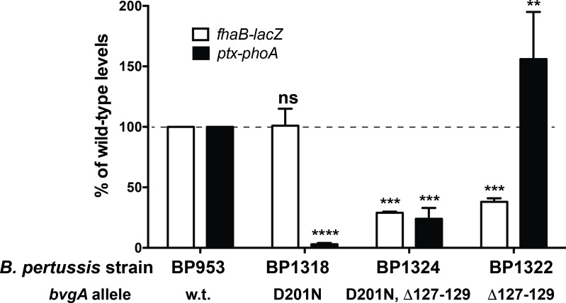
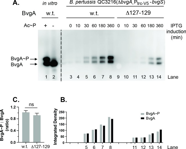
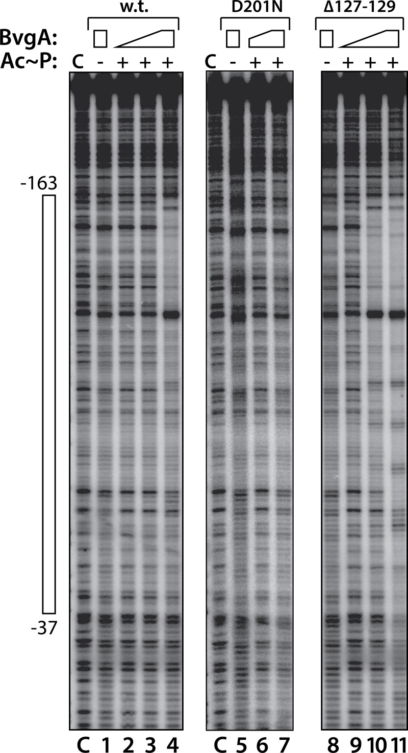
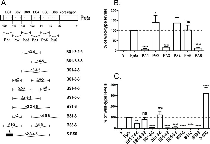
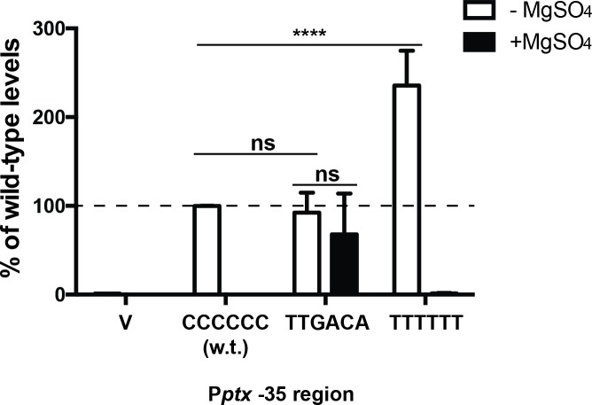
Pertussis toxin is the preeminent virulence factor and major protective antigen produced by Bordetella pertussis, the human respiratory pathogen and etiologic agent of whooping cough. Genes for its synthesis and export are encoded by the 12 kb ptx-ptl operon, which is under the control of the pertussis promoter, Pptx. Expression of this operon, like that of all other known protein virulence factors, is regulated by the BvgAS two-component global regulatory system. Although Pptx has been studied for years, characterization of its promoter architecture vis-à-vis BvgA-binding has lagged behind that of other promoters, mainly due to its lower affinity for BvgA~P. Here we take advantage of a mutant BvgA protein (Δ127-129), which enhances ptx transcription in B. pertussis and also demonstrates enhanced binding affinity to Pptx. By using this mutant protein labeled with FeBABE, binding of six head-to-head dimers of BvgA~P was observed, with a spacing of 22 bp, revealing a binding geometry similar to that of other BvgA-activated promoters carrying at least one strong binding site. All of these six BvgA-binding sites lack sequence features associated with strong binding. A genetic analysis indicated the degree to which each contributes to Pptx activity. Thus the weak/medium binding affinity of Pptx revealed in this study explains its lower responsiveness to phosphorylated BvgA, relative to other promoters containing a high affinity binding site, such as that of the fha operon.


Conflict of interest statement

The authors have declared that no competing interests exist.

Figures

Fig 1
Fig 1. Activity of gene fusions in BP953 and derivatives.
BP953 contains two transcriptional fusions, fha-lacZ and ptx-phoA. The results of beta-galactosidase and alkaline phosphatase enzymatic assays are shown, with w.t. values set to 100% for comparison. Also shown are the results of assays of the derivatives, BP1318, BP1324, and BP1322 harboring the bvgA alleles indicated, normalized to those for BP953. Isolation of these alleles is described in the text. Values were normalized to fha-lacZ and ptx-phoA expressed in BP953 and data from at least four assays were used in the calculation of means, standard deviations, as indicated by error bars, and statistical analysis by one-way ANOVA. Outcomes of the latter analysis are presented using the symbols: ns, P > 0.05; **, P ≤ 0.01; ***, P ≤ 0.001; ****, P ≤ 0.0001.
Fig 2
Fig 2. Kinetics of in vivo phosphorylation of wild type and Δ127–129 BvgA.
A. B. pertussis strain QC3216 harboring plasmids conferring expression of wild-type BvgA (pSS4893, lanes 3–8) and BvgAΔ127–129 (pQC1894, lane 9–14), respectively, were grown in PLB liquid media, induced with 1 mM IPTG and sampled at various time points post induction. The collected samples were analyzed by Phos-tag gel electrophoresis, followed by Western blot with anti-BvgA detection, as previously described [9]. Control lanes contained 1 pmol of purified BvgA incubated in the presence (+, lane 1) or absence (-, lane 2) of acetyl phosphate as described previously [9]. B. The intensities of BvgA (black bar) and BvgA~P (grey bar) for lanes 5–8 and 11–14 were quantified and reported as integrated density using ImageJ software. C. The quantitative intensities derived from four IPTG-induction time points (30 min, 60 min, 180 min and 360 min) in panel B were used to calculate the ratios of BvgA~P to BvgA for the wild type and the mutant BvgA, respectively, and to obtain the means, standard deviations, as indicated by error bars, and statistical analysis by one-way ANOVA. Outcome of the latter analysis is presented using the symbol: ns, P > 0.05.
Fig 3
Fig 3. DNase footprinting of BvgA and derivatives on the pertussis toxin promoter.
DNase footprints were performed as described in Materials and Methods. BvgA proteins were wild-type (lanes 1–4), BvgAΔ201N (lanes 5–7), and BvgAΔ127–129 (lanes 8–11). Lanes designated “C” show the labeled fragment, digested with DNase I, in the absence of any added protein. Lanes 1, 5, and 8 show the digestion pattern obtained when BvgA was added but Ac~P was not. Acetyl phosphate was added to the reactions shown in the remaining lanes, as indicated. Concentrations of BvgA proteins used were 16 nM (lanes 2 and 9), 32 nM (lanes 3, 6, and 10) and 65 nM (lanes 1, 4, 5, 7, 8, and 11). The open bar to the left shows the maximal region protected in lane 11.
Fig 4
Fig 4. Details of BvgA-binding to the pertussis toxin promoter.
A. BvgAΔ127–129 derivatives in which both naturally occurring cysteine residues had been replaced by alanine and in which either the valine at position 148 or the threonine at position 194 had been replaced with cysteine, were subjected to derivatization with FeBABE and used to reveal the locations of BvgA-binding to the ptx promoter using methods previously described for a similar analysis of the fha promoter [12]. The protein modified at the 148 position produces cleavages at the outer boundaries of a bound dimer of BvgA~P, while the 194 derivative produces closely spaced cleavages corresponding to the location of the inter-monomer interface. Maxam and Gilbert A + G reactions of the same 32P end-labelled Pptx DNA fragment were run in parallel for orientation. B. DNA sequences of the ptx promoter showing the sites of binding of BvgA~P derived from the analysis in panel A. In addition, each heptameric half-site has been scored according to the algorithm presented in panel C, with the scores given above the arrows indicating the binding half-sites. Nucleotides in red indicate core promoter elements, with the consensus sequence shown above. In a similar fashion, Pfha and Pfim3 are shown for comparison. Green arrows below the Pptx sequence indicate two 21 bp imperfect direct repeats and two inverted heptameric imperfect repeats previously cited as potential BvgA binding sites. C. Algorithm for predicting binding strength of BvgA-binding half-sites. This algorithm was derived from a study examining the ability of systematically mutated derivatives of the Pfha primary binding site to bind BvgA~P and to activate transcription [14].
Fig 5
Fig 5. Contribution of Pptx BvgA-binding sites to Pptx function.
A. Diagram of BvgA-binding sites within Pptx and its derivatives. Boxes delineate the extents of 22-bp deletions removing each of the six binding sites, with the binding sites themselves within each box indicated by a double-headed arrow. Below this schematic are shown the different combinations of these 22 bp segments in different derivatives. The black bar in derivative S-BS6 indicates the presence of the primary binding site from Pfha (TAAGAAATTTCCTA). B & C. Luciferase activity of B. pertussis strain BP536 carrying ectopically integrated plasmids. Values for the empty pSS3967 control (V) and for promoter-lux fusion derivatives harboring the wild type (Pptx) and the deletion derivatives shown in panel A are presented. Strains were grown on BG agar at 37°C for 2 days and assayed as described in Materials and Methods. Values were normalized to wild-type Pptx and data from at least four assays were used in the calculation of means, standard deviations, as indicated by error bars, and statistical analysis by one-way ANOVA. Outcomes of the latter analysis are presented using the symbols: ns, P > 0.05; *, P ≤ 0.05; **, P ≤ 0.01; ***, P ≤ 0.001; ****, P ≤ 0.0001.
Fig 6
Fig 6. A non-consensus -35 region is required for BvgA-regulated Pptx activity.
Luciferase activity of B. pertussis BP536 carrying ectopically integrated plasmids. Values for the empty pSS3967 control (V) and for promoter-lux fusion derivatives harboring the wild type (Pptx) and -35 substitution derivatives are presented. Strains were grown on BG agar at 37°C for 2 days and assayed as described in Materials and Methods. Values were normalized to wild-type Pptx and data from at least four assays were used in the calculation of means, standard deviations, as indicated by error bars, and statistical analysis by one-way ANOVA. Outcomes of the latter analysis are presented using the symbols: ns, P > 0.05; *, P ≤ 0.05; ****, P ≤ 0.0001.
Fig 7
Fig 7. Comparing the relative strengths of Pfha and Pptx by transcriptional fusion.
Promoter-lux (A & C) and promoter-rfp (B & D) transcriptional fusions were constructed and integrated into the BP536 chromosome. Fusions to the lux operon used either pSS3967, to integrate at an ectopic location (A), or pSS4162, to integrate in situ (C). Similarly, transcriptional fusions to rfp used either pQC2241 for ectopic insertion (B) or pQC2319 for in situ insertion (D). For the two ectopic constructs “V” indicates insertion of the vector alone. This control is not possible for the in situ insertions. The extent of the promoter sequences cloned in each construct are provided as nucleotide coordinates relative to the transcriptional start. B. pertussis strains carrying these constructs were grown on BG agar at 37°C for 2 days and analyzed for and luciferase and RFP activity as described in Materials and Methods. In each panel activity is reported relative to the Pfha-promoter fusion and the results of at least four assays were used in the calculation of standard deviations and statistical analysis by an unpaired two-tailed t test between two samples. Statistical symbols are: **, P ≤ 0.01; ***, P ≤ 0.001; ****, P ≤ 0.0001.

Similar articles

Cited by

References

    1. Steffen P, Goyard S, Ullmann A. Phosphorylated BvgA is sufficient for transcriptional activation of virulence-regulated genes in Bordetella pertussis. EMBO J. 1996;15(1):102–9. - PMC - PubMed
    1. Boucher PE, Murakami K, Ishihama A, Stibitz S. Nature of DNA binding and RNA polymerase interaction of the Bordetella pertussis BvgA transcriptional activator at the fha promoter. J Bacteriol. 1997;179(5):1755–63. 10.1128/jb.179.5.1755-1763.1997 - DOI - PMC - PubMed
    1. Boucher PE, Yang MS, Schmidt DM, Stibitz S. Genetic and biochemical analyses of BvgA interaction with the secondary binding region of the fha promoter of Bordetella pertussis. J Bacteriol. 2001;183(2):536–44. 10.1128/JB.183.2.536-544.2001 - DOI - PMC - PubMed
    1. Boulanger A, Moon K, Decker KB, Chen Q, Knipling L, Stibitz S, et al. Bordetella pertussis fim3 gene regulation by BvgA: phosphorylation controls the formation of inactive vs. active transcription complexes. Proc Natl Acad Sci U S A. 2015;112(6):E526–35. 10.1073/pnas.1421045112 - DOI - PMC - PubMed
    1. Kinnear SM, Boucher PE, Stibitz S, Carbonetti NH. Analysis of BvgA activation of the pertactin gene promoter in Bordetella pertussis. J Bacteriol. 1999;181(17):5234–41. - PMC - PubMed

Publication types

MeSH terms