

Personalized iPSC-Derived Dopamine Progenitor Cells for Parkinson's Disease
- PMID: 32402162
- PMCID: PMC7288982
- DOI: 10.1056/NEJMoa1915872
Personalized iPSC-Derived Dopamine Progenitor Cells for Parkinson's Disease
Abstract
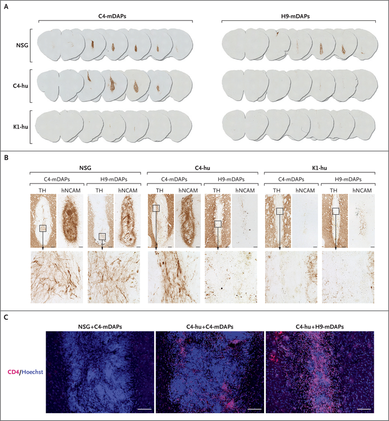
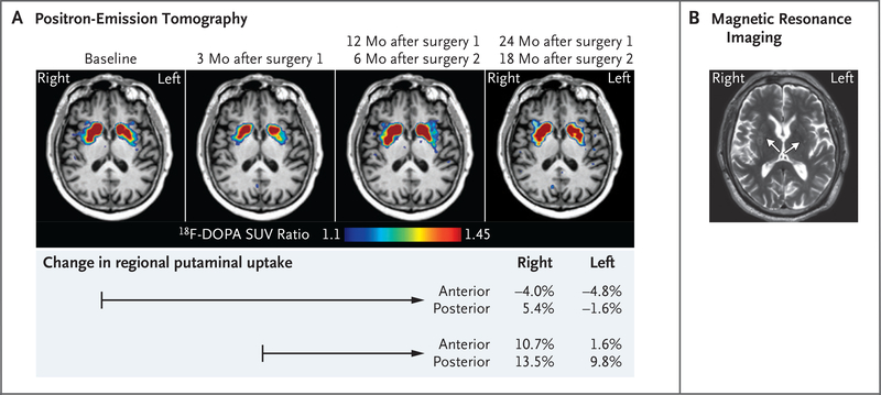
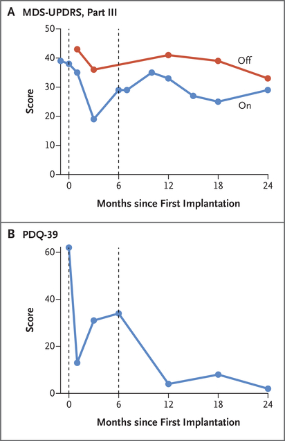
We report the implantation of patient-derived midbrain dopaminergic progenitor cells, differentiated in vitro from autologous induced pluripotent stem cells (iPSCs), in a patient with idiopathic Parkinson's disease. The patient-specific progenitor cells were produced under Good Manufacturing Practice conditions and characterized as having the phenotypic properties of substantia nigra pars compacta neurons; testing in a humanized mouse model (involving peripheral-blood mononuclear cells) indicated an absence of immunogenicity to these cells. The cells were implanted into the putamen (left hemisphere followed by right hemisphere, 6 months apart) of a patient with Parkinson's disease, without the need for immunosuppression. Positron-emission tomography with the use of fluorine-18-L-dihydroxyphenylalanine suggested graft survival. Clinical measures of symptoms of Parkinson's disease after surgery stabilized or improved at 18 to 24 months after implantation. (Funded by the National Institutes of Health and others.).
Copyright © 2020 Massachusetts Medical Society.
Figures
Comment in
-
Stem Cells: Scientific and Ethical Quandaries of a Personalized Approach to Parkinson's Disease.Mov Disord. 2020 Aug;35(8):1312-1314. doi: 10.1002/mds.28187. Epub 2020 Jul 7. Mov Disord. 2020. PMID: 32529662 No abstract available.
-
From Skin to Brain: A Parkinson's Disease Patient Transplanted with His Own Cells.Cell Stem Cell. 2020 Jul 2;27(1):8-10. doi: 10.1016/j.stem.2020.06.008. Cell Stem Cell. 2020. PMID: 32619520
-
Letter: Stem Cell Transplantation for Parkinson Disease: Déjà Vu All Over Again?Neurosurgery. 2021 Jan 13;88(2):E216-E217. doi: 10.1093/neuros/nyaa487. Neurosurgery. 2021. PMID: 33289520 No abstract available.
-
Toward a Personalized Approach to Parkinson's Cell Therapy.Mov Disord. 2020 Nov;35(11):2119-2120. doi: 10.1002/mds.28328. Mov Disord. 2020. PMID: 33463754 No abstract available.
References
-
- Lindvall O, Backlund EO, Farde L, et al. Transplantation in Parkinson’s disease: two cases of adrenal medullary grafts to the putamen. Ann Neurol 1987;22:457–68. - PubMed
-
- Goetz CG, Olanow CW, Koller WC, et al. Multicenter study of autologous adrenal medullary transplantation to the corpus striatum in patients with advanced Parkinson’s disease. N Engl J Med 1989;320:337–41. - PubMed
-
- Lindvall O, Brundin P, Widner H, et al. Grafts of fetal dopamine neurons survive and improve motor function in Parkinson’s disease. Science 1990;247:574–7. - PubMed
-
- Freed CR, Breeze RE, Rosenberg NL, et al. Survival of implanted fetal dopamine cells and neurologic improvement 12 to 46 months after transplantation for Parkinson’s disease. N Engl J Med 1992;327:1549–55. - PubMed
-
- Spencer DD, Robbins RJ, Naftolin F, et al. Unilateral transplantation of human fetal mesencephalic tissue into the caudate nucleus of patients with Parkinson’s disease. N Engl J Med 1992;327:1541–8. - PubMed