. 2020 Jul 7;28(7):810-819.e5.
doi: 10.1016/j.str.2020.04.012. Epub 2020 May 12.

Activation of Phospholipase C β by Gβγ and Gαq Involves C-Terminal Rearrangement to Release Autoinhibition

Affiliations

Activation of Phospholipase C β by Gβγ and Gαq Involves C-Terminal Rearrangement to Release Autoinhibition

Isaac J Fisher et al. Structure. .

Abstract

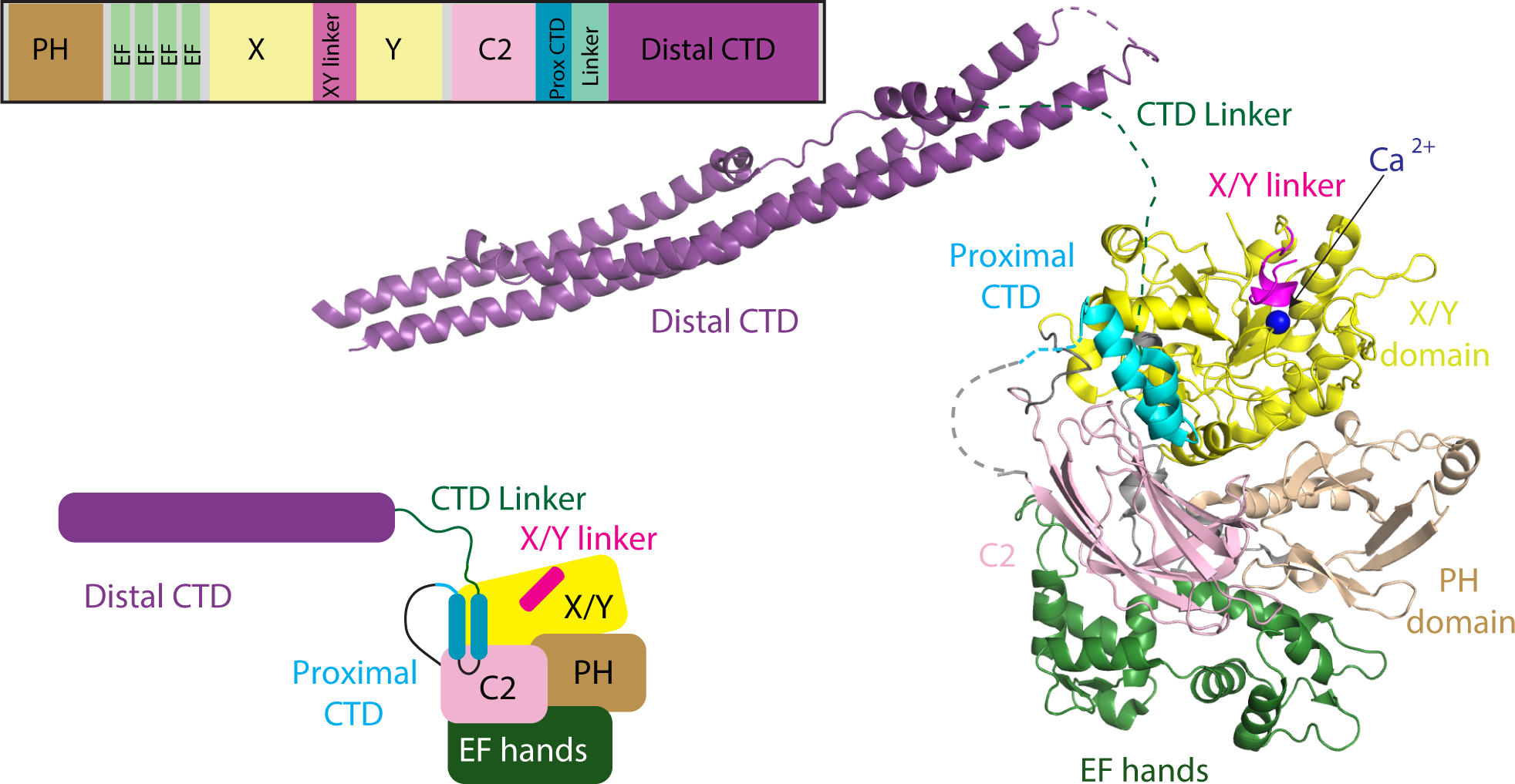
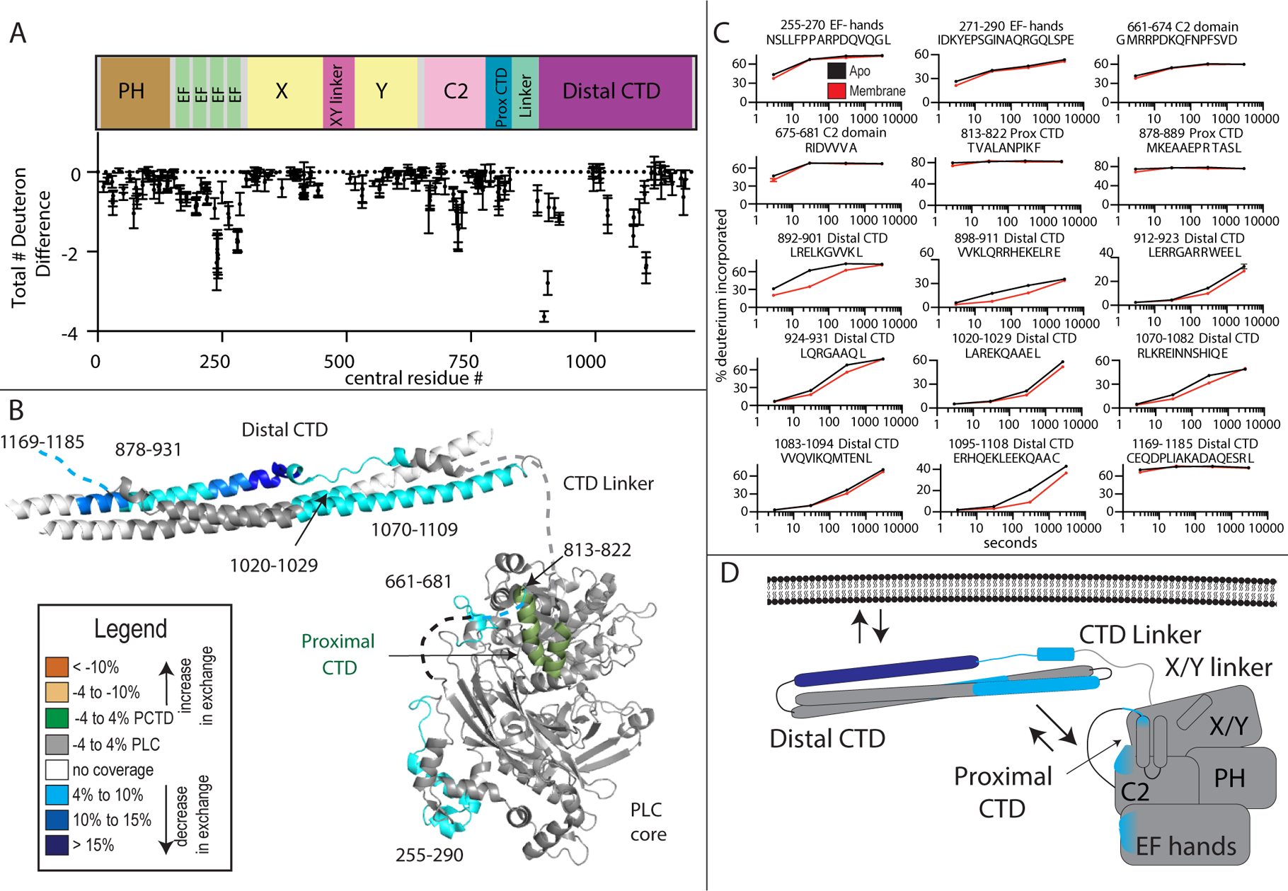
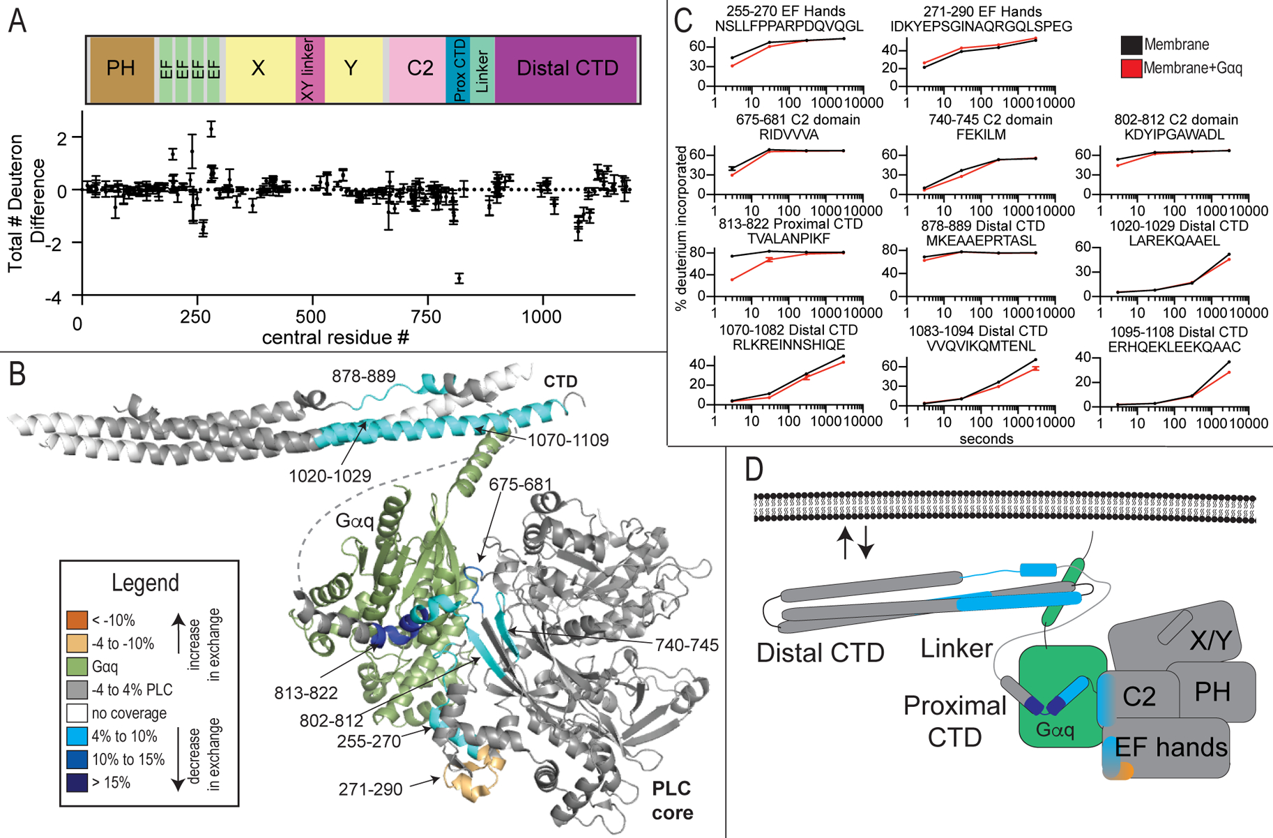
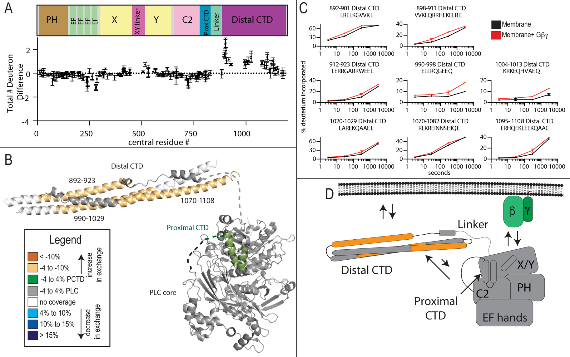
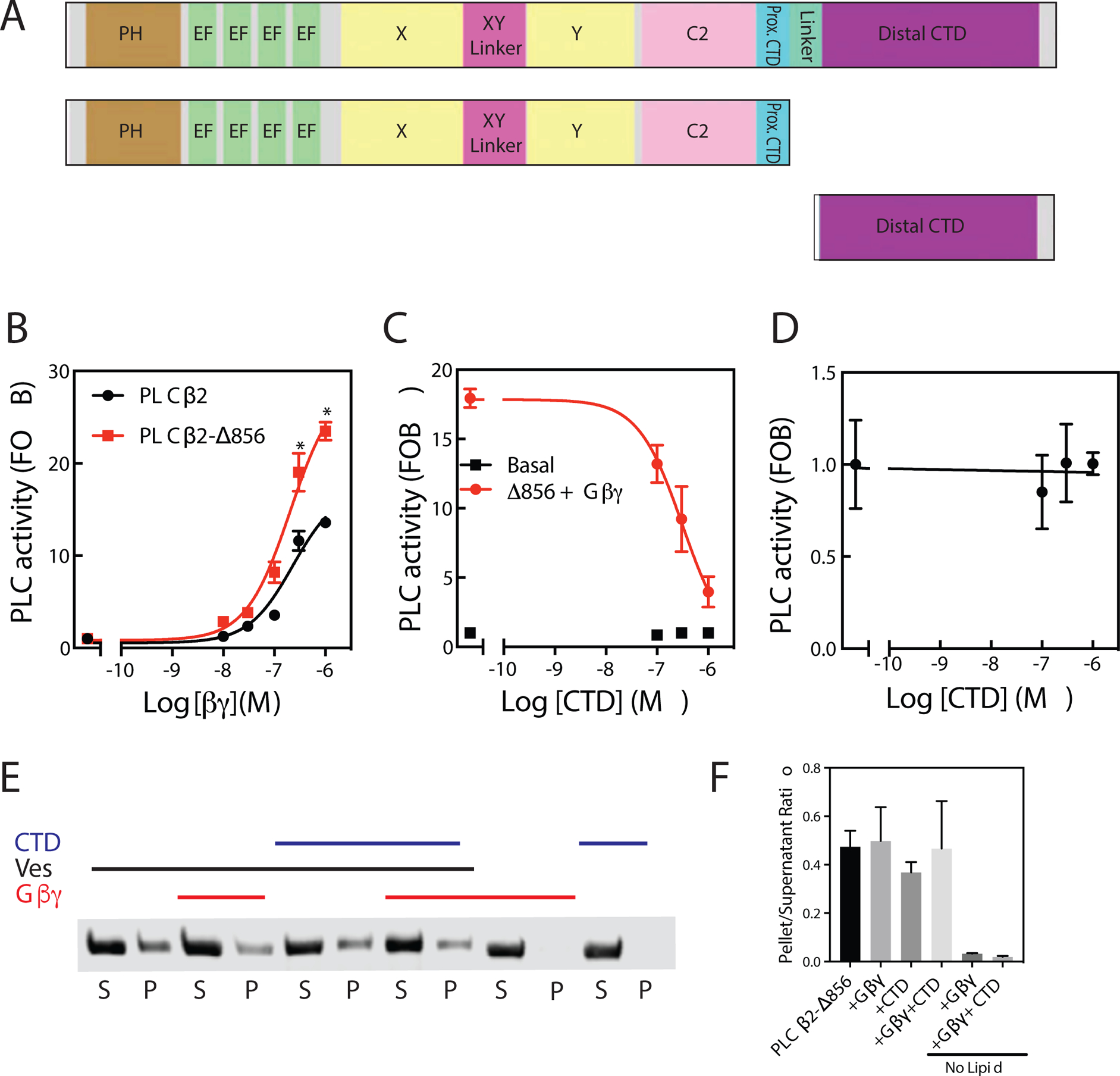
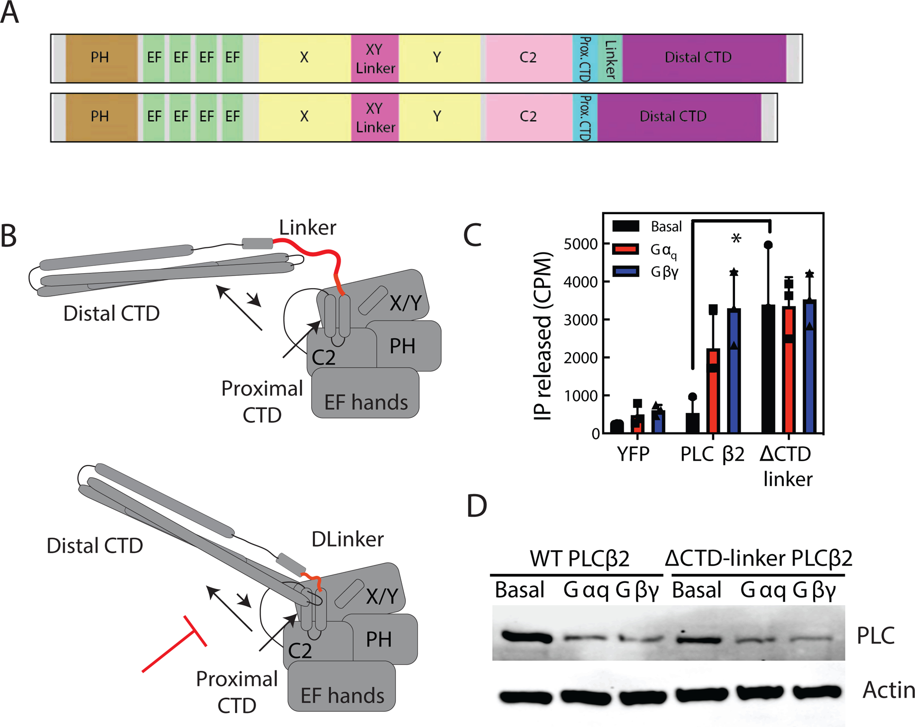
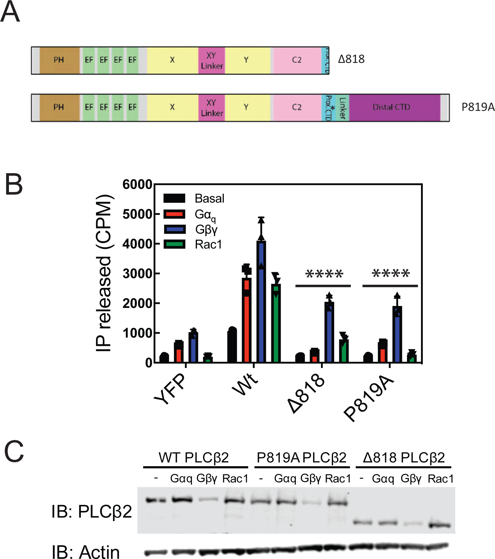
Phospholipase C (PLC) enzymes hydrolyze phosphoinositide lipids to inositol phosphates and diacylglycerol. Direct activation of PLCβ by Gαq and/or Gβγ subunits mediates signaling by Gq and some Gi coupled G-protein-coupled receptors (GPCRs), respectively. PLCβ isoforms contain a unique C-terminal extension, consisting of proximal and distal C-terminal domains (CTDs) separated by a flexible linker. The structure of PLCβ3 bound to Gαq is known, however, for both Gαq and Gβγ; the mechanism for PLCβ activation on membranes is unknown. We examined PLCβ2 dynamics on membranes using hydrogen-deuterium exchange mass spectrometry (HDX-MS). Gβγ caused a robust increase in dynamics of the distal C-terminal domain (CTD). Gαq showed decreased deuterium incorporation at the Gαq binding site on PLCβ. In vitro Gβγ-dependent activation of PLC is inhibited by the distal CTD. The results suggest that disruption of autoinhibitory interactions with the CTD leads to increased PLCβ hydrolase activity.

Keywords: G protein; G-protein-coupled receptor; GPCR; Gαq; Gβγ; HDX-MS; hydrogen-deuterium exchange; lipids; membrane interactions; phospholipase C; protein dynamics; signal transduction.


Conflict of interest statement

Declaration of Interests The authors declare no competing interests.

Figures

Figure 1:
Figure 1:. Domain Architecture and model of PLCβ2 architecture
A model of PLCβ2 based on a monomer of PLCβ3 from the co-crystal structure of Gαq-PLCβ3 (PDB:4GNK), and the resolved portion of the proximal CTD from the structure of S. officinalis PLC21 (PDB: 3QR0). This model is oriented such that the active site (as indicated by the catalytic Ca2+ ion) is oriented towards the top of the figure. Domains are colored as in the domain schematics in the top and bottom left and the domain boundaries are (PLCβ2 numbering system) PH domain: 15–135; EF hands: 143–296; X: 321–466; XY Linker: 466–537; Y: 537–662; C2: 672–803; Prox CTD: 809–837; CTD Linker: 838–874; Distal CTD: 875–1150. The Gαq binding Helix-loop-Helix (amino acids: 810–827) is disordered in the structures solved in the apo PLCβ models with no bound Gαq. A model of the structure is shown on the right, with this used as a template for describing all HDX-MS data.
Figure 2.
Figure 2.. Membrane dependent changes in PLCβ2 dynamics.
(A) The total number of deuteron difference between apo and membrane bound states for all peptides analyzed over the entire deuterium exchange time course for in PLCβ2. Every point represents the centroid of an individual peptide, with the domain schematic present above. Error bars represent S.D. (n=3). (See Table S1 for HDX data characterization) (B) Peptides with significant changes in deuterium incorporation (both >0.4 Da and >4% and T-test p < 0.05 at time point) in the presence of membrane are mapped on a structural model of PLCβ2 as described in Fig. 1. Differences are mapped according to the legend. (C) Representative PLCβ2 peptides displaying significant increases or decreases in exchange in the presence of membrane are shown. For all panels, error bars show SD (n=3), with most smaller than the size of the point. The full list of all peptides and their deuterium incorporation is shown in Data S1. For dynamic peptides with changes in exchange at only the earliest time point (813–822, 878–889, and 1169–1185) there are additional graphs in Supplemental Fig 1B that show these differences more clearly. (D) Cartoon schematic displaying deuterium exchange differences, and potential mechanisms for altered protein dynamics.
Figure 3.
Figure 3.. Gαq dependent changes in PLCβ2 dynamics
(A) The total number of deuteron difference between membrane and membrane-q states for all peptides analyzed over the entire deuterium exchange time course for PLCβ2. Every point represents the centroid of an individual peptide. Error bars represent S.D. (n=3). (B) Peptides with significant changes in deuterium incorporation (both >0.4 Da and >4% and T-test p < 0.05 at time point) in the presence of Gαq are mapped on a structural model of PLCβ2 as described in Fig. 1. Differences are mapped according to the legend. (C) Representative PLCβ2 peptides displaying significant increases or decreases in exchange in the presence of Gαq are shown. For all peptides, error bars show SD (n=3), with most smaller than the size of the point. The full list of all peptides and their deuterium incorporation is shown in Data S1. (D) Cartoon schematic displaying deuterium exchange differences on a model of Gαq binding.
Figure 4.
Figure 4.. Gβγ binding to PLCβ2 causes conformational changes in the distal CTD domain.
(A)The total number of deuteron difference between membrane and membrane- Gβγ states for all peptides analyzed over the entire deuterium exchange time course for PLCβ2. Every point represents the centroid of an individual peptide. Error bars represent S.D. (n=3). (See also Figure S2) (B) Peptides with significant changes in deuterium incorporation (both >0.4 Da and >4% T-test p < 0.05 at any time point) in the presence of membrane are mapped on a structural model of PLCβ2 as described in Fig. 1. Differences are mapped according to the legend. (C) Representative PLCβ2 peptides displaying significant increases or decreases in exchange in the presence of Gβγ are shown. For all panels, error bars show SD (n=3), with most smaller than the size of the point. (D) Cartoon schematic displaying deuterium exchange differences upon Gβγ, and potential mechanisms for altered protein dynamics.
Figure 5:
Figure 5:. The Distal CTD inhibits Gβγ activation of PLCβ2.
(A) Schematic of PLCβ2 constructs. (B) Assay of PLC enzymatic activity for the indicated purified PLC protein with the indicated concentrations of purified Gβγ subunits performed at least 3 times in duplicate. The data shown are mean ± S.D. and analysed by 2-way ANOVA with multiple comparisons post-test. *, p < 0.05 versus Full length PLCβ2 at equal concentration of Gβγ. (C) Assay of PLC enzymatic activity for the PLCβ2-ΔCTD with 250 nM Gβγ subunits and incubated with the indicated concentrations of purified CTD performed at least 3 times in duplicate. The data shown are mean ± S.D. (D) Expanded version of panel C to emphasize the PLCβ2-ΔCTD basal activity. The data shown are mean ± S.D. (Basal CPM = 234±15, total CPM = 6700 in the assay) (See also Figure S3). (E) Representative western blot image of PLCβ following ultracentrifugation in the presence (+) or absence (−) of PE/PIP2 liposomes, 1 μM CTD and/or purified 500ng Gβγ as indicated. (F) The ratio of protein present in the supernatant samples versus the respective total protein control samples is shown. No significant differences in lipid binding for PLCβ2-ΔCTD in the presence of CTD or Gβγ was detected. Data represent at least three independent experiments. The data shown are mean ± S.D.
Figure 6:
Figure 6:. Restriction of Distal CTD movement increases PLC activity.
(A) Schematic of CTD-linker deletion (amino acids 839–872 deleted). (B) Cartoon diagram of model of CTD-linker deletion effects. By preventing C-terminal domain rearrangement, CTD linker would prevent autoinhibition. (C) Total inositol phosphate assay. COS-7 cells were transfected with 250 ng PLCβ2 WT or PLCβ2-ΔCTD-linker in the presence or absence of 200 ng Gβ1 and 200 ng Gγ2 or 200 ng Gαq, and total [3H] inositol phosphate accumulation was measured. The data shown are mean ± S.D. for at least three independent experiments and analysed by 2-way ANOVA with multiple comparisons. *, p < 0.05. (D) Western blot for PLCβ2 wt and PLCβ2-ΔCTD-linker mutant expressed in C, and actin as a loading control.
Figure 7:
Figure 7:. The Proximal CTD is involved PLC activation by Gαq and Rac1.
(A) Schematic of mutants and deletions of PLCβ2. (B) COS-7 cells were transfected with 250ng of WT PLCβ2, P819A PLCβ2, or Δ818-PLCβ2 in the presence or absence of 200ng Gβ1 and 200 ng Gγ2 or 300 ng Rac1G12V or 200ng wild type Gαq and total [3H] inositol phosphate accumulation was measured. The data shown are mean ± S.D. for at least three independent experiments and analysed by 2-way ANOVA with multiple comparisons. ****, p < 0.001 statistically different that wt PLC transfection. (C) Western blot for PLCβ2 wild type and mutants in B and C with an actin loading control.

Similar articles

Cited by

References

    1. Adjobo-Hermans MJ, Crosby KC, Putyrski M, Bhageloe A, van Weeren L, Schultz C, Goedhart J, and Gadella TW Jr. (2013). PLCβ isoforms differ in their subcellular location and their CT-domain dependent interaction with Gαq. Cell Signal 25, 255–263. - PubMed
    1. Berridge MJ (1987). Inositol tisphosphate and diacylglycerol: two interacting second messengers. Ann Rev Biochem 56, 159–193. - PubMed
    1. Bianchi E, Norcini M, Smrcka A, and Ghelardini C (2009). Supraspinal Gβγ-dependent stimulation of PLCβ originating from G inhibitory protein-μ opioid receptor-coupling is necessary for morphine induced acute hyperalgesia. J.Neurochem 111, 171–180. - PMC - PubMed
    1. Bonacci TM, Ghosh M, Malik S, and Smrcka AV (2005). Regulatory interactions between the amino terminus of G-protein βγ subunits and the catalytic domain of PLCβ2. J Biol Chem 280, 10174–10181. - PubMed
    1. Charpentier TH, Waldo GL, Barrett MO, Huang W, Zhang Q, Harden TK, and Sondek J (2014). Membrane-induced Allosteric Control of Phospholipase C-β Isozymes. J Biol Chem 289, 29545–29557. - PMC - PubMed

Publication types

MeSH terms

Substances

LinkOut - more resources