. 2020 May 12;31(6):107642.
doi: 10.1016/j.celrep.2020.107642.

Maternal Anti-Dengue IgG Fucosylation Predicts Susceptibility to Dengue Disease in Infants

Affiliations

Maternal Anti-Dengue IgG Fucosylation Predicts Susceptibility to Dengue Disease in Infants

Natalie K Thulin et al. Cell Rep. .

Abstract

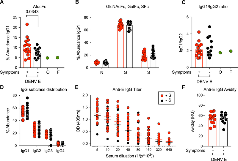
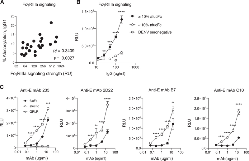
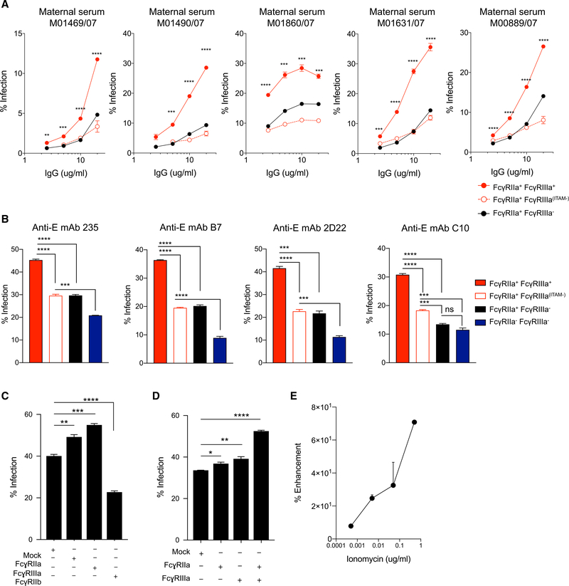
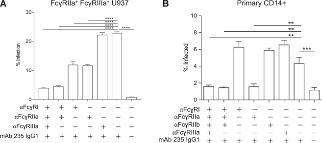
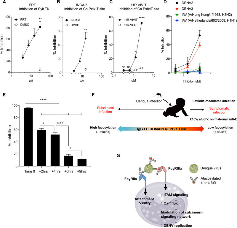
Infant mortality from dengue disease is a devastating global health burden that could be minimized with the ability to identify susceptibility for severe disease prior to infection. Although most primary infant dengue infections are asymptomatic, maternally derived anti-dengue immunoglobulin G (IgGs) present during infection can trigger progression to severe disease through antibody-dependent enhancement mechanisms. Importantly, specific characteristics of maternal IgGs that herald progression to severe infant dengue are unknown. Here, we define ≥10% afucosylation of maternal anti-dengue IgGs as a risk factor for susceptibility of infants to symptomatic dengue infections. Mechanistic experiments show that afucosylation of anti-dengue IgGs promotes FcγRIIIa signaling during infection, in turn enhancing dengue virus replication in FcγRIIIa+ monocytes. These studies identify a post-translational modification of anti-dengue IgGs that correlates with risk for symptomatic infant dengue infections and define a mechanism by which afucosylated antibodies and FcγRIIIa enhance dengue infections.

Keywords: CD16; FcγRIIIa; IgG fucosylation; antibody glycosylation; infant dengue; severe dengue.


Conflict of interest statement

Declaration of Interests The authors declare no competing interests.

Figures

Figure 1.
Figure 1.. Maternal Anti-E IgG Characterization
Anti-E Fc glycoforms were characterized. (A) Fucosylation of Fc glycoforms was significantly different in abundance between mothers of infants with symptomatic (red) and asymptomatic (black) dengue infections, with afucosylated Fc glycans significantly increased in mothers of infants with symptomatic dengue infections. ≥10% afucosylated Fc glycans was predictive of elevated risk for symptomatic infant dengue infections with a positive predictive value of 88%. (B) No significant differences in other Fc glycoforms, bisected (N), galactosylated (G), or sialylated (S), were observed. (C–F) There were also no differences in the maternal anti-E (C) IgG1/IgG2 subclass ratio, (D) IgG subclass distribution, (E) IgG titer on dengue-infected cells, or (F) IgG avidity between mothers of infants who developed symptomatic or asymptomatic primary dengue infections. Each symbol (A–F) represents an individual donor. Data are representative of one experiment per donor (A–D) or three experiments (E and F); (A–C and F) horizontal bars represent mean of clinical samples. Significance was assessed by unpaired Student’s t test, where p < 0.05 was considered significant; *p < 0.05.
Figure 2.
Figure 2.. Anti-E Fucosylation in Dengue Virus Immune Complexes Determines FcγRIIIa Signaling by Effector Cells
Dengue immune complexes were generated from maternal serum IgGs. (A) FcγRIIIa signaling was linearly correlated with the abundance of afucosylated Fc glycoforms of anti-DENV E IgGs within immune complexes, n = 29. (B) Dengue immune complexes generated from pooled maternal IgGs with ≥ 10% anti-E afucosylation triggered significantly more FcγRIIIa signaling when compared with immune complexes generated from pooled maternal IgGs with <10% anti-E afucosylation. Both pools were generated from sera of mothers of infants who experienced symptomatic primary dengue infections. (C) Fucosylation of distinct anti-E mAbs modulates FcγRIIIa signaling. Fucosylated (black circles) or afucosylated (white circles) anti-DENV E mAbs with distinct Fab specificities were tested for the ability to trigger FcγRIIIa signaling. Enhanced FcγRIIIa signaling by afucosylated immune complexes was observed in all cases. GRLR is a non-FcγR-binding Fc variant. Each point in (A) represents an individual donor. Data shown in (B) and (C) are representative of three experiments. *p<0.05, **p < 0.01, ***p < 0.001, ****p < 0.0001 (unpaired Student’s t test). Data in (A)–(C) are representative of a minimum of three experiments. Error bars (B and C) represent the standard error of three technical replicates. Replicates were performed at all points; where error bars are not present, the standard error was smaller than the width of the symbol marking the data point.
Figure 3.
Figure 3.. FcγRIIIa Signaling Enhances Antibody-Dependent Infection of U937 Monocytes
(A) Infection by dengue immune complexes generated with maternal IgGs was significantly enhanced in FcγRIIIa+ monocytes, and FcγRIIIa signaling was required for enhancement in all cases. Red line: FcγRIIa+FcγRIIIa+ cells; black line: FcγRIIa+FcγRIIIa cells; broken red line, unfilled circles: FcγRIIa+FcγRIIIa(ITAM–) cells. (B) The role of FcγRIIIa signaling in ADE was evaluated by comparison of infection in FcγRIIa+FcγRIIIa+ cells (red), FcγRIIIa-signaling null FcγRIIa+FcγRIIIa(ITAM–) cells (white), and FcγRIIa+FcγRIIIa (black) or FcγRIIaFcγRIIIa (blue) cells. Immune complexes were generated from dengue virus and afucosylated anti-envelope mAbs 235, B7, 2D22, or C10, which have distinct anti-E binding specificities. In all cases, FcγRIIIa signaling was required for maximal infection. (C) Signaling induced by crosslinking of FcγRIIa or FcγRIIIa promoted infection of FcγRIIa+FcγRIIIa+ U937 cells, whereas crosslinking the inhibitory FcγR, FcγRIIb, reduced infectibility of cells. (D) Combined signaling by activating FcγRIIa and FcγRIIIa during infection resulted in maximal infectibility of FcγRIIa+FcγRIIIa+ U937 cells. (E) Increasing cellular calcium flux promoted infection of FcγRIIa+FcγRIIIa+ U937 cells. *p < 0.05, **p < 0.01, ***p < 0.001, ***p < 0.0001 (unpaired Student’s t test). Data are representative of a minimum of three experiments. Error bars represent the standard error of three technical replicates. Replicates were performed at all points; where error bars are not present, the standard error was smaller than the width of the symbol marking the data point.
Figure 4.
Figure 4.. FcγRIIIa Does Not Mediate Virus Immune Complex Attachment during ADE in Monocytes
(A) Individual or combined FcγRs were blocked during the viral attachment step of infection in FcγRIIa+ FcγRIIIa+ U937 monocytes. Blocking FcγRIla and/or FcγRI during immune complex attachment significantly reduced infection, whereas blocking FcγRIIIa did not impact infection. (B) Blocking FcγRIIa alone during attachment in primary monocytes prevented their infection, whereas blocking FcγRI, FcγRIIb, or FcγRIIIa did not impact infection. **p < 0.01, ***p < 0.001, ****p < 0.0001 (unpaired Student’s t test). Data are representative of a minimum of three experiments. Error bars represent the standard error of three technical replicates.
Figure 5.
Figure 5.. Inhibition of Dengue Infection through Targeted Inhibition of ITAM Signaling Pathway Elements
(A) A small molecule that targets Syk tyrosine kinase (Syk TK), PRT, inhibited dengue infection in FcγRNa+FcγRNIa+ U937 monocytes. (B and C) A small-molecule inhibitor, INCA-6 (B), and a peptide inhibitor, 11R-VIVIT (VIVIT) (C), which act by blocking calcineurin-substrate interactions at the calcineurin PxIxIT site, inhibited dengue infections. A scrambled peptide containing the amino acids found in VIVIT (11R VEET) did not inhibit infection (open circles). (D) Inhibition of dengue viruses (DENV2, DENV3), but not H1N1 or H3N2 influenza viruses (IAVs), was observed using the VIVIT inhibitor (solid lines); scrambled VEET peptide in dashed lines. (E) Treatment of cells with VIVIT up to 6 h after infection by dengue immune complexes significantly inhibited infection. (F) Model for susceptibility to clinically significant dengue infections in infants. Infants of mothers who are dengue-immune and have ≥10% afucosylated anti-E IgGs are at elevated risk for symptomatic primary dengue infections due to modulation of FcγRIIIa-expressing cells during infection. (G) Model for the roles of FcγRIIa, FcγRIIIa, and afucosylated dengue immune complexes in ADE. While FcγRIIa supports a majority of viral attachment and entry, FcγRIIIa signaling increases cellular ITAM signaling. ITAM signaling, in turn, triggers calcium flux and modulates the calcineurin signaling network, which supports dengue virus replication. *p < 0.05, **p < 0.01, ****p < 0.0001 (unpaired Student’s t test). Data are representative of aminimum of three experiments. Error bars represent the standard error of three technical replicates. Replicates were performed at all points; where error bars are not present, the standard error was smaller than the width of the symbol marking the data point.

References

    1. Alkan Ozdemir S, Ozer EA, Kose S, Ilhan O, Ozturk C, and Sutcuoglu S (2016). Reference values of serum IgG and IgM levels in preterm and term new-borns. J. Matern. Fetal Neonatal Med. 29, 972–976. - PubMed
    1. Anderson KB, Gibbons RV, Cummings DA, Nisalak A, Green S, Libraty DH, Jarman RG, Srikiatkhachorn A, Mammen MP, Darunee B, et al. (2014). A shorter time interval between first and second dengue infections is associated with protection from clinical illness in a school-based cohort in Thailand. J. Infect. Dis. 209, 360–368. - PMC - PubMed
    1. Anthony RM, Kobayashi T, Wermeling F, and Ravetch JV (2011). Intravenous gammaglobulin suppresses inflammation through a novel T(H)2 pathway. Nature 475, 110–113. - PMC - PubMed
    1. Aramburu J, Yaffe MB, López-Rodríguez C, Cantley LC, Hogan PG, and Rao A (1999). Affinity-driven peptide selection of an NFAT inhibitor more selective than cyclosporin A. Science 285, 2129–2133. - PubMed
    1. Aye KS, Charngkaew K, Win N, Wai KZ, Moe K, Punyadee N, Thiemmeca S, Suttitheptumrong A, Sukpanichnant S, Prida M, and Halstead SB (2014). Pathologic highlights of dengue hemorrhagic fever in 13 autopsy cases from Myanmar. Hum. Pathol. 45, 1221–1233. - PubMed

Publication types