. 2020 Jul:131:155115.
doi: 10.1016/j.cyto.2020.155115. Epub 2020 May 3.

Therapeutic potential of Nitazoxanide against Newcastle disease virus: A possible modulation of host cytokines

Affiliations

Therapeutic potential of Nitazoxanide against Newcastle disease virus: A possible modulation of host cytokines

Ferrin Antony et al. Cytokine. 2020 Jul.

Abstract

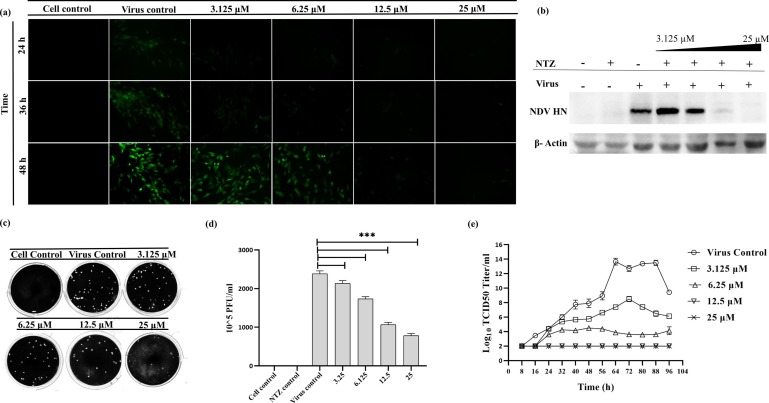
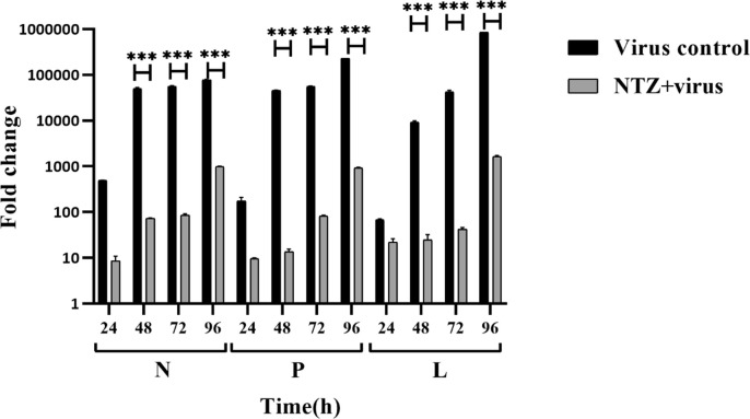
Newcastle disease (ND) is prevalent among the domesticated and the wild birds and is caused by the avian paramyxovirus serotype-I (APMV-I). It is commonly known to affect chicken, pheasant, ostrich, pigeon and waterfowl. Depending on the virulence, the velogenic NDV strains cause severe respiratory and nervous disorders with a high mortality rate. The live and killed vaccines are available for the prevention of infection in the market, but the drug for the treatment is not available. Nitazoxanide (NTZ), a member of thiazolides, is an antiparasitic drug. In the present study, the effect of NTZ on the NDV replication was explored. The experiments were conducted in chicken fibroblast cells (DF-1), PBMC, embryonated chicken eggs, and two-week old chickens. The inhibition of the NDV was observed upon post-treatment of NTZ at a concentration of ~12.5 μM. Cytokine profiling of the DF-1, PBMC, and chicken embryonic tissue treated with NTZ revealed significant upregulation in all the cytokines studied except for IL-1β in DF-1 cells. It is plausible that NTZ is involved in causing immune-modulatory effects in poultry. NTZ treatment in two weeks old chicken showed significant reduction in NDV replication in trachea, and lungs, respectively, at 72 h post-infection. Encouraging results from the present study warrants repurposing NTZ as a drug for the treatment of viral infection in poultry. It will also pave the way towards understanding of similar effect against other animal pathogens.

Keywords: Avian paramyxovirus; Cytokines; In vitro; Nitazoxanide; Repurposing.


Conflict of interest statement

Declaration of Competing Interest The authors declare that they have no known competing financial interests or personal relationships that could have appeared to influence the work reported in this paper.

Figures

Fig. 1
Fig. 1
Cytotoxicity of NTZ in DF-1 cells using MTT assay. Graph depicting the percentage cell viability after 24 h treatment with increasing concentrations of NTZ (0.3–100 µM) was plotted (a). Linear regression graph was plotted to determine the IC50 value of NTZ (~71.6 μM) after 24 h (b). Graph showing the percentage cell viability after 48 h treatment with increasing concentrations of NTZ (0.3–100 µM) was plotted (c). Linear regression graph was plotted to determine the IC50 value of NTZ (~63.3 μM) after 48 h (d). DF-1 cell images were taken in an inverted microscope after 24 h and 48 h treatment with NTZ (12.5 μM, 25 μM and 50 μM) (e).
Fig. 2
Fig. 2
Time of addition assay in DF-1 cells to determine the anti-NDV effect of NTZ. Schematic representation of the time of addition assay performed (a). DF-1 cell images were taken in an inverted fluorescence microscope after infection with rNDV/GFP and pre, co and post-treatment of NTZ (b). Mock-infected DF-1 cells were used as a control. Growth kinetics of NDV was determined after pre, co and post-treatment of NTZ using TCID50 assay (c). NDV reduction was observed when treated with NTZ (pre, co and post-treatment) using plaque assay (d). The graph was plotted from the number of plaques observed after NDV infection and pre, co, and post-treatment of NTZ (e). Statistical significance difference was determined by using ordinary one way ANOVA (Dunnett’s multiple comparisons test, GraphPad Prism 8). A p-value less than 0.01 is flagged with one star (**), and less than 0.001 is flagged with three stars (***).
Fig. 3
Fig. 3
Dose-dependent anti-NDV effect of NTZ. DF-1 cell images were taken in an inverted fluorescence microscope after infection with rNDV/GFP and post-treatment of NTZ with varying concentrations (3.125 μM, 6.25 μM, 12.5 μM and 25 μM) (a). Mock-infected DF-1 cells were used as a control. Reduction in NDV protein HN after dose-dependent treatment of NTZ was observed using western blotting (b). β-Actin was used as a loading control. NDV reduction was observed after dose-dependent treatment with NTZ using plaque assay (c). The graph was plotted from the number of plaques observed after NDV infection and dose-dependent treatment of NTZ. Statistical significance difference was determined by using ordinary one way ANOVA (Dunnett’s multiple comparisons test, GraphPad Prism 8). A p-value less than 0.001 is flagged with three stars (***) (d). Growth kinetics of NDV was determined after treatment with varying concentrations of NTZ using TCID50 assay (e).
Fig. 4
Fig. 4
Reduction of NDV gene expression after treatment with NTZ in vitro. A comparison of the expression of NDV non-structural genes (N, P, and L) was done using real-time PCR. Graph depicting the fold change in NDV gene expression in the presence and absence of NTZ at different time points (24, 48, 72, and 96 h) was plotted. Normalization was done with mock-infected cells. GAPDH was used as an endogenous gene expression control. Statistical significance difference was determined by using two way ANOVA (Sidak’s multiple comparisons test, GraphPad Prism 8). A p-value less than 0.001 is flagged with three stars (***). Bars that are not flagged are not significant.
Fig. 5
Fig. 5
In ovo experiment showing the anti-NDV effect of NTZ. Nine-day-old embryos after 48 h of NDV infection in the presence and absence of NTZ (~12.5 μM) was observed (a). Visible gross lesions indicated NDV infection. Quantification of NDV from the allantoic fluid and the tissue collected from the embryos using plaque assay (b). The graph was plotted from the number of plaques observed from the allantoic fluid and the tissue collected from the embryos (c).
Fig. 6
Fig. 6
Cytokine expression profiling (IL18, TLR7, TNF-α, IL-1β, NLRP3, IFN-α, and IFN-β) of DF-1, PBMC and chicken embryo tissue treated with NTZ was performed using qPCR (a, b, c). Normalization was done with the mock-treated controls. GAPDH was used as an endogenous gene expression control. Statistical significance difference was determined by using two way ANOVA (Tukey’s multiple comparisons test, GraphPad Prism 8). A p-value less than 0.05 is flagged with one star (*), less than 0.01 is flagged with two stars (**), and less than 0.001 is flagged with three stars (***). Bars that are not flagged are not significant.
Fig. 7
Fig. 7
In vivo experiment showing the anti-NDV effect of NTZ. NDV viral titer in trachea, lung, and spleen in six different groups of chicken, namely, mock treated control group, NTZ only treated group, virus alone infected group, Pre-treatment NTZ with virus infected group, Co-treatment NTZ with virus infected group, and Post-treatment NTZ with virus infected group. All the birds were harvested 72 h post virus infection, and various tissues were collected, triturated and subjected to TCID50 assay. Statistical significance difference was determined by using two way ANOVA (Tukey’s multiple comparisons test, GraphPad Prism 8). A p-value less than 0.001 is flagged with three star (***).

References

    1. R.A. Lamb, G.D. Parks, Paramyxoviridae: The Viruses and their Replication, 5th ed., Lippincott, Williams, and Wilkins, 2007.
    1. Czegledi A., Ujvari D., Somogyi E., Wehmann E., Werner O., Lomniczi B. Third genome size category of avian paramyxovirus serotype 1 (Newcastle disease virus) and evolutionary implications. Virus Res. 2006;120(1–2):36–48. - PubMed
    1. de Leeuw O., Peeters B. Complete nucleotide sequence of Newcastle disease virus: evidence for the existence of a new genus within the subfamily Paramyxovirinae. J. Gen. Virol. 1999;80(Pt 1):131–136. - PubMed
    1. Huang Y., Wan H.Q., Liu H.Q., Wu Y.T., Liu X.F. Genomic sequence of an isolate of Newcastle disease virus isolated from an outbreak in geese: a novel six nucleotide insertion in the non-coding region of the nucleoprotein gene. Brief Rep. Arch. Virol. 2004;149(7):1445–1457. - PubMed
    1. Krishnamurthy S., Samal S.K. Nucleotide sequences of the trailer, nucleocapsid protein gene and intergenic regions of Newcastle disease virus strain Beaudette C and completion of the entire genome sequence. J. Gen. Virol. 1998;79(Pt 10):2419–2424. - PubMed

Publication types

MeSH terms