Case Reports
. 2020 Aug;88(2):332-347.
doi: 10.1002/ana.25772. Epub 2020 Jun 18.

GGPS1 Mutations Cause Muscular Dystrophy/Hearing Loss/Ovarian Insufficiency Syndrome

Affiliations
Case Reports

GGPS1 Mutations Cause Muscular Dystrophy/Hearing Loss/Ovarian Insufficiency Syndrome

A Reghan Foley et al. Ann Neurol. 2020 Aug.

Abstract

Objective: A hitherto undescribed phenotype of early onset muscular dystrophy associated with sensorineural hearing loss and primary ovarian insufficiency was initially identified in 2 siblings and in subsequent patients with a similar constellation of findings. The goal of this study was to understand the genetic and molecular etiology of this condition.

Methods: We applied whole exome sequencing (WES) superimposed on shared haplotype regions to identify the initial biallelic variants in GGPS1 followed by GGPS1 Sanger sequencing or WES in 5 additional families with the same phenotype. Molecular modeling, biochemical analysis, laser membrane injury assay, and the generation of a Y259C knock-in mouse were done.

Results: A total of 11 patients in 6 families carrying 5 different biallelic pathogenic variants in specific domains of GGPS1 were identified. GGPS1 encodes geranylgeranyl diphosphate synthase in the mevalonate/isoprenoid pathway, which catalyzes the synthesis of geranylgeranyl pyrophosphate, the lipid precursor of geranylgeranylated proteins including small guanosine triphosphatases. In addition to proximal weakness, all but one patient presented with congenital sensorineural hearing loss, and all postpubertal females had primary ovarian insufficiency. Muscle histology was dystrophic, with ultrastructural evidence of autophagic material and large mitochondria in the most severe cases. There was delayed membrane healing after laser injury in patient-derived myogenic cells, and a knock-in mouse of one of the mutations (Y259C) resulted in prenatal lethality.

Interpretation: The identification of specific GGPS1 mutations defines the cause of a unique form of muscular dystrophy with hearing loss and ovarian insufficiency and points to a novel pathway for this clinical constellation. ANN NEUROL 2020;88:332-347.


Conflict of interest statement

T.V. reports personal fees from DZHK, Antisense Therapeutics, Biophytis, Capricor, Italfarmaco, Santhera, Servier, Sarepta, Solid Biosciences, Dynacure, DiNAQOR, and Catabasis outside the submitted work.

Figures
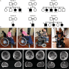
FIGURE 1
FIGURE 1
Autosomal recessive inheritance of GGPS1 mutations and phenotypic characteristics of GGPS1‐related muscular dystrophy. (A) Patient pedigrees demonstrating biallelic GGPS1 pathogenic variants in 6 separate families. (B) Prominent lordosis (following spinal fixation surgery) and hip flexion, knee flexion, and elbow flexion contractures seen in Patients P1 and P2, who both demonstrate diaphragm compressing maneuvers, including rocking back and forth or increasing intra‐abdominal pressure on expiration by hand (a–d). Patient P1 has a tracheostomy for ventilation (a and b), whereas Patient P2 uses noninvasive ventilation in the form of bilevel positive airway pressure. Severe neck extension contracture is evident in Patient P2 (c), necessitating support of the neck with the hand to promote upright head posture (d). (C) Muscle magnetic resonance imaging (MRI) performed in Family 3 (Patients P6 [a and d], P4 [b and e], and P5 [c and f]) shows variably increased T1 signal in muscles, indicative of fatty infiltration. There is evidence of relative sparing of the rectus femoris, sartorius, and gracilis muscles (a, b, and c) and relative involvement of the soleus muscle (d, e, and f). There is evidence of considerably asymmetric involvement with abnormal T1 signal in the lateral aspect of the vastus lateralis in Patient P4 on the right only (b, arrow) and abnormal T1 signal of the adductor longus on the left only (arrowhead). Patient P5 (c) has asymmetric involvement of the hamstrings with notable involvement of the hamstrings in the right leg and relative sparing of the hamstrings in the left leg.
FIGURE 2
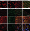
FIGURE 2
Histological, ultrastructural, and immunostaining characteristics of muscle in GGPS1‐related muscular dystrophy. (A–C) Muscle from a deltoid biopsy performed in Patient P1 at 21 years demonstrates nuclear internalization and a rimmed vacuole (arrow) on Gömöri Trichrome staining (A). Nicotinamide adenine dinucleotide (NADH; B) and cytochrome oxidase (COX; C) stains reveal focally irregular staining, including core‐like regions (C). (D–F) Muscle from a deltoid biopsy performed in Patient P4 at 39 years demonstrates a ragged red fiber (asterisk) and myophagocytosis (arrow) on Gömöri trichrome staining (D). NADH (E) and COX (F) staining demonstrate mildly irregular staining and a COX‐negative fiber (asterisk; F). (G) Electron microscopy (EM) performed on Patient P1's deltoid muscle biopsy at 21 years reveals autophagic material including myeloid bodies. (H, I) EM of Patient P2's deltoid biopsy at 19 years shows findings suggestive of mitophagy (H) and accumulation of subsarcolemmal mitochondria (I). (J–L) Patient P8's quadriceps biopsy at 14 years reveals ultrastructural evidence of elongated mitochondria (J) and an accumulation of autophagic debris (K). Further evidence of disordered autophagy is seen by positive staining for the autophagy marker LC3B (L).
FIGURE 3
FIGURE 3
Geranylgeranyl diphosphate synthase (GGPPS): mutation localization, conformation, and the mevalonate pathway. (A) Alignment of human GGPPS with GGPPS from other species. Red shaded boxes indicate the position of the mutations. Blue boxes show residues involved in protein/protein interactions; orange boxes show conserved catalytic residues. Red cylinders show α‐helices of human GGPPS. Whereas the catalytic domain is conserved across animals and plants, the mutated residues are conserved in all animals including drosophila, but not in plant species, suggesting a specific role for this domain in animal cells. (B) The molecular weight of GGPPS produced in the bacterial expression system in solution was measured using gel filtration. The GGPPS mutants eluted essentially at a similar point to the wild‐type (WT) enzyme, with estimated molecular size of about 240 to 255kD, suggesting they are presented as a hexamer. There was no indication of any smaller enzyme unit such as the dimer, but a small amount of aggregated protein was apparent for mutants Y259C, R261H, and R261G (arrow). Blue = WT, red = P15S, brown = F257C, green = Y259C, pink = R261H, cyan = R261G. (C) The mevalonate pathway. Mevalonate is the precursor of farnesyl pyrophosphate (FPP) and geranylgeranyl pyrophosphate (GGPP), which are the substrates for prenylation enzymes. Various inhibitors in the pathway are indicated: digeranyl bisphosphonate (DGBP), farnesyl protein transferase inhibitor (FTI), and geranylgeranyl transferase inhibitor (GGTI). FPPS = farnesyl pyrophosphate synthase; FTase = farnesyltransferase; GGTase = geranylgeranyl transferase; GPP = geranyl pyrophosphate; LXR = liver X receptor.
FIGURE 4
FIGURE 4
Geranylgeranyl diphosphate synthase (GGPPS) modeling. (A) GGPPS hexamer overview showing the position of the mutations. Each dimer is composed of a blue and a yellow monomer; the whole molecule is made up of three dimers. F257, Y259, and R261 are shown on monomer A (blue); P15 is shown on monomer D (yellow). The C‐terminus and active site of monomer A are arrowed. The N‐terminus of monomer D is also annotated. Mutated residues at the C‐terminus (F257, Y259, and R261) are located at an externally facing orientation of the 11th α‐helix and outside of the barrel of the hexamer where the catalytic activity of GGPPS is located. The P15S mutation, on the other hand, maps to the first helical domain, which is in close interaction with residues 226–254 (region C in Fig 3A) of monomer A on the adjacent dimer, consistent with its purported role in assembly of the hexameric structure. (B–F) GGPPS mutant sites molecular model. Structural data were analyzed and figures were drawn using Molsoft Browser Pro and PyMOL 2. (B) P15 is inducing a kink in the helix. The side chain of the amino acid is in a relatively large pocket, which contains a water molecule (not shown). This area forms the interaction zone between monomers A (dark blue) and D (yellow) of dimers A/B and C/D. The mutant protein has a serine in this location. The side chain volume of serine is smaller than that of proline, and so modeling a serine residue here using PyMOL 2 shows no steric clashes. (C) F257 (gray balls) sits just buried below the surface of GGPPS. It is surrounded by mainly hydrophobic residues: Y180, H194, V248, and L251 (transparent blue balls). R261 (not shown) fills the gap above it. The van de Waals volume of phenylalanine is 135Å, so it is possible to substitute with the 86Å volume of cysteine, resulting in no steric hinderances. (D) Y259 sits on the surface with several interactions (F215, H219). The hydroxyl of Y259 sits closely with the oxygen (red) atoms of glutamic acid E141 and possibly forms an interaction. Replacing Y259 with C259 presents no steric clashes but probably abolishes any interaction with E141. (E) R261 sits on the outer surface of the GGPPS dimer facing the solvent. The side chain is shown as gray spheres for carbon atoms and blue for nitrogen, and the main chain is in green. (F) Modeling the replacement of this residue with a histidine highlights steric clashes with K265 and F294; however, K265 can rotate to minimize this clash. Replacing R261 with a glycine does not appear to cause a structural clash.
FIGURE 5
FIGURE 5
Geranylgeranyl diphosphate synthase (GGPPS) in human dermal fibroblast and skeletal muscle. (A) In human dermal fibroblasts, GGPPS showed a rather uniform cytoplasmic expression, along with signals concentrated in some organelles and in the perinuclear region. No significant difference was observed between patient‐derived fibroblasts (Patient P1, with GGPS1 compound heterozygous mutations: p.Y259C; R261G) and normal control fibroblasts. (B) GGPPS showed a prominent Z disc localization (Z disc labeled with desmin antibody) in the longitudinal section of normal human muscle biopsy. Scale bar = 2μm. (C) As shown in the human muscle cross section, an increased GGPPS immunoreactive signal was observed in the muscle of patients (Patients P1 and P8) compared with normal control, with some focal, subsarcolemmal accumulation, which partially colocalizes with the mitochondrial marker adenosine triphosphate synthase beta. Scale bars = 15μm.
FIGURE 6
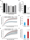
FIGURE 6
Geranylgeranyl diphosphate synthase (GGPPS) enzyme activity and kinetics of human myoblast/myotube membrane repair following laser injury. (A, B) GGPPS catalytic enzyme activity (A) in wild‐type (WT) and mutant GGPPS expressed in a bacterial expression system, using 10μM farnesyl pyrophosphate (FPP)/C14‐isopentenyl pyrophosphate (C14‐IPP) as a substrate, showed the P15S mutant had similar activity as wild‐type; however, the other mutants (F257C, Y259C, R261G, and R261H) consistently showed reduced activity (range about 70~85% of wild‐type activity; n = 3, technical repeats). (B) In human MyoD‐converted myoblasts, the GGPPS enzymatic activity in pooled patients' cells (Patients P1, P2, P6, and P8) decreased to about 50% of the activity in pooled control cells (p = 0.1127). (C–F) Patient myoblasts and myotubes have poor membrane repair ability. Patient myoblasts (C) and myotubes (E) were focally injured by laser, and the traces show the kinetics of the FM 1‐43 dye fluorescence in patient myoblasts (n = 93) and control myoblasts (n = 105), indicating a delay in membrane repair in patient myoblasts (t test, p = 1.9E‐24). (D) Bar graph shows a greater fraction of the injured patient myoblasts failed to repair (30%) compared with control myoblasts (15%; t test, p = 0.026). (E) Similarly, differentiated myotubes showed significantly greater FM 1‐43 dye entry in patient myotubes (n = 23) compared with control myotubes (n = 31; t test, p = 3.1E‐15). Again, a higher proportion of the injured patient myotubes failed to repair (60%) compared with normal control (30%; t test, p = 0.038). (F) Note that myotubes have less efficient repair at baseline compared to myoblasts, hence the difference to the mutant line, although still significant, is less obvious. [CPM = counts per minute; * = statistically significant (p < 0.05)]
FIGURE 7
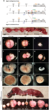
FIGURE 7
Generation of homozygous knock‐in of Y259C mice results in delayed embryonic growth and embryonic lethality. (A) Schematic representation of the targeting strategy for creation of a knock‐in Y259C mutant mouse. Hatched rectangles represent Ggps1 coding sequences; gray rectangles indicate noncoding exon portions. The neomycin‐positive selection cassette is driven by a phosphoglycerate kinase promoter (pGK) as indicated, and the loxP sites are represented by blue triangles and flippase recognition target (FRT) sites by double red triangles. The initiation (ATG) and the Stop (Stop) codon used for isoform 3 where the Y259C mutation is present are indicated. In vivo Flp‐mediated excision of the neomycin cassette is depicted. The diagram is not depicted to scale. (B) Photograph of a representative uterus from a heterozygous female at embryonic day (ED)10.5 following mating to a heterozygous male. Arrows indicate the embryos in C–E while in the uterus. (C–E) Photographs of embryos for each genotype enveloped by their yolk sac with placentas following dissection from the uterus (B). (F–H) Embryos and placentas isolated at ED10.5. Delayed growth in the homozygous knock‐in embryos is shown in H. The knock‐in embryo reflects the development typically observed at ED9.5 in wild‐type (WT) mice. (I–K) Recovery of all genotypes at ED12.5 from heterozygous breeders. Delayed embryonic growth in the homozygous embryo at ED12.5 (K) is reflective of the development of a WT embryo at ED10. (L–N) Placentas from the ED12.5 embryos above. The placenta from the homozygous knock‐in embryo is reduced in size and is developmentally delayed compared to litter controls. (O) Photograph of a uterus from a heterozygous female at ED13.5 showing obvious differences in conceptus size. (P) Representative photograph of embryos in their yolk sac with placentas following dissection from the uterus for each genotype. A homozygous knock‐in embryo in an advanced state of reabsorption is indicated with an asterisk. A WT and a heterozygous embryo are also observed to be undergoing reabsorption in this pregnancy. An embryo that we were unable to extract DNA from for genotyping is indicated with a diamond. Homo = homozygous; Het = heterozygous.

Similar articles

Cited by

References

    1. Mercuri E, Bonnemann CG, Muntoni F. Muscular dystrophies. Lancet 2019;394:2025–2038. - PubMed
    1. Tawil R. Facioscapulohumeral muscular dystrophy. Handb Clin Neurol 2018;148:541–548. - PubMed
    1. Demain LA, Urquhart JE, O'Sullivan J, et al. Expanding the genotypic spectrum of Perrault syndrome. Clin Genet 2017;91:302–312. - PubMed
    1. Goldstein JL, Brown MS. Regulation of the mevalonate pathway. Nature 1990;343:425–430. - PubMed
    1. Xu N, Shen N, Wang X, et al. Protein prenylation and human diseases: a balance of protein farnesylation and geranylgeranylation. Sci China Life Sci 2015;58:328–335. - PubMed

Publication types

MeSH terms

Substances