. 2020 May 11;9(5):409.
doi: 10.3390/antiox9050409.

The Role of Sirtuin 3 in Radiation-Induced Long-Term Persistent Liver Injury

Affiliations

The Role of Sirtuin 3 in Radiation-Induced Long-Term Persistent Liver Injury

Francesca V LoBianco et al. Antioxidants (Basel). .

Abstract

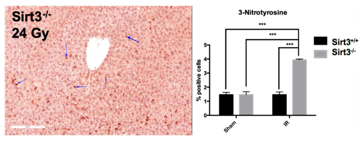
In patients with abdominal region cancers, ionizing radiation (IR)-induced long-term liver injury is a major limiting factor in the use of radiotherapy. Previously, the major mitochondrial deacetylase, sirtuin 3 (SIRT3), has been implicated to play an important role in the development of acute liver injury after total body irradiation but no studies to date have examined the role of SIRT3 in liver's chronic response to radiation. In the current study, ten-month-old Sirt3-/- and Sirt3+/+ male mice received 24 Gy radiation targeted to liver. Six months after exposure, irradiated Sirt3-/- mice livers demonstrated histopathological elevations in inflammatory infiltration, the loss of mature bile ducts and higher DNA damage (TUNEL) as well as protein oxidation (3-nitrotyrosine). In addition, increased expression of inflammatory chemokines (IL-6, IL-1β, TGF-β) and fibrotic factors (Procollagen 1, α-SMA) were also measured in Sirt3-/- mice following 24 Gy IR. The alterations measured in enzymatic activities of catalase, glutathione peroxidase, and glutathione reductase in the livers of irradiated Sirt3-/- mice also implied that hydrogen peroxide and hydroperoxide sensitive signaling cascades in the absence of SIRT3 might contribute to the IR-induced long-term liver injury.

Keywords: hydroperoxide; inflammation; ionizing radiation; liver; sirtuin 3.


Conflict of interest statement

The authors declare no conflict of interest. The funders had no role in the design of the study; in the collection, analyses, or interpretation of data; in the writing of the manuscript, or in the decision to publish the results.

Figures

Figure 1
Figure 1
(A) Experimental timeline for the irradiation and tissue harvest from Sirt3+/+ and Sirt3−/− male mice. (B) Image guided irradiation of the liver using Small Animal Radiation Research Platform (SARRP).
Figure 2
Figure 2
Increased numbers of inflammatory cells were seen in irradiated Sirt3−/− mice. H&E scoring: − (none), + (mild/focal), and ++ (moderate). All sections were normalized to the Sham Sirt3+/+ group and presented as number of animals with marker per animals in each group (n = 4–6). Sham groups underwent the same procedures without receiving 24 Gy IR (irradiation).
Figure 3
Figure 3
Expression of inflammatory markers determined by quantitative real-time PCR. IL6 and TGFβ were significantly increased in Sirt3−/− mice after irradiation compared to sham irradiated Sirt3+/+ or Sirt3−/− as well as Sirt3+/+ irradiated groups. IL1β was significantly increased in irradiated Sirt3−/− mice compared to irradiated Sirt3+/+ livers (n = 4–6; * p < 0.05, *** p < 0.001).
Figure 4
Figure 4
Sirt3−/− irradiated mice showed decreased number of bile ducts. Liver sections were stained and counted with anti-cytokeratin 19 to determine the presence of bile ducts. Scale bars: 200 µm. (8–10 fields were scored per mouse, n = 6; * p < 0.01).
Figure 5
Figure 5
Expression of profibrotic markers were increased in all Sirt3−/− mice after irradiation compared to sham irradiated Sirt3+/+ or Sirt3−/− as well as Sirt3+/+ irradiated groups (n = 4–6; ** p < 0.01, *** p < 0.001).
Figure 6
Figure 6
Protein oxidation, determined by 3-nitrotyrosine staining (indicated by the arrows), was increased in irradiated Sirt3−/− mice compared to sham irradiated Sirt3+/+ or Sirt3−/− as well as Sirt3+/+ irradiated groups. Scale bar: 200 µm (15 fields scored per mouse, n = 4–6; *** p < 0.001).
Figure 7
Figure 7
The number of TUNEL positive cells were significantly increased in Sirt3−/− mice after irradiation compared to sham irradiated Sirt3+/+ or Sirt3−/− as well as Sirt3+/+ irradiated groups. Scale bar: 100 µm (n = 4–6; * p < 0.05 ** p < 0.01, *** p < 0.001).
Figure 8
Figure 8
Exposure to 24 Gy liver only irradiation did not significantly alter enzymatic activity of MnSOD in Sirt3+/+ or Sirt3−/− mice (n = 4–6).
Figure 9
Figure 9
Glutathione Peroxidase (GPx) and catalase (CAT) activity were significantly increased while Glutathione Reductase (GR) enzymatic activity was decreased in irradiated Sirt3−/− mice compared to sham irradiated Sirt3+/+ or Sirt3−/− as well as Sirt3+/+ irradiated groups (n = 4–6; * p < 0.05, ** p < 0.01, *** p < 0.001).

References

    1. Siegel R.L., Miller K.D., Jemal A. Cancer statistics. CA Cancer J. Clin. 2020 doi: 10.3322/caac.21590. - DOI - PubMed
    1. Kim J., Jung Y. Radiation-induced liver disease: Current understanding and future perspectives. Exp. Mol. Med. 2017 doi: 10.1038/emm.2017.85. - DOI - PMC - PubMed
    1. Benson R., Madan R., Kilambi R., Chander S. Radiation induced liver disease: A clinical update. J. Egypt Natl. Canc. Inst. 2016 doi: 10.1016/j.jnci.2015.08.001. - DOI - PubMed
    1. Pan C.C., Kavanagh B.D., Dawson L.A., Li X.A., Das S.K., Miften M., Ten Haken R.K. Radiation-Associated Liver Injury. Int. J. Radiat. Oncol. Biol. Phys. 2010;76 doi: 10.1016/j.ijrobp.2009.06.092. - DOI - PMC - PubMed
    1. Ben-Josef E., Normolle D., Ensminger W.D., Walker S., Tatro D., Ten Haken R.K., Knol J., Dawson L.A., Pan C., Lawrence T.S. Phase II trial of high-dose conformal radiation therapy with concurrent hepatic artery floxuridine for unresectable intrahepatic malignancies. J. Clin. Oncol. 2005 doi: 10.1200/JCO.2005.01.5354. - DOI - PubMed

LinkOut - more resources