Super-Resolution Localisation of Nuclear PI(4)P and Identification of Its Interacting Proteome
- PMID: 32403279
- PMCID: PMC7291030
- DOI: 10.3390/cells9051191
Super-Resolution Localisation of Nuclear PI(4)P and Identification of Its Interacting Proteome
Abstract
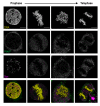
Phosphoinositides are glycerol-based phospholipids, and they play essential roles in cellular signalling, membrane and cytoskeletal dynamics, cell movement, and the modulation of ion channels and transporters. Phosphoinositides are also associated with fundamental nuclear processes through their nuclear protein-binding partners, even though membranes do not exist inside of the nucleus. Phosphatidylinositol 4-phosphate (PI(4)P) is one of the most abundant cellular phosphoinositides; however, its functions in the nucleus are still poorly understood. In this study, we describe PI(4)P localisation in the cell nucleus by super-resolution light and electron microscopy, and employ immunoprecipitation with a specific anti-PI(4)P antibody and subsequent mass spectrometry analysis to determine PI(4)P's interaction partners. We show that PI(4)P is present at the nuclear envelope, in nuclear lamina, in nuclear speckles and in nucleoli and also forms multiple small foci in the nucleoplasm. Nuclear PI(4)P undergoes re-localisation to the cytoplasm during cell division; it does not localise to chromosomes, nucleolar organising regions or mitotic interchromatin granules. When PI(4)P and PI(4,5)P2 are compared, they have different nuclear localisations during interphase and mitosis, pointing to their functional differences in the cell nucleus. Mass spectrometry identified hundreds of proteins, including 12 potentially novel PI(4)P interactors, most of them functioning in vital nuclear processes such as pre-mRNA splicing, transcription or nuclear transport, thus extending the current knowledge of PI(4)P's interaction partners. Based on these data, we propose that PI(4)P also plays a role in essential nuclear processes as a part of protein-lipid complexes. Altogether, these observations provide a novel insight into the role of PI(4)P in nuclear functions and provide a direction for further investigation.
Keywords: PI(4)P; nucleus; phosphoinositides.
Conflict of interest statement
The authors declare no conflict of interest.
Figures


Similar articles
-
Nuclear Phosphoinositides as Key Determinants of Nuclear Functions.Biomolecules. 2023 Jun 28;13(7):1049. doi: 10.3390/biom13071049. Biomolecules. 2023. PMID: 37509085 Free PMC article. Review.
-
Nanoscale mapping of nuclear phosphatidylinositol phosphate landscape by dual-color dSTORM.Biochim Biophys Acta Mol Cell Biol Lipids. 2021 May;1866(5):158890. doi: 10.1016/j.bbalip.2021.158890. Epub 2021 Jan 27. Biochim Biophys Acta Mol Cell Biol Lipids. 2021. PMID: 33513445
-
Limited Proteolysis-Coupled Mass Spectrometry Identifies Phosphatidylinositol 4,5-Bisphosphate Effectors in Human Nuclear Proteome.Cells. 2021 Jan 4;10(1):68. doi: 10.3390/cells10010068. Cells. 2021. PMID: 33406800 Free PMC article.
-
A role for β-dystroglycan in the organization and structure of the nucleus in myoblasts.Biochim Biophys Acta. 2013 Mar;1833(3):698-711. doi: 10.1016/j.bbamcr.2012.11.019. Epub 2012 Dec 4. Biochim Biophys Acta. 2013. PMID: 23220011
-
The Dynamic Nature of the Nuclear Envelope.Curr Biol. 2018 Apr 23;28(8):R487-R497. doi: 10.1016/j.cub.2018.01.073. Curr Biol. 2018. PMID: 29689232 Review.
Cited by
-
Apolipoprotein-L Functions in Membrane Remodeling.Cells. 2024 Dec 20;13(24):2115. doi: 10.3390/cells13242115. Cells. 2024. PMID: 39768205 Free PMC article. Review.
-
Nuclear phosphoinositide signaling promotes YAP/TAZ-TEAD transcriptional activity in breast cancer.EMBO J. 2024 May;43(9):1740-1769. doi: 10.1038/s44318-024-00085-6. Epub 2024 Apr 2. EMBO J. 2024. PMID: 38565949 Free PMC article.
-
Exploring Genetic Interactions with Telomere Protection Gene pot1 in Fission Yeast.Biomolecules. 2023 Feb 15;13(2):370. doi: 10.3390/biom13020370. Biomolecules. 2023. PMID: 36830739 Free PMC article. Review.
-
The Many Faces of Lipids in Genome Stability (and How to Unmask Them).Int J Mol Sci. 2021 Nov 29;22(23):12930. doi: 10.3390/ijms222312930. Int J Mol Sci. 2021. PMID: 34884734 Free PMC article. Review.
-
Heterochromatin Networks: Topology, Dynamics, and Function (a Working Hypothesis).Cells. 2021 Jun 23;10(7):1582. doi: 10.3390/cells10071582. Cells. 2021. PMID: 34201566 Free PMC article. Review.
References
Publication types
MeSH terms
Substances
LinkOut - more resources
Full Text Sources
Research Materials
Miscellaneous

