. 2020 Sep 2;11(5):1324-1347.
doi: 10.1080/19490976.2020.1754714. Epub 2020 May 13.

Rotavirus infection induces glycan availability to promote ileum-specific changes in the microbiome aiding rotavirus virulence

Affiliations

Rotavirus infection induces glycan availability to promote ileum-specific changes in the microbiome aiding rotavirus virulence

Melinda A Engevik et al. Gut Microbes. .

Abstract

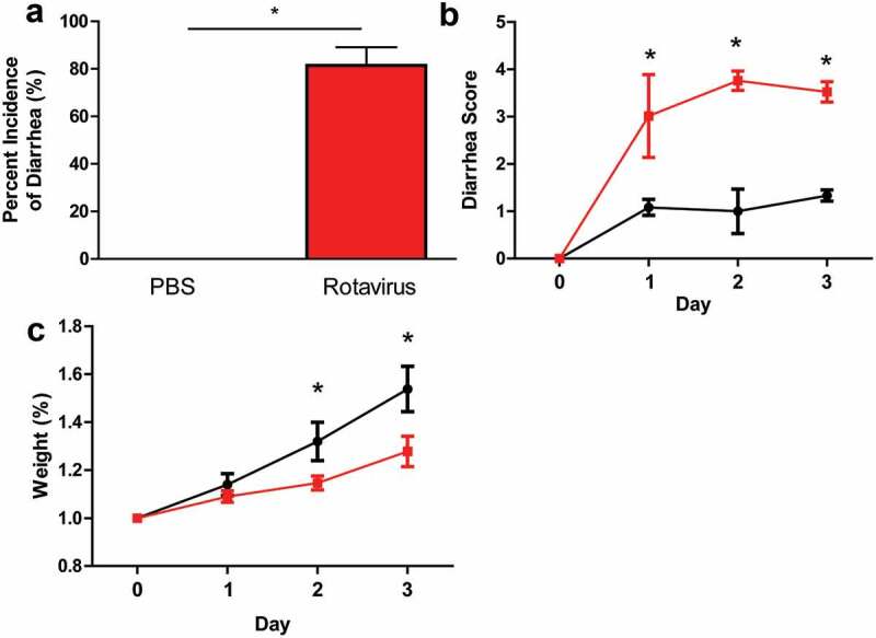
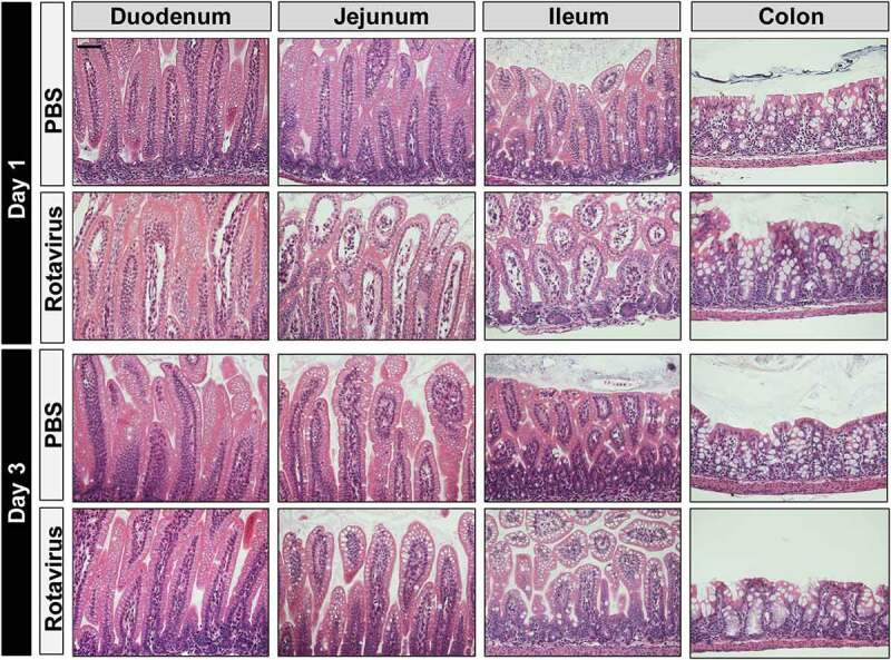
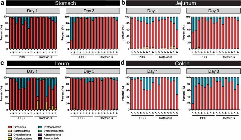
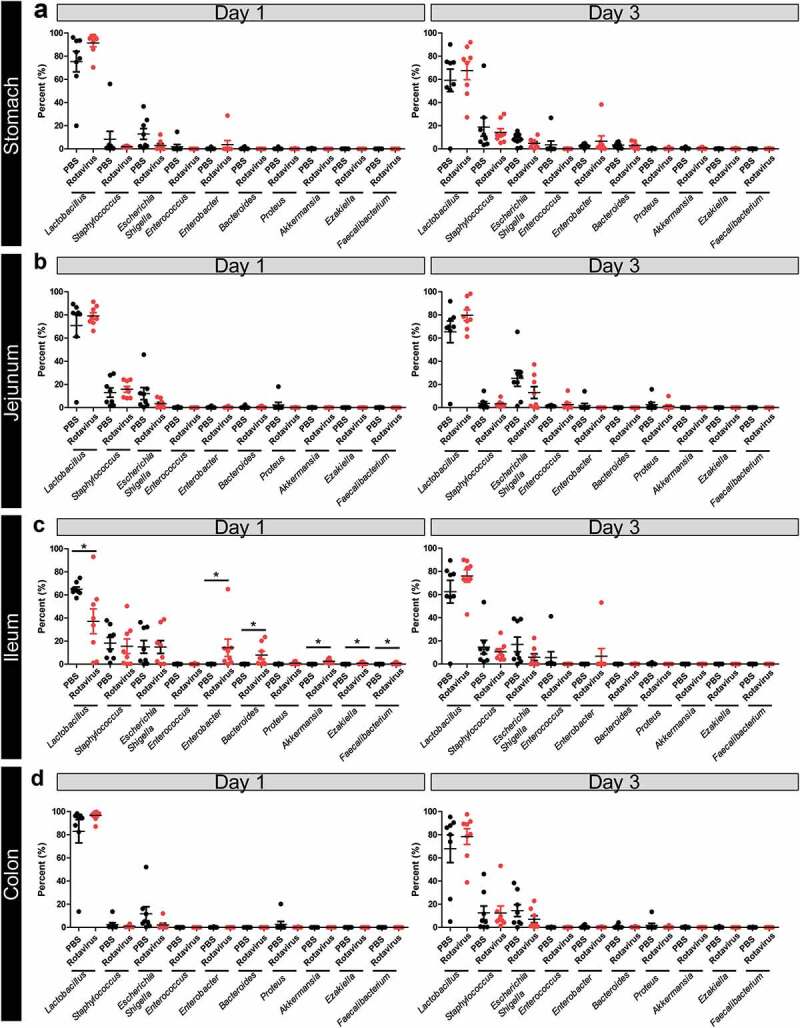
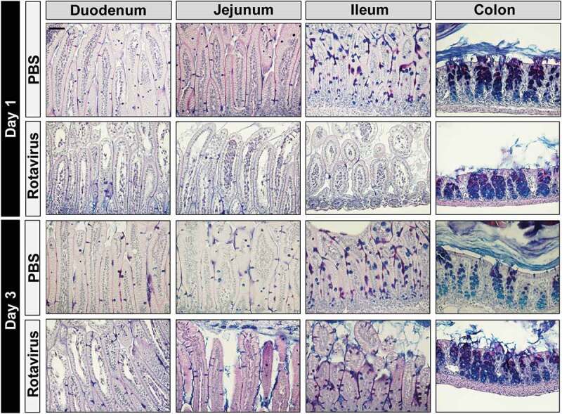
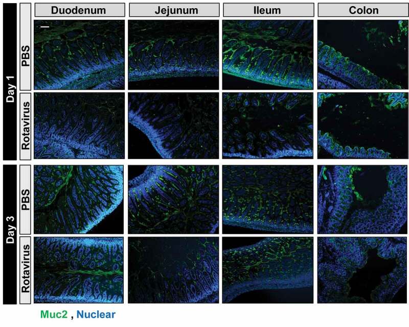
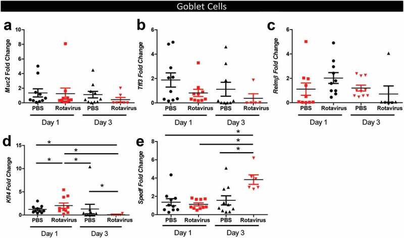
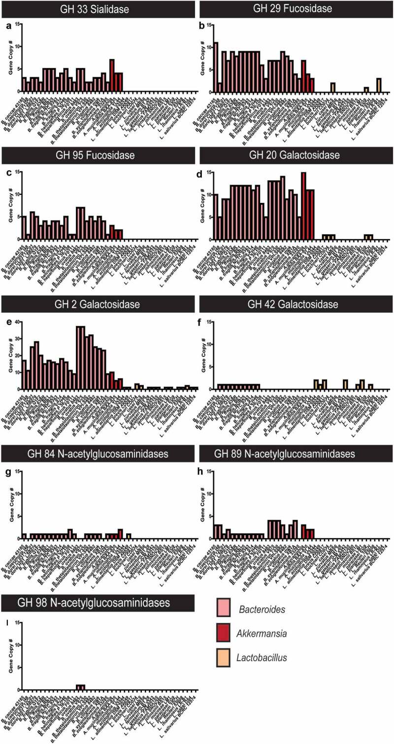
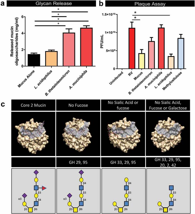
Multiple studies have identified changes within the gut microbiome in response to diarrheal-inducing bacterial pathogens. However, examination of the microbiome in response to viral pathogens remains understudied. Compounding this, many studies use fecal samples to assess microbiome composition; which may not accurately mirror changes within the small intestine, the primary site for most enteric virus infections. As a result, the functional significance of small intestinal microbiome shifts during infection is not well defined. To address these gaps, rotavirus-infected neonatal mice were examined for changes in bacterial community dynamics, host gene expression, and tissue recovery during infection. Profiling bacterial communities using 16S rRNA sequencing suggested significant and distinct changes in ileal communities in response to rotavirus infection, with no significant changes for other gastrointestinal (GI) compartments. At 1-d post-infection, we observed a loss in Lactobacillus species from the ileum, but an increase in Bacteroides and Akkermansia, both of which exhibit mucin-digesting capabilities. Concomitant with the bacterial community shifts, we observed a loss of mucin-filled goblet cells in the small intestine at d 1, with recovery occurring by d 3. Rotavirus infection of mucin-producing cell lines and human intestinal enteroids (HIEs) stimulated release of stored mucin granules, similar to in vivo findings. In vitro, incubation of mucins with Bacteroides or Akkermansia members resulted in significant glycan degradation, which altered the binding capacity of rotavirus in silico and in vitro. Taken together, these data suggest that the response to and recovery from rotavirus-diarrhea is unique between sub-compartments of the GI tract and may be influenced by mucin-degrading microbes.

Keywords: Akkermansia; Bacteroides; Muc2; Rotavirus; glycans; microbiome; mucus.


Figures

Figure 1.
Figure 1.
Rotavirus-infected BALB/c pups exhibit hallmarks of rotavirus-associated disease. a. The average percentage of BALB/c pups with diarrhea as recorded every 24 h for PBS-treated (n = 11; black bar) or rotavirus-infected (n = 12; red bar). One-Way ANOVA, *p < .05. b. Diarrhea scores over a 3-d period from PBS-treated (black) and rotavirus-infected (red) mice. One-Way Repeated Measures ANOVA, *p < .05. c. The average weight change of PBS-treated or rotavirus-infected BALB/c pups as measured every 24 h. All data presented as the average ± standard deviation. One-Way Repeated Measures ANOVA, *p < .05.
Figure 2.
Figure 2.
Rotavirus-infected BALB/c Pups Exhibit Histopathology in the Small Intestine. H&E staining of intestinal segments (duodenum, jejunum, ileum, and colon) from PBS-treated or rotavirus-infected at 1 or 3 dpi (day post-infection). H&E images were acquired on an upright Nikon Eclipse 90i microscope using a 20X Plan Apo (NA 0.75) DIC objective with a DS-Fi1-U2 camera and Nikon Elements software. Scale bar = 50 µm.
Figure 3.
Figure 3.
Phylum shifts occur in the ileum at day 1 post-rotavirus infection. 16S rRNA sequencing analysis of the bacterial community at the level of phylum. Eight phyla are presented: Actinobacteria (purple), Bacteroidetes (orange), Cyanobacteria (peach), Deferribacteres (yellow), Firmicutes (red), Fusobacteria (black), Proteobacteria (blue), and Verrucomicrobia (green). Data is represented for the stomach (a), jejunum (b), ileum (c), and colon (d) of PBS-treated (black) and rotavirus-infected (red) BALB/c pups at 1 and 3 dpi. Only statistical significance was reached in the ileum at 1 dpi (p < .05) One-Way ANOVA.
Figure 4.
Figure 4.
Shifts in bacterial genera are observed in the ileum at 1 dpi. Relative abundances of the top 10 genera present in the expressed contents of the stomach (a), jejunum (b), ileum (c), and colon (d) of PBS-treated (black) and rotavirus-infected (red) BALB/c pups at 1 and 3 dpi as determined by 16S rRNA gene sequencing. Only statistical significance was reached in the ileum at 1 dpi (p < .05) One-Way ANOVA.
Figure 5.
Figure 5.
Infection with rotavirus depletes mucus-filled goblet cells. Periodic Acid-Schiffs Alcian Blue (PAS-AB) stains at 1 and 3 dpi of intestinal segments (stomach, jejunum, ileum, and colon) from PBS-treated or rotavirus-infected mice. Acidic mucins are denoted by their purple staining. PAS-AB images were acquired on an upright Nikon Eclipse 90i microscope using a 20X Plan Apo (NA 0.75) DIC objective with a DS-Fi1-U2 camera and Nikon Elements software. Scale bar = 50 µm.
Figure 6.
Figure 6.
Infection with rotavirus depletes Muc2-filled goblet cells. Muc2 immunostaining at 1 and 3 dpi of intestinal segments (stomach, jejunum, ileum, and colon) from PBS-treated or rotavirus-infected mice (Muc2: green, nuclear Hoechst: blue). Fluorescent images were acquired on an upright Nikon Eclipse 90i microscope using a 10X Plan Fluor (NA 0.3) phase contrast objective with an ORCA-Flash 4.0 sCMOS camera and Nikon Elements software. Scale bar = 100 µm.
Figure 7.
Figure 7.
Quantitative Real-time PCR (qPCR) analysis reveals no changes in goblet cell secreted factors. qPCR of goblet cell-secreted factors Mucin 2 (Muc2) (a), Trefoil factor 3 (Tff3) (b), Relm-β (c), and goblet cell transcription factors Krüppel-like factor 4 (Kfl4) (d) and SAM pointed domain-containing Ets transcription factor (SPEDF) (e). All data is presented for PBS-treated (black) and rotavirus-infected (red) mice on 1 and 3 dpi. One-Way ANOVA *p < .05.
Figure 8.
Figure 8.
Rotavirus stimulates release of stored mucin-granules in vitro. MUC2-producing human cell lines, HT29-MX and T84 cells, as well human intestinal enteroids (HIEs) derived from jejunum were infected with Ito rotavirus in serum-free DMEM in the presence of 10 µg/mL Worthington’s Trypsin MA104 lysates. Supernatant was collected after overnight infection (16 h) and mucin-content was examined by PAS staining. Rotavirus infection (red) stimulated release of mucin compared to mock treatment (black). One-Way ANOVA *p < .05.
Figure 9.
Figure 9.
Bacteroides and Akkermansia species harbor extensive glycosyl hydrolases for mucin glycan degradation. All annotated Bacteroides and Akkermanisa genomes as well as selected intestinal Lactobacilli genomes were screened for glycosyl hydrolases using the CAZY database. Data presented in terms of copy number for glycosyl hydrolase (GH) families predicted to be involved in O-linked (A–I) mucin degradation. Glycosyl hydrolases include: a. sialidase GH 33, b. Fucosidase GH 29, c. Fucosidase GH 95, d.Galactosidase GH 20, e. Galactosidase GH 2, f. Galactosidase GH 42, g.N-acetylglucosaminidase GH 84, h. N-acetylglucosaminidase GH 89, and I. N-acetylglucosaminidase GH 98.
Figure 10.
Figure 10.
Bacteroides and Akkermanisa degrades mucin glycans in a manner which diminishes the binding capacity of rotavirus VP4. a. Incubation of Bacteroidetes thetaiotaomicron and Akkermansia mucinphila with purified germ-free cecal Muc2 result in release mucin oligosaccharides, as determined by the phenol-sulfuric acid method. Incubation of Muc2 with Lactobacillus acidophilus had no effect on oligosaccharide concentration. b. Plaque Assays quantification (plaque-forming units per milliliter of virus (PFU/ml)) of rotavirus added to MA104 African green monkey kidney in the presence of 10 mg/ml intact Muc2 or Muc2 pre-treated with B. thetaiotaomicron, A. mucinphila or L. acidophilus. Methylcellulose, which mirrors the viscosity of mucus was added as another control. One-Way ANOVA *p < .05. c. In silico analysis of the VP8 domain of the spike protein of rotavirus VP4 binding to mucin core 2 glycans (constructed using SWEET II software).

References

    1. Qin J, Li R, Raes J, Arumugam M, Burgdorf KS, Manichanh C, Nielsen T, Pons N, Levenez F, Yamada T, et al. A human gut microbial gene catalogue established by metagenomic sequencing. Nature. 2010;464(7285):59–65. doi:10.1038/nature08821. - DOI - PMC - PubMed
    1. Norman JM, Handley SA, Virgin HW.. Kingdom-agnostic metagenomics and the importance of complete characterization of enteric microbial communities. Gastroenterology. 2014;146(6):1459–1469. doi:10.1053/j.gastro.2014.02.001. - DOI - PMC - PubMed
    1. Brown EM, Sadarangani M, Finlay BB.. The role of the immune system in governing host-microbe interactions in the intestine. Nat Immunol. 2013;14(7):660–667. doi:10.1038/ni.2611. - DOI - PubMed
    1. Engevik MA, Aihara E, Montrose MH, Shull GE, Hassett DJ, Worrell RT. Loss of NHE3 alters gut microbiota composition and influences bacteroides thetaiotaomicron growth. Am J Physiol Gastrointest Liver Physiol. 2013;305(10):G697–711. doi:10.1152/ajpgi.00184.2013. - DOI - PMC - PubMed
    1. Engevik MA, Faletti CJ, Paulmichl M, Worrell RT. Prebiotic properties of galursan HF 7K on mouse gut microbiota. Cell Physiol Biochem. 2013;32(7):96–110. doi:10.1159/000356631. - DOI - PubMed

Publication types

MeSH terms

LinkOut - more resources