Case Reports
. 2020 May 13;20(1):342.
doi: 10.1186/s12879-020-05063-x.

Subacute pericardial abscess after aortic valve replacement: a case report

Affiliations
Case Reports

Subacute pericardial abscess after aortic valve replacement: a case report

Ilenia Magnani et al. BMC Infect Dis. .

Abstract

Background: Purulent pericarditis is an infectious disease, frequently caused by gram-positive bacteria, that is rarely observed in healthy individuals, and is often associated with predisposing conditions.

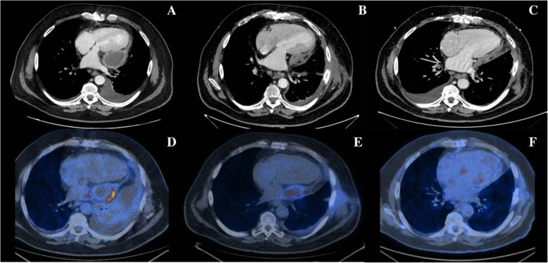
Case presentation: Here, we present the case of an Escherichia coli post-surgical localized purulent pericarditis complicated by transient constrictive pericarditis and its diagnostic and therapeutic management.

Conclusions: Our case report focuses on the importance of imaging-guided treatment of purulent pericardial diseases, in particular on the emerging role of 18 F-labelled 2-fluoro-2-deoxy-D-glucose Positron Emission Tomography/Computed Tomography in pericardial diseases and on the management of transient constrictive pericarditis, often seen after thoracic surgery.

Keywords: Constrictive pericarditis; Escherichia coli; Pericardial abscess; Purulent pericarditis.


Conflict of interest statement

The authors declare that they have no competing interests.

Figures

Fig. 1
Fig. 1
Thoracic computed tomography and 18 F-labeled 2-fluoro-2-deoxy-D-glucose positron emission tomographic/computer tomography imaging. In the upper series of figures, thoracic computed tomography scan sections acquired in February 2018 (a), April 2018 (b) and October 2018 (c), respectively, are shown. (a) Severe loculated pericardial effusion localized to the lateral wall of the left ventricle (51 mm) and bilateral pleural effusion. (b) Paracardiac egg-shaped fluid collection (38 × 27 mm) with peripheric hypercaptation of the tracer. (c) Mild diffuse postero-lateral pericardial effusion (maximal thickness: 11 mm). No evidence of persistence of localized fluid collection. In the lower series, 18 F-labeled 2-fluoro-2-deoxy-D-glucose positron emission tomographic/computer tomography (18F-FDG PET/CT) sections acquired in February 2018 (d), April 2018 (e) and April 2019 (f) are presented. d Pathological hypercaptation at the walls of the pericardial effusion (SUV max = 11). e In comparison with previous 18F-FDG PET/CT, the area of tracer hypercaptation in left paracardiac position is reduced in dimension, but stable in intensity (SUV max = 9.6). d Normalization of the area of metabolic hypercaptation reported in the previous exams
Fig. 2
Fig. 2
Histological and immunohistochemical analysis. In a there is evidence of chronic inflammation with fibrosis, mesothelial hyperplasia and mixed cellular infiltrate rich in macrophages, neutrophils, eosinophils and plasma cells. b shows the immunohistochemical analysis for Calretinin that highlights mesothelial cells and their hyperplasia. c we can observed the immunohistochemical analysis for CD 68, a specific surface marker of macrophages

References

    1. Adler Y, Charron P, Imazio M, et al. 2015 ESC guidelines for the diagnosis and management of pericardial diseases: the task force for the diagnosis and Management of Pericardial Diseases of the European Society of Cardiology (ESC) endorsed by: the European Association for Cardio-Thoracic Surgery (EACTS) Eur Heart J. 2015;36:2921–2964. doi: 10.1093/eurheartj/ehv318. - DOI - PMC - PubMed
    1. Parikh SV, Memon N, Echols M, Shah J, McGuire DK, Keeley EC. Purulent pericarditis: report of 2 cases and review of the literature. Medicine (Baltimore) 2009;88:52–65. doi: 10.1097/MD.0b013e318194432b. - DOI - PubMed
    1. Mayosi BM, Burgess LJ, Doubell AF. Tuberculous pericarditis. Circulation. 2005;112:3608–3616. doi: 10.1161/CIRCULATIONAHA.105.543066. - DOI - PubMed
    1. Horii S, Yada H, Ito K, et al. A rare case of rush progression of purulent pericarditis by Escherichia coli in a patient with malignant lymphoma. Int Heart J. 2018;59:655–659. doi: 10.1536/ihj.17-238. - DOI - PubMed
    1. Gelijns AC, et al. Management practices and major infections after cardiac surgery. J Am Coll Cardiol. 2014;64:372–381. doi: 10.1016/j.jacc.2014.04.052. - DOI - PMC - PubMed

Publication types

MeSH terms