. 2020 Jul 10;295(28):9409-9420.
doi: 10.1074/jbc.RA120.013590. Epub 2020 May 13.

The bacterial metalloprotease NleD selectively cleaves mitogen-activated protein kinases that have high flexibility in their activation loop

Affiliations

The bacterial metalloprotease NleD selectively cleaves mitogen-activated protein kinases that have high flexibility in their activation loop

Lihi Gur-Arie et al. J Biol Chem. .

Abstract

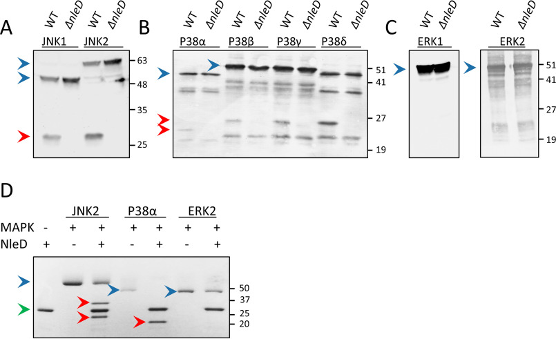
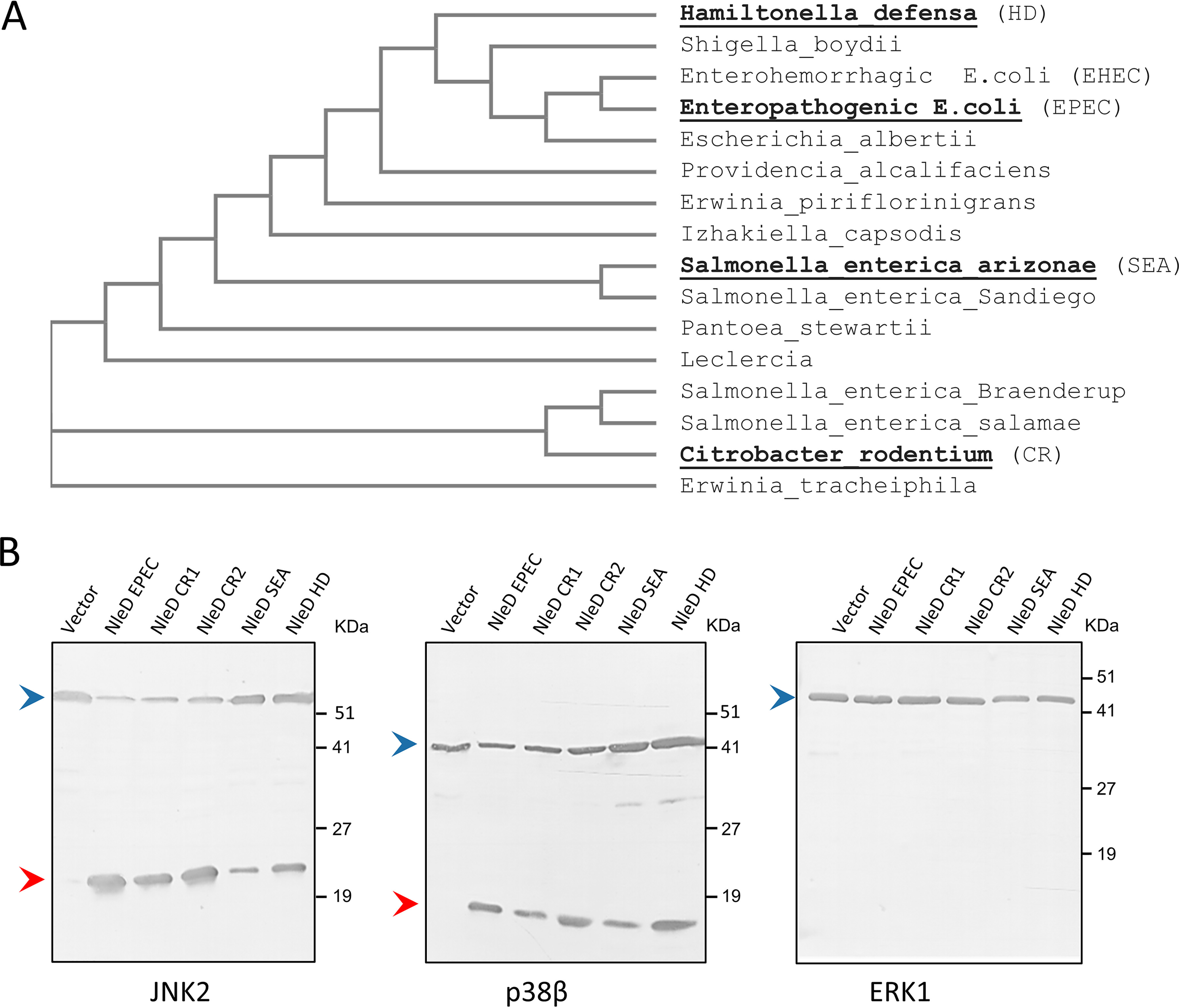
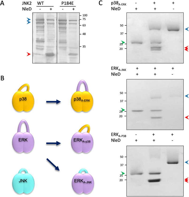
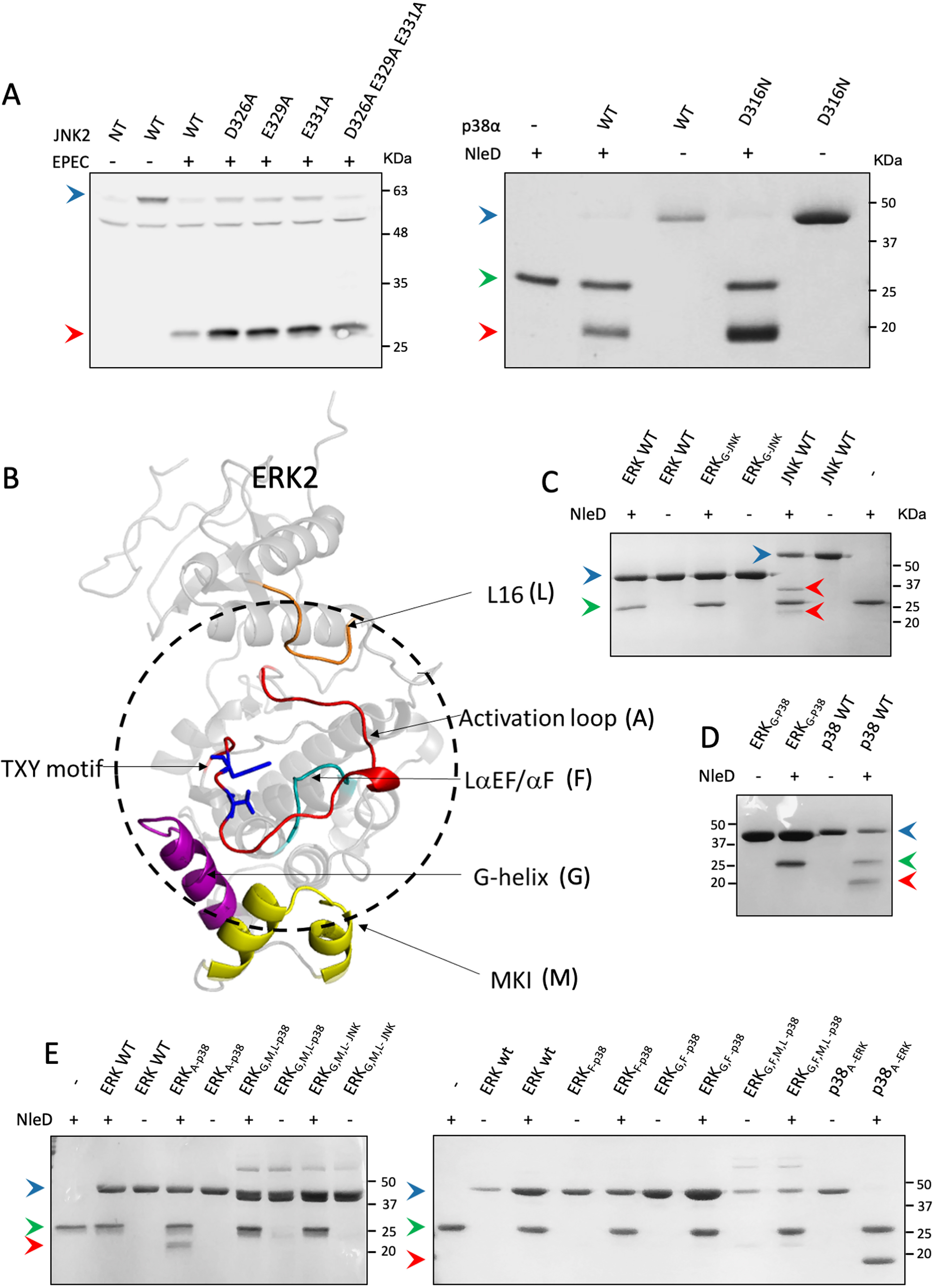
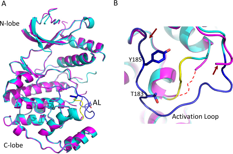
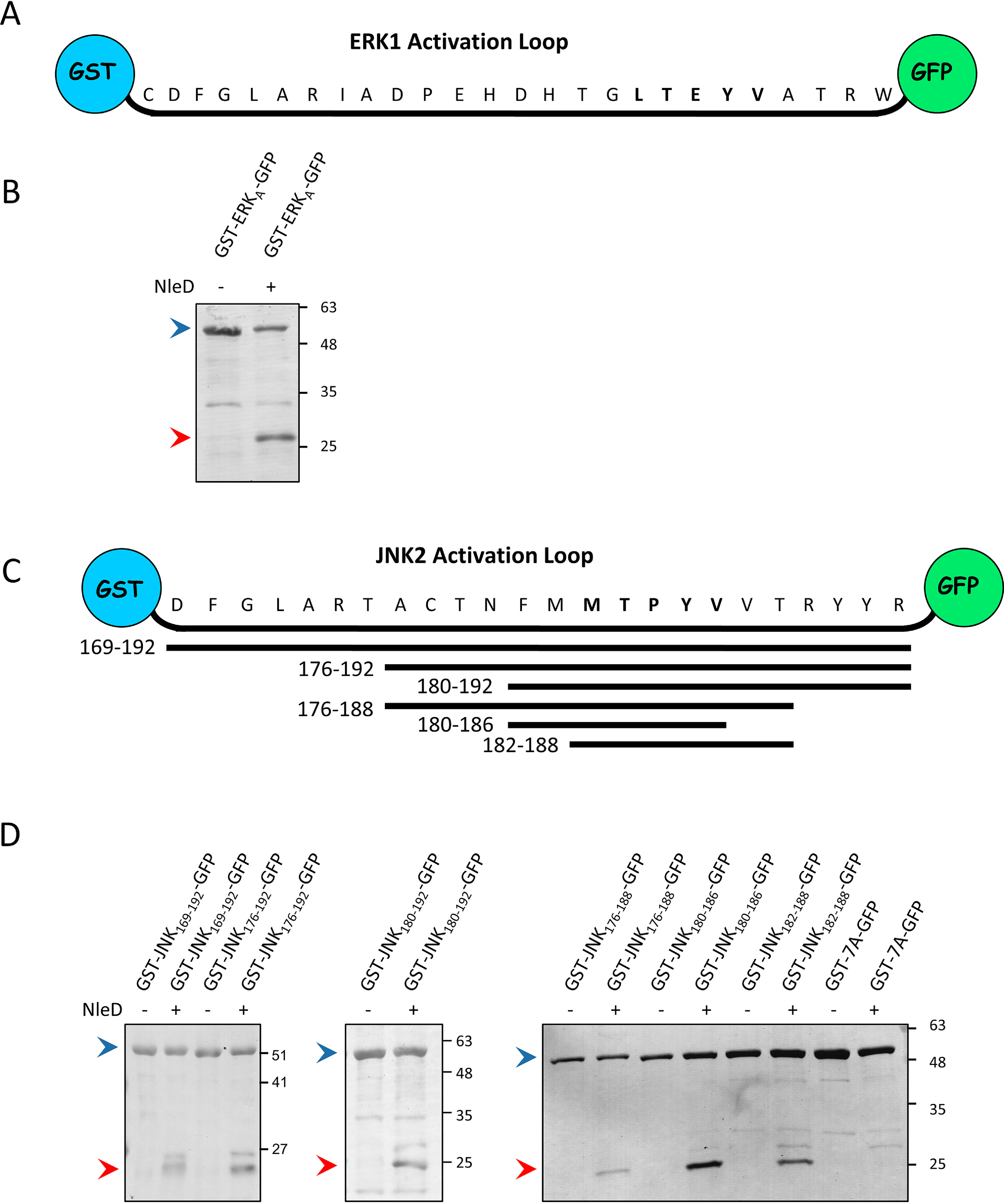
Microbial pathogens often target the host mitogen-activated protein kinase (MAPK) network to suppress host immune responses. We previously identified a bacterial type III secretion system effector, termed NleD, a metalloprotease that inactivates MAPKs by specifically cleaving their activation loop. Here, we show that NleDs form a growing family of virulence factors harbored by human and plant pathogens as well as insect symbionts. These NleDs disable specifically Jun N-terminal kinases (JNKs) and p38s that are required for host immune response, whereas extracellular signal-regulated kinase (ERK), which is essential for host cell viability, remains intact. We investigated the mechanism that makes ERK resistant to NleD cleavage. Biochemical and structural analyses revealed that NleD exclusively targets activation loops with high conformational flexibility. Accordingly, NleD cleaved the flexible loops of JNK and p38 but not the rigid loop of ERK. Our findings elucidate a compelling mechanism of native substrate proteolysis that is promoted by entropy-driven specificity. We propose that such entropy-based selectivity is a general attribute of proteolytic enzymes.

Keywords: Jun N-terminal kinase (JNK); NleD; bacterial pathogen; bacterial pathogenesis; crystallography; effector protein; extracellular signal-regulated kinase (ERK); metalloprotease; mitogen-activated protein kinase (MAPK); p38; protein structure; proteolysis; structural biology; type III secretion system (T3SS).


Conflict of interest statement

Conflict of interest—The authors declare that they have no conflicts of interest with the contents of this article.

Figures

Figure 1.
Figure 1.
Cleavage of MAPK isoforms by NleDEPEC. A–C, HEK293T cells, transiently expressing different epitope-tagged isoforms of JNK (A), p38 (B), and ERK (C), were infected with WT EPEC (WT) or the EPEC ΔnleD mutant, as indicated above the lanes. Proteins were extracted from the cells and subjected to Western blot analysis using anti-HA (HA-JNKs and HA-ERK2) or anti-His antibody (His-p38s and His-ERK1). D, Recombinant purified NleDEPEC was incubated with recombinant p38a, JNK2, or ERK2. Proteins were then resolved using SDS-PAGE and visualized by Coomassie staining. The cleavage of p38a results in a single product band, as the two fragments exhibit almost identical molecular masses of 20.8 and 20.4 kDa. JNK2, however, has a C-terminal extension region that is 49 residues larger than that of p38a; thus, its cleavage results in two distinct bands. Intact and fragmented MAPKs are indicated by blue and red arrowheads, respectively, and a green arrowhead indicates NleD. The incomplete cleavage of JNK and p38 proteins is likely due to the presence of misfolded subpopulations of the MAPKs.
Figure 2.
Figure 2.
NleD orthologues cleave JNK and p38 but not ERK. A, An NleD orthologue phylogenetic tree was created with Clustal Omega. Proteins selected for further analysis are highlighted in boldface. B, NleD orthologues of C. rodentium (NleDCR1 and NleDCR2), S. enterica serovar Arizonae (NleDSEA), and H. defensa (NleDHD) were coexpressed in E. coli BL21 together with hexahistidine-tagged MAP kinases (i.e. p38β, JNK2, or ERK1). Proteins were then extracted from the bacteria, and MAPK cleavage was probed by Western blot analysis using the anti-6His antibody. Intact and fragmented MAPKs are indicated by blue and red arrowheads, respectively.
Figure 3.
Figure 3.
The impact of the X residue within the TXY motif and the activation loop context on NleD substrate specificity. A, JNK2 WT or JNK2-P184E mutant was coexpressed in E. coli BL21 with NleD (+) or vector control (−). Proteins were extracted from the expressing bacteria and subjected to Western blot analysis using an anti-JNK antibody. Intact and cleaved JNKs are indicated by blue and red arrowheads, respectively. B, Schematic view of native p38a, ERK2, and JNK2 and the activation loop swapped variants. C, Purified proteins variants were incubated with purified NleDEPEC, and, as negative controls, the MAPK variants and NleD were incubated separately. For cleavage analysis, proteins were subjected to SDS-PAGE and visualized by Coomassie staining. Intact and cleaved MAPKs are indicated by blue and red arrowheads, respectively. A green arrowhead indicates NleD protein.
Figure 4.
Figure 4.
Region replacements between ERK and p38/JNK did not alter cleavage preferences of NleD. A, HEK293T cells were transfected, or not (NT), with plasmids expressing WT JNK2 carrying an HA tag (WT) or JNK2 mutated in critical CD domain residues, as indicated above the lanes (left). The cells were then infected with WT EPEC and subjected to Western blot analysis using an anti-HA antibody. (Right) In addition, WT p38a (WT) or p38 mutated in a critical CD domain residue (D316N) was purified and incubated in vitro with NleDEPEC. Samples were subjected to SDS-PAGE and visualized by Coomassie staining. B, Structure model of ERK2 (PDB entry 4S31). The activation loop is colored red, and threonine and tyrosine of the TXY motif are highlighted in blue. The dashed circle highlights the regions that are in close proximity to the activation loop. Regions that were replaced in the variants include MKI (M), colored yellow, G-helix (G), colored purple, L16 (L), colored orange, and LaEF/aF (F), colored cyan. C–E, Purified MAPK variants were incubated with or without purified NleDEPEC and then resolved by SDS-PAGE, followed by Coomassie staining. The variants used are indicated above the respective lanes and include WT ERK2 (ERKwt), WT JNK2 (JNKwt), WT p38α (p38wt), ERK2 with G-helix of JNK2 or p38α (ERKG-JNK and ERKG-p38, respectively), ERK2 with the p38α activation loop (ERKA-p38), p38α with the ERK2 activation loop (p38A-ERK), ERK2 with the LαEF/αF region of p38α (ERKF-p38), double mutant ERK2 with G-helix and LαEF/αF regions of p38α (ERKG,F-p38), triple mutant ERK2 with G-helix, MKI, and L16 regions of p38α (ERKG,M,l-p38), and quadruple mutant ERK2 with G-helix, LαEF/αF, MKI, and L16 regions of p38α (ERKG,F,M,l-p38). The presence (+) or absence (−) of NleD is indicated. Intact and fragmented MAPKs are indicated by blue and red arrowheads, respectively. A green arrowhead indicates NleD protein.
Figure 5.
Figure 5.
Structure comparison between WT MAPKs and activation loop-swapped variants. Structural superposition of the ERK molecules resolved in this study, including overall structures (A) and view of the activation loop vicinity (B). The overall structures of ERKG-p38 (cyan and blue) and ERKA-p38 (magenta) remain similar without significant conformation changes other than in the activation loops. The activation loop (blue) in ERKG-p38 and the αEF/αF loop (yellow) maintain the conformation of the WT structure (not shown). The activation loop of ERKG-p38 is disordered, and its edges are indicated in panel B by red arrows. The αEF/αF loop of ERKG-p38, known to be conjugated with the contour of the activation loop, is also disordered (shown by a dotted red line in panel B).
Figure 6.
Figure 6.
Activation loops of JNK2 and ERK1 are recognized and cleaved by NleD when inserted between GST and GFP. A, Schematic presentation of the ERK1 activation loop (ERKA) cloned between the GST and GFP proteins. B, The ERK1 activation loop flanked by GST and GFP (GST-ERKA-GFP) was coexpressed in E. coli BL21 with or without NleDEPEC. The extracted proteins were pulled down using GSH beads and subjected to SDS-PAGE, followed by Coomassie staining. Intact and fragmented GST-ALERK-GFP proteins are indicated by blue and red arrowheads, respectively. C, Schematic view of the JNK2 activation loop (residues 169 to 192) cloned between GST and GFP proteins. Below are shown the different fragments of the JNK2 activation loop and the corresponding coordinates, which were cloned between the GST and GFP proteins. D, GST-GFP bridged by different segments of the JNK2 activation loop shown in panel C or by seven alanine residues (7A) were coexpressed in E. coli BL21 with or without NleDEPEC. The extracted proteins were pulled down using GSH beads and subjected to SDS-PAGE, followed by Coomassie staining. The variants used and the presence or absence of NleD are indicated above the lanes. Intact and fragmented proteins are indicated by blue and red arrowheads, respectively.

References

    1. Arbibe L., Kim D. W., Batsche E., Pedron T., Mateescu B., Muchardt C., Parsot C., and Sansonetti P. J. (2007) An injected bacterial effector targets chromatin access for transcription factor NF-kappaB to alter transcription of host genes involved in immune responses. Nat. Immunol. 8, 47–56 10.1038/ni1423 - DOI - PubMed
    1. Arthur J. S., and Ley S. C. (2013) Mitogen-activated protein kinases in innate immunity. Nat. Rev. Immunol. 13, 679–692 10.1038/nri3495 - DOI - PubMed
    1. Peti W., and Page R. (2013) Molecular basis of MAP kinase regulation. Protein Sci. 22, 1698–1710 10.1002/pro.2374 - DOI - PMC - PubMed
    1. Zarubin T., and Han J. (2005) Activation and signaling of the p38 MAP kinase pathway. Cell Res. 15, 11–18 10.1038/sj.cr.7290257 - DOI - PubMed
    1. Stofega M. R., Yu C. L., Wu J., and Jove R. (1997) Activation of extracellular signal-regulated kinase (ERK) by mitogenic stimuli is repressed in v-Src-transformed cells. Cell Growth Differ. 8, 113–119 - PubMed

Publication types

MeSH terms

Substances