. 2020 Jul 10;295(28):9392-9408.
doi: 10.1074/jbc.RA119.012335. Epub 2020 May 13.

A single mutation increases heavy-chain heterodimer assembly of bispecific antibodies by inducing structural disorder in one homodimer species

Affiliations

A single mutation increases heavy-chain heterodimer assembly of bispecific antibodies by inducing structural disorder in one homodimer species

Cian Stutz et al. J Biol Chem. .

Abstract

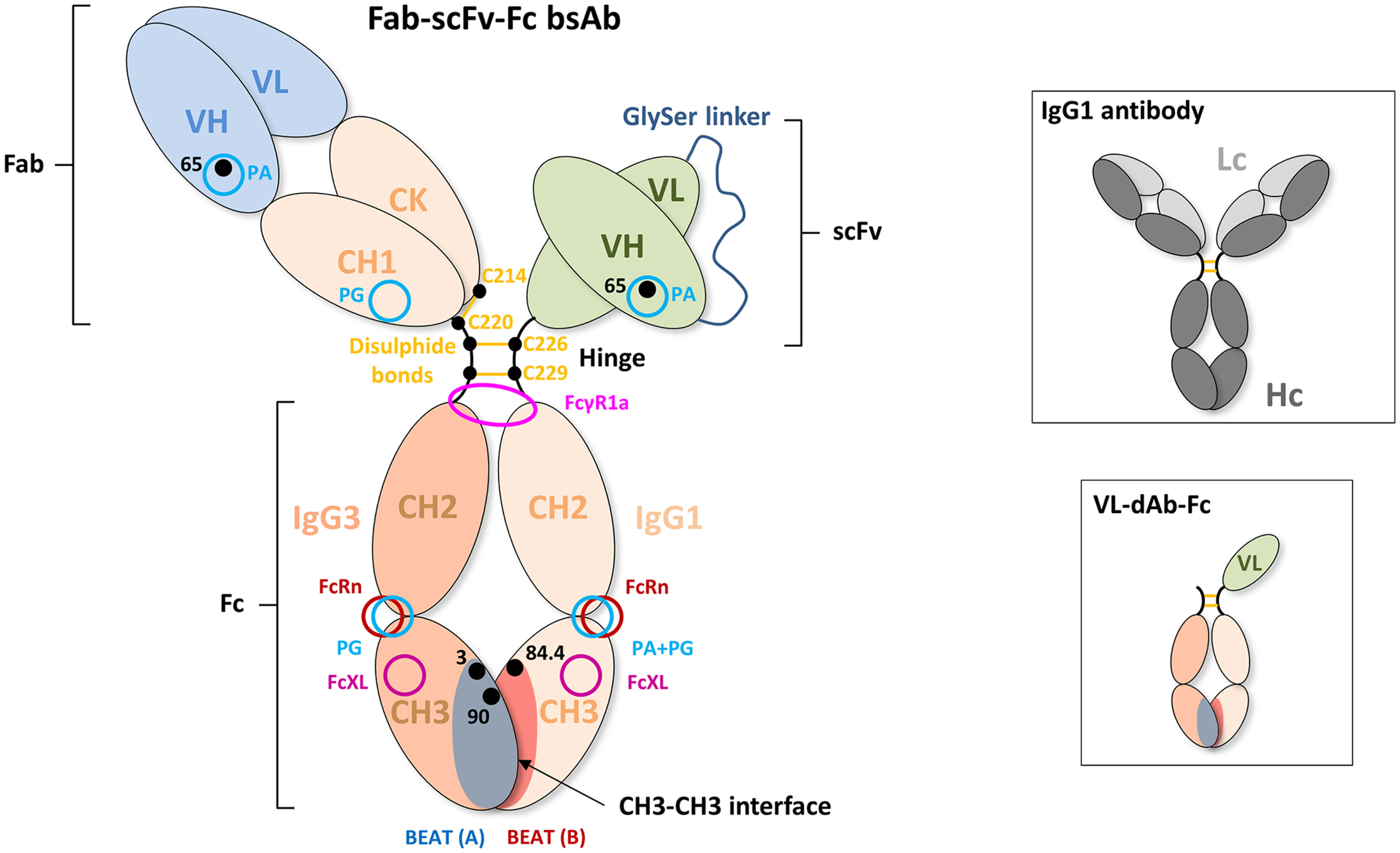
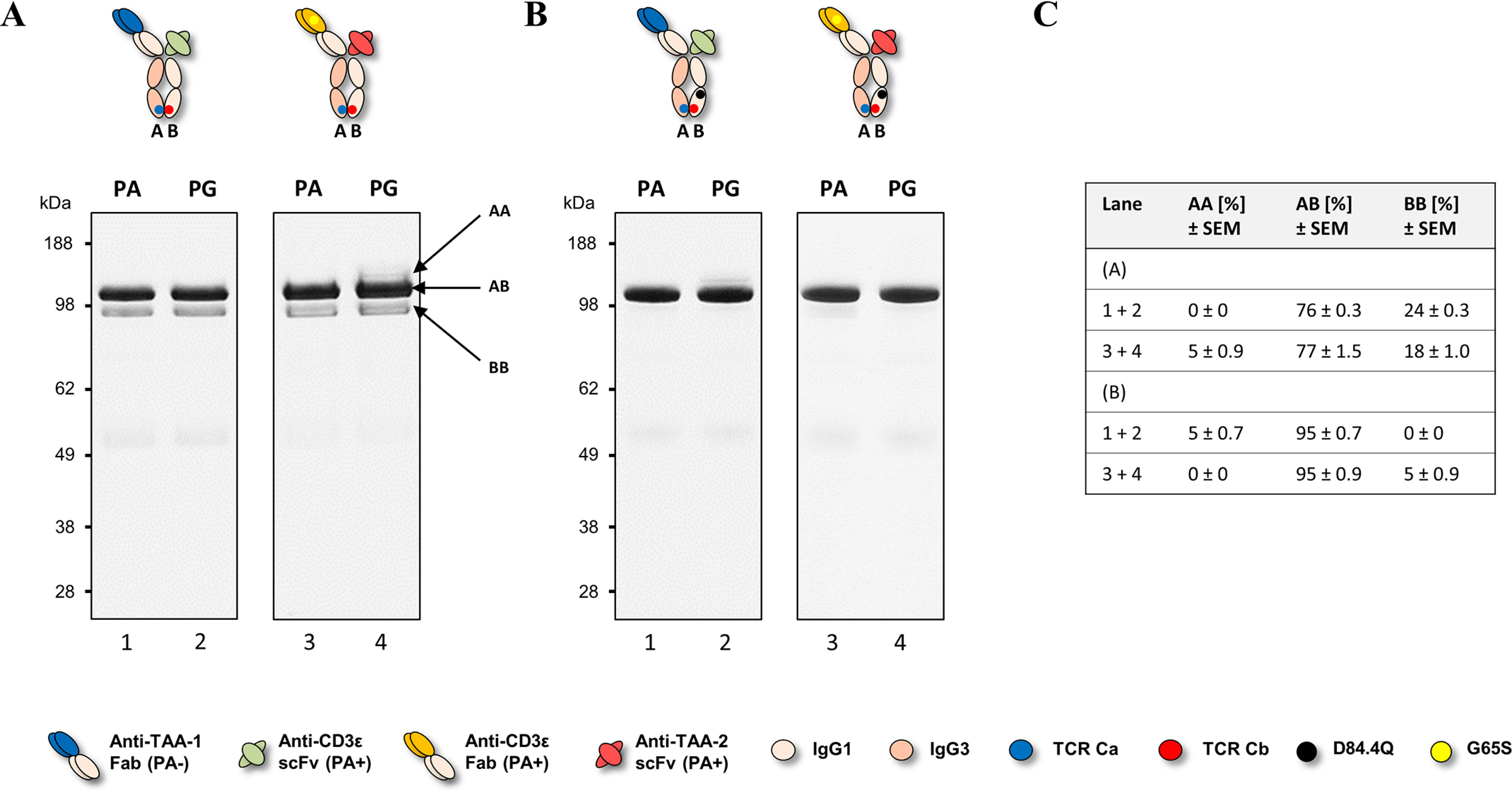
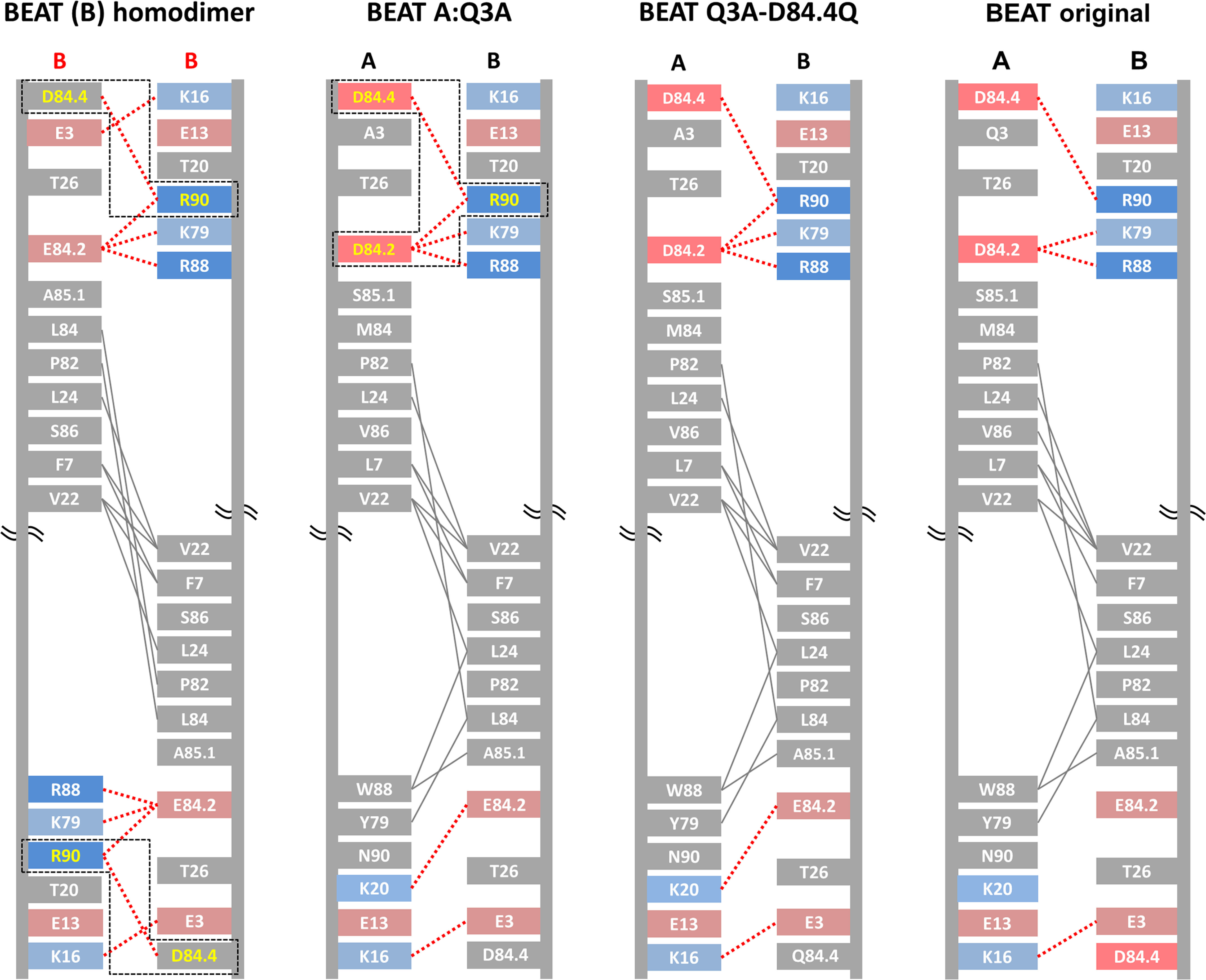
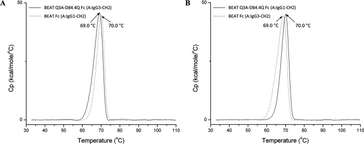
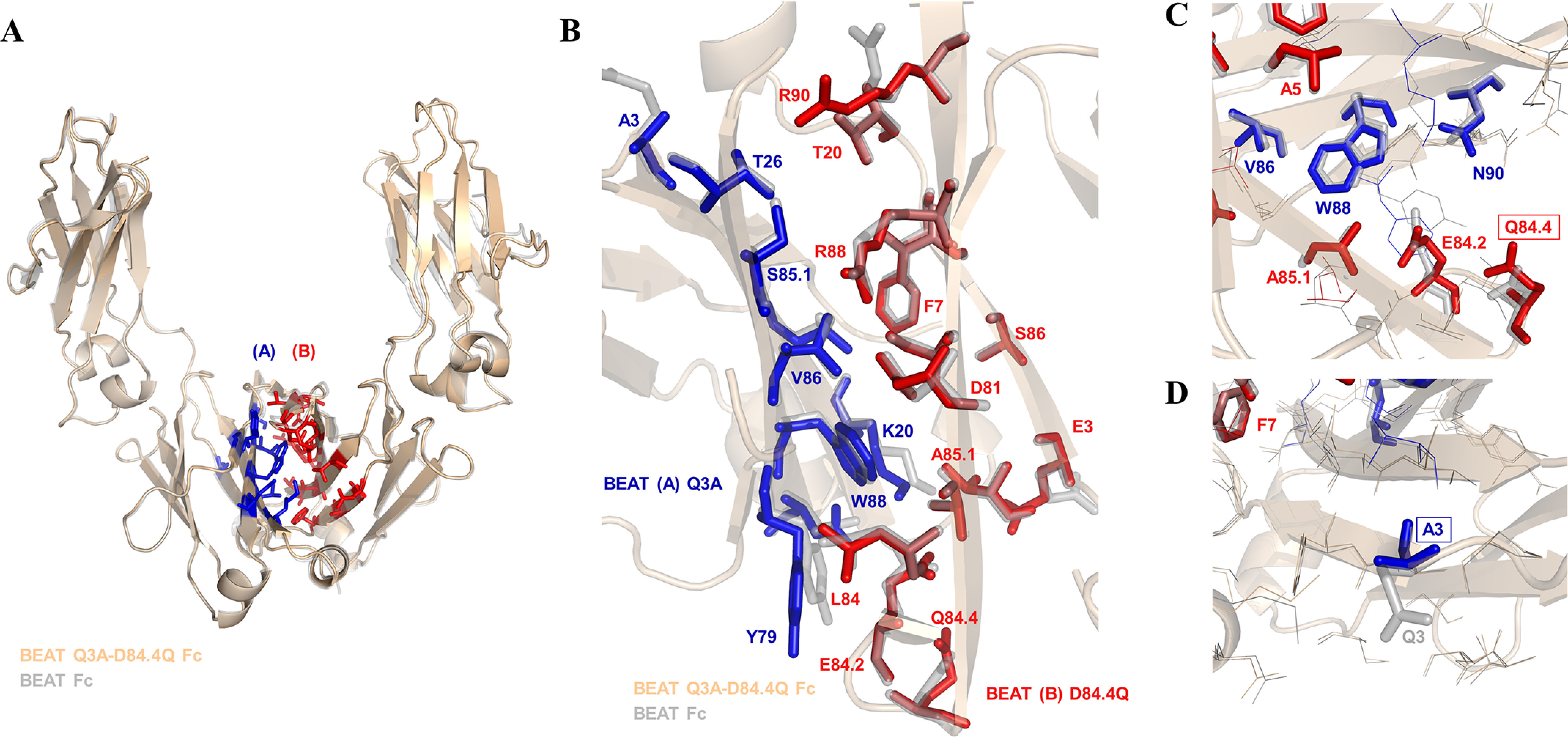
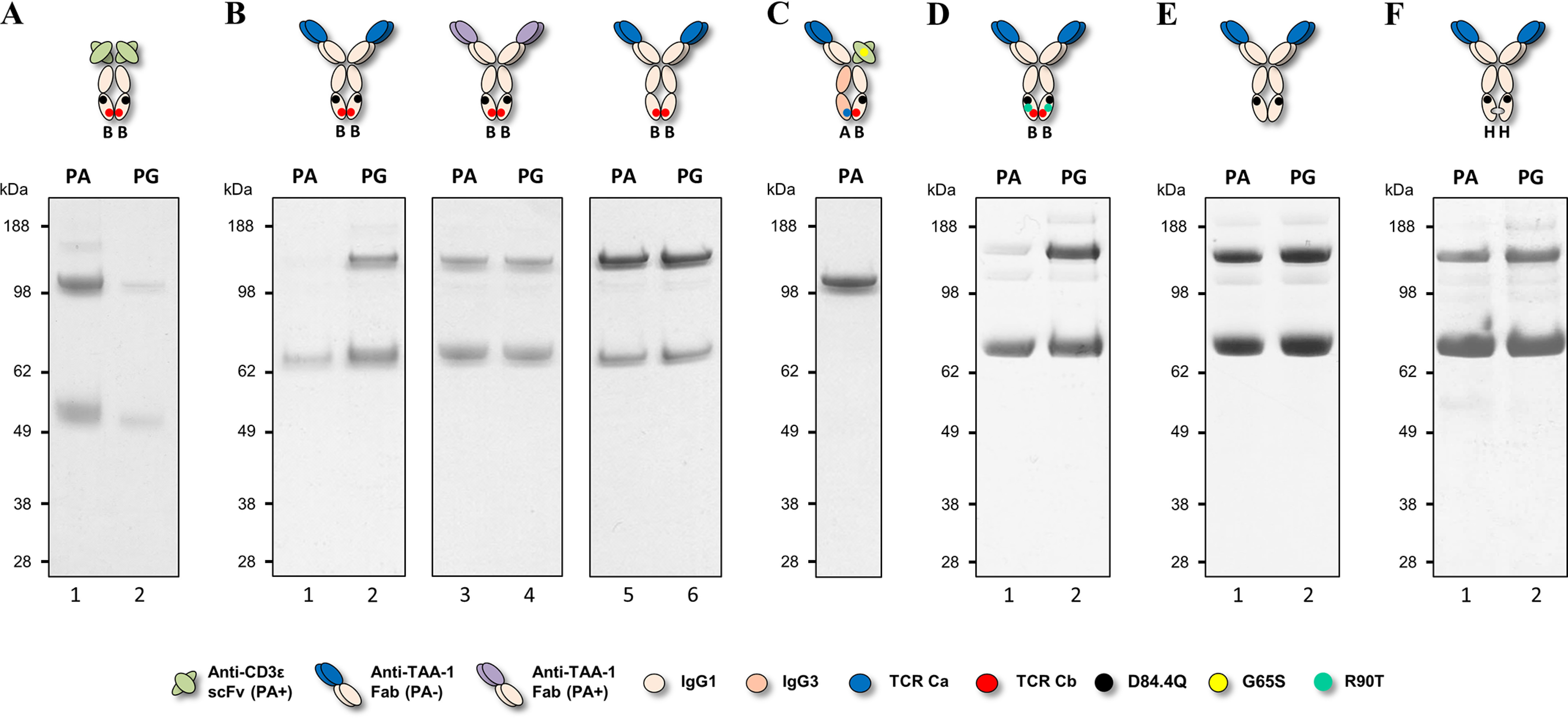
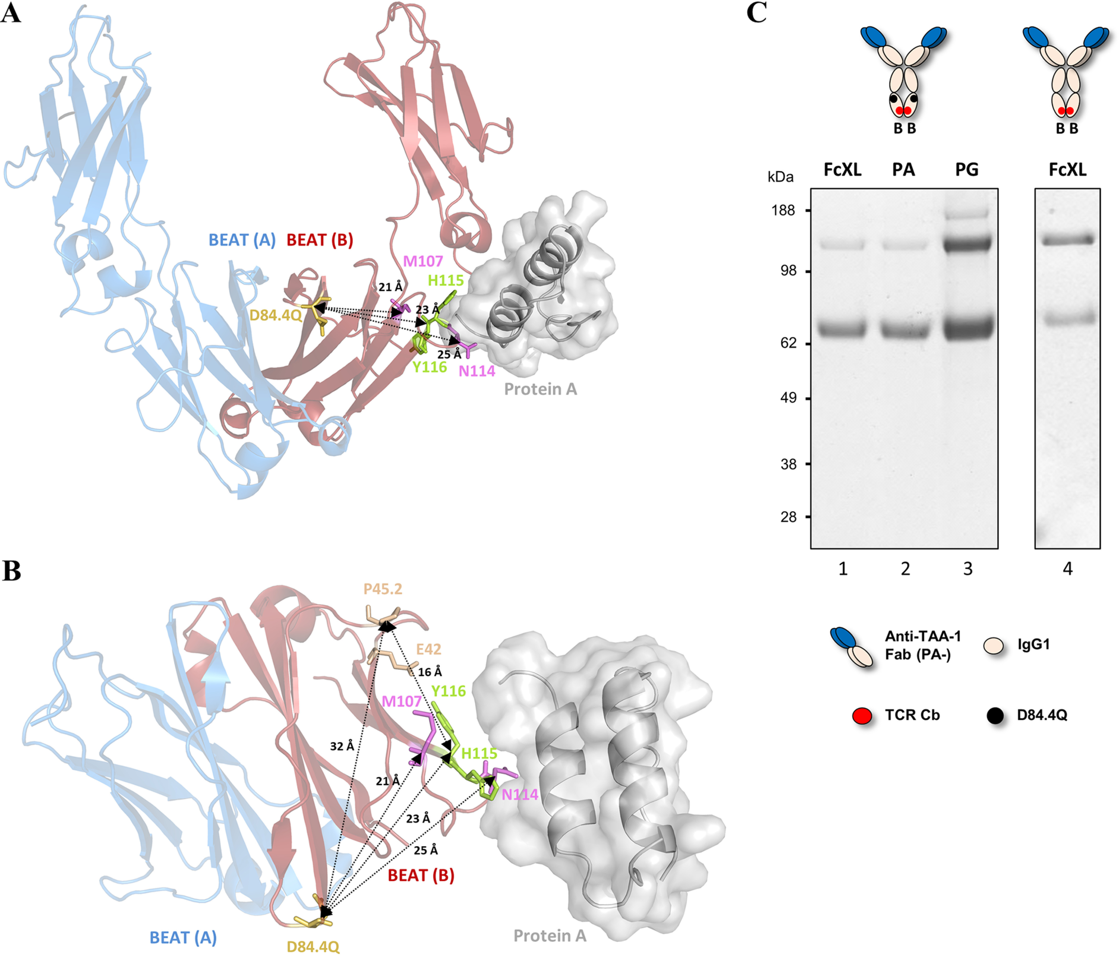
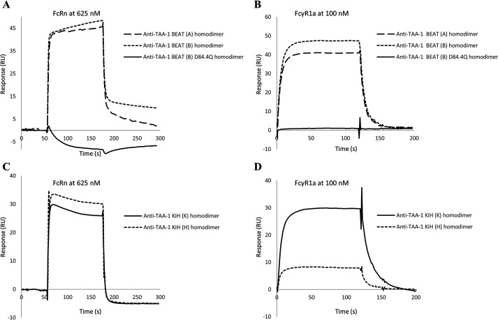
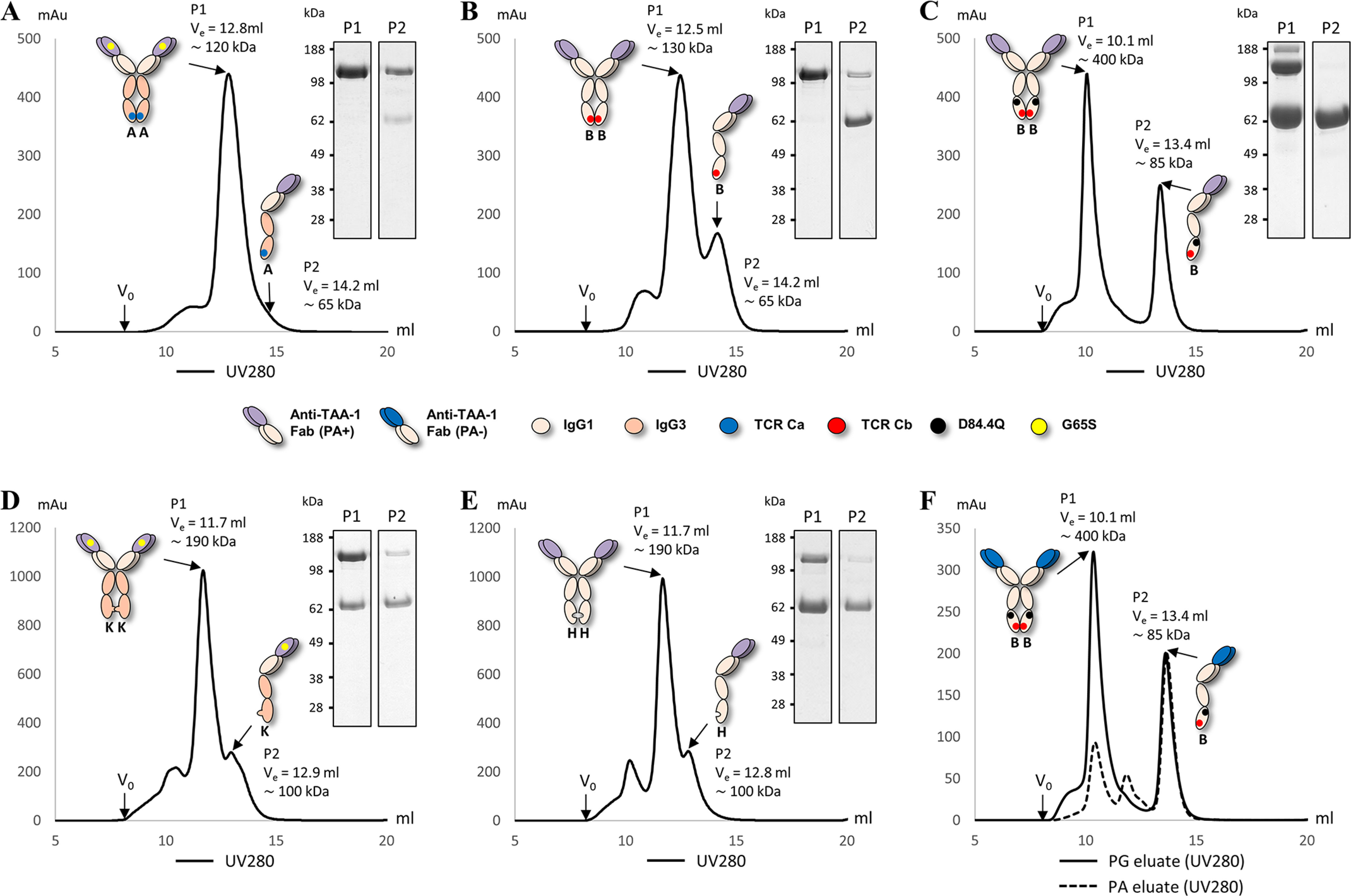
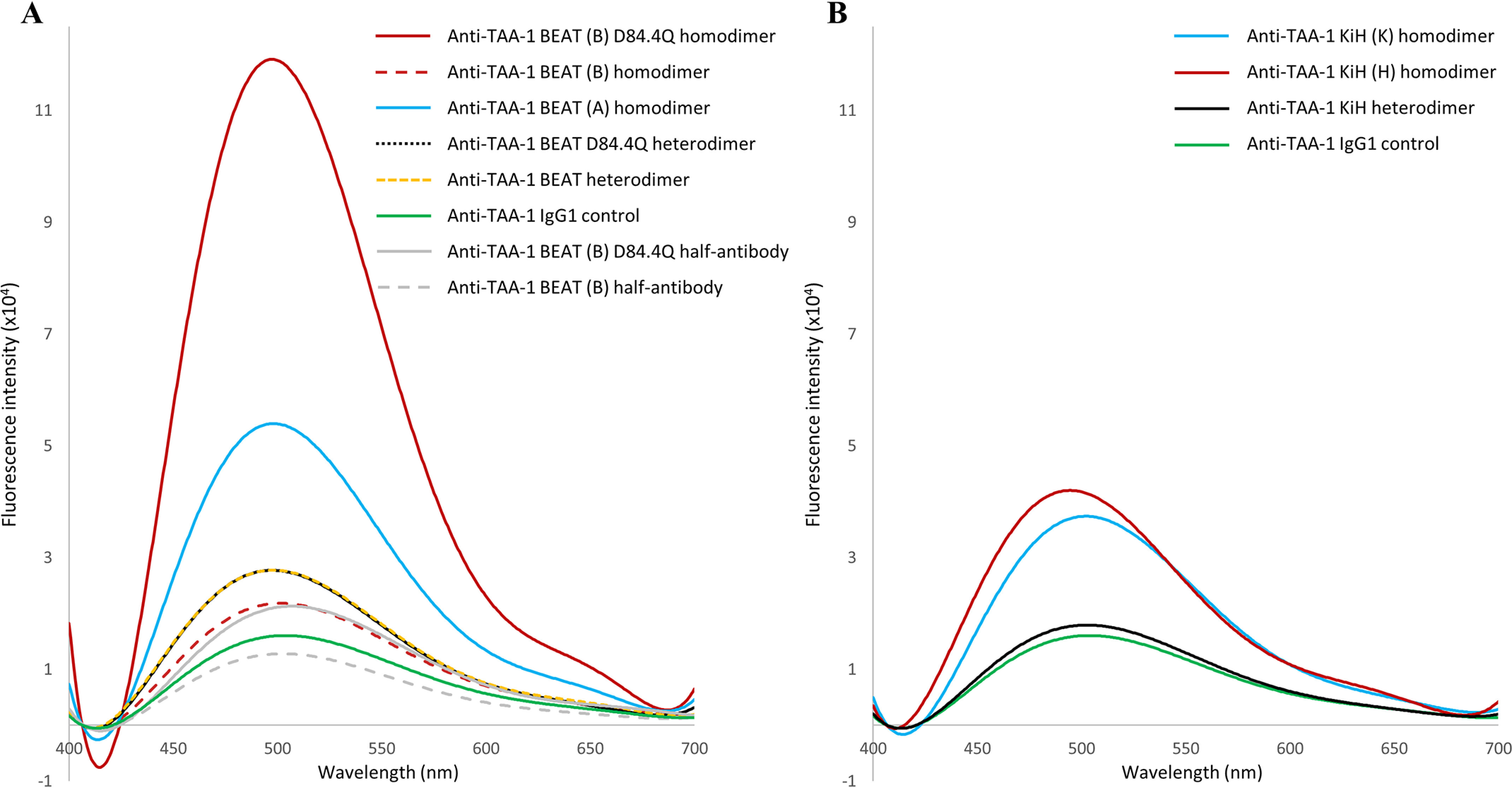
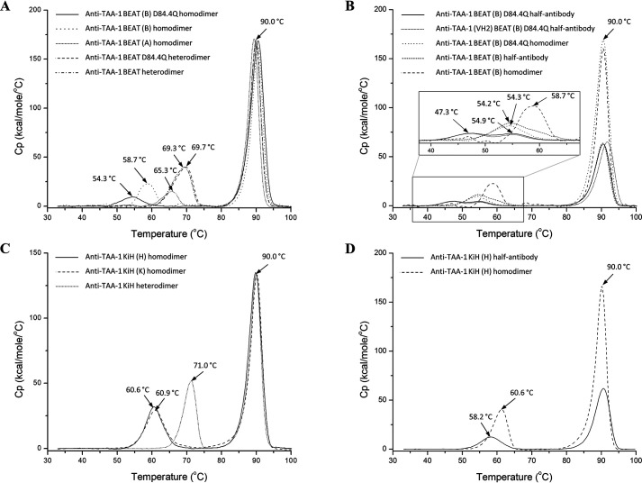
We previously reported efficient heavy-chain assembly of heterodimeric bispecific antibodies by exchanging the interdomain protein interface of the human IgG1 CH3 dimer with the protein interface of the constant α and β domains of the human T-cell receptor, a technology known as bispecific engagement by antibodies based on the T-cell receptor (BEAT). Efficient heterodimerization in mammalian cell transient transfections was observed, but levels were influenced by the nature of the binding arms, particularly in the Fab-scFv-Fc format. In this study, we report a single amino acid change that significantly and consistently improved the heterodimerization rate of this format (≥95%) by inducing partial disorder in one homodimer species without affecting the heterodimer. Correct folding and assembly of the heterodimer were confirmed by the high-resolution (1.88-1.98 Å) crystal structure presented here. Thermal stability and 1-anilinonaphthalene-8-sulfonic acid-binding experiments, comparing original BEAT, mutated BEAT, and "knobs-into-holes" interfaces, suggested a cooperative assembly process of heavy chains in heterodimers. The observed gain in stability of the interfaces could be classified in the following rank order: mutated BEAT > original BEAT > knobs-into-holes. We therefore propose that the superior cooperativity found in BEAT interfaces is the key driver of their greater performance. Furthermore, we show how the mutated BEAT interface can be exploited for the routine preparation of drug candidates, with minimal risk of homodimer contamination using a single Protein A chromatography step.

Keywords: CH3; antibody; bispecific engagement by antibodies based on the T-cell receptor (BEAT); heterodimer; knobs-into-holes (KiH); protein engineering; protein folding; protein stability; protein structure; structural disorder.


Conflict of interest statement

Conflict of interest—The authors declare that they were all employees of Ichnos Sciences S.A. at the time the work presented here was conducted.

Figures

Figure 1.
Figure 1.
Schematic drawing of a Fab–scFv–Fc bsAb. The first Hc noncovalently interacts with the second Hc via the CH3 domains that form the CH3–CH3 interface. Mutations that promote Hc HD, such as those composing the BEAT interface, are generally found in this portion of the antibody. The CH3 domains are connected to the CH2 domains, which together form the antibody Fc portion. The purification resins PA and PG bind at the interface between the CH2 and CH3 domains of the human IgG1 isotype. PA does not bind the IgG3 isotype. The FcRn receptor (neonatal Fc receptor), which promotes long serum half-life, also binds at the CH2–CH3 interface. The CH2 domains are connected to the hinge region. The hinge consists of the lower, middle, and upper hinge, wherein the lower hinge is coded by the CH2 exon. FcγR1a binds asymmetrically across the N-terminal region of the CH2 domains and lower hinge. The middle hinge of the IgG1 isotype contains two cysteine residues (Cys226 and Cys229) that form intermolecular disulfide bonds with the same cysteines of the second Hc. The N terminus of the upper hinge is connected to the CH1 domain followed by the VH domain. The CH1 domain interacts noncovalently with the constant domain of the Lc, which is termed constant κ (CK) or constant λ (Cλ), depending on the class of Lc. An intermolecular disulfide bond is formed between Hc and Lc, as shown here between Cys220 of the upper hinge of the IgG1 isotype and Cys214 of the CΚ domain. The VH domain interacts noncovalently with the VL domain. The CH1 domain contains a PG-binding site, and VH domains of the VH3 subclass contain a PA-binding site. PA binding in VH domains of the VH3 subclass can be abrogated using the G65S mutation. To circumvent Lc mispairing, one of the Fab domains can be converted into a scFv by genetic fusion of the VH domain to the VL domain via a flexible linker (generally (Gly4-Ser)3). The resulting domain is then fused to the hinge region. In the case of the VL–dAb–Fc format (inset), a VL-dAb is fused directly to the N terminus of the hinge of one of the Hcs. Cysteine residues are numbered according to the Eu numbering system, VH residues are numbered according to the Kabat numbering system, and CH3 residues are numbered according to IMGT.
Figure 2.
Figure 2.
Non-reduced SDS-PAGE analysis of hetero- and homodimer content of two different BEAT constructs. bsAbs were transiently expressed in HEK293-EBNA, purified by PA or PG chromatography, and analyzed by SDS-PAGE. PA+ indicates the presence of a PA-binding site, and PA− indicates the absence of a PA-binding site. For engineering, the G65S mutation was used to abrogate PA binding in VH3-type variable domains when needed. A, lanes 1 and 2, anti–TAA-1 × anti-CD3ε bsAb. Lanes 3 and 4, anti-CD3ε × anti–TAA-2 bsAb. Bands for homodimers (AA and BB) and heterodimers (AB) are annotated with arrows. B, the same bsAbs as in A but carrying the D84.4Q substitution in the CH3 domain of the BEAT (B) chain. C, summary of heterodimer content. Percentages were derived from the combined analysis of the PA and PG pulldowns.
Figure 3.
Figure 3.
Schematic diagrams depicting the CH3 interfaces of the BEAT (B) homodimer, the BEAT heterodimer containing the additional Q3A substitution in BEAT (A), the BEAT Q3A-D84.4Q heterodimer, and the original BEAT heterodimer. Interdomain interactions were calculated from the homology models for the first two and from the crystal structures for the latter two (PDB codes 6G1E and 5M3V, respectively). Homology models were generated based on the crystal structure of the original BEAT Fc. The IMGT numbering is used. Charged residues are colored in red (negative) or blue (positive). Hydrophobic interactions are in gray lines, and electrostatic interactions are in dashed red lines. The symmetric electrostatic interactions between Asp84.4 and Arg90 in the BEAT (B) homodimer as well as the asymmetric interactions between Asp84.4, Asp84.2, and Arg90 in the BEAT Q3A heterodimer are boxed in dashed black lines, and the residues are colored in yellow.
Figure 4.
Figure 4.
Thermal stability by DSC showing that D84.4Q has no impact on stability in the heterodimeric context. A, an overlay of BEAT Q3A-D84.4Q Fc (solid line) and BEAT Fc (dashed line) is shown. The first was built with an IgG3 CH2 domain in the BEAT (A) chain, whereas the latter had an IgG1 CH2 domain. The peak corresponds to the melting transitions (Tm) of both the CH2 and CH3 domain as the transitions overlap. The Tm values were ∼69.0 and ∼70.0 °C, respectively. B, similar to before but BEAT Q3A-D84.4Q Fc (solid line) encompassed an IgG1 CH2 domain in the BEAT (A) chain, whereas BEAT Fc (dashed line) had an IgG3 CH2 domain. The Tm values were ∼70.0 and ∼69.0 °C, respectively.
Figure 5.
Figure 5.
Crystal structure of the BEAT Q3A-D84.4Q Fc. A, ribbon diagram. Structural alignment of the original BEAT Fc structure (PDB code 5M3V) with that of BEAT Q3A-D84Q Fc (PDB code 6G1E). BEAT substitutions are in blue and red. B, close-up view of the same structural alignment. Side chains of the original BEAT Fc are in gray. A significant conservation of side-chain conformations could be observed (IMGT numbering). C, close-up showing position Gln84.4 and residues in its immediate environment, highlighting that no significant structural changes were induced by the mutation. D, close-up showing position A3 and residues in its immediate surroundings.
Figure 6.
Figure 6.
Non-reduced SDS-PAGE analysis of BEAT (B) homodimers. Antibodies were transiently expressed, purified by PA or PG chromatography, and analyzed by SDS-PAGE. A, anti-CD3ε BEAT (B) D84.4Q homodimer produced for analytical purposes. The band just above the 98-kDa molecular mass marker corresponds to the homodimer, and the band immediately above the 49-kDa molecular mass marker corresponds to the half-antibody. B, lanes 1 and 2 show the anti–TAA-1 BEAT (B) D84.4Q homodimer wherein the variable domain is of the VH2 subclass. In lanes 3 and 4, the VH2-type anti–TAA-1 was replaced with a VH3-type anti–TAA-1. In lanes 5 and 6, the D84.4Q substitution was removed in the VH2-type anti–TAA-1 BEAT (B) homodimer. The upper bands above 98 kDa correspond to homodimers, and the bands above 62 kDa correspond to half-antibodies. C, anti–TAA-1 × anti-CD3ε bsAb wherein both variable domains did not bind PA, which confirmed that the PA-binding site in the Fc of the BEAT (B) D84.4Q chain was functional in the context of an heterodimer. D, VH2-type anti–TAA-1 BEAT (B) D84.4Q homodimer containing the back-to-WT mutation R90T. E, VH2-type anti–TAA-1 homodimeric WT IgG1 antibody with the D84.4Q mutation. F, VH2-type anti–TAA-1 KiH (H) homodimer with the D84.4Q mutation.
Figure 7.
Figure 7.
Molecular details of PA, PG, and FcXL binding sites in the BEAT Fc. A, ribbon diagram of the structure of BEAT Q3A-D84.4Q Fc superimposed with the structure of PA–human Fc complex (PDB code 1FC2; the Fc is not shown). Front view of CH2 and CH3 domains. BEAT (A) is in blue, and BEAT (B) in red. PA is in gray. Position 84.4 is in yellow, and residues His115 and Tyr116 (His435 and Tyr436, respectively, according to Eu numbering), which make important interactions with PA, are in green. PG-binding residues are in pink. Distances between Cα of positions 84.4 and Cα 107, 115, and 116 are indicated. B, top view of the protein complexes with the CH2 domains hidden. Residues mediating FcXL binding are colored in wheat. Distances between Cα atoms are indicated. C, BEAT (B) homodimers with and without D84.4Q were transiently expressed, affinity-purified by FcXL, PA, or PG chromatography and analyzed by non-reduced SDS-PAGE. For the BEAT (B) D84.4Q homodimer, purification by PA confirmed the same lack of binding as with the FcXL resin (lanes 1 and 2), whereas PG purification confirmed expression of the homodimer (lane 3). Lane 4 shows that a BEAT (B) homodimer lacking the D84.4Q substitution was able to bind the FcXL resin.
Figure 8.
Figure 8.
SPR analysis of FcRn and FcγR1a binding to anti–TAA-1 BEAT and KiH homodimers. A, FcRn injected at 625 nm onto a sensor chip covalently coupled with BEAT (A), BEAT (B), or BEAT (B) D84.4Q homodimers. B, FcγR1a injected at 100 nm onto a PA-coupled sensor chip wherein BEAT homo- or heterodimers were previously captured via their VH3-type variable domains. C, FcRn injected at 625 nm onto a sensor chip covalently coupled with KiH homo- or heterodimers. D, FcγR1a injected at 100 nm onto a PA-coupled sensor chip where KiH homo- or heterodimers were previously captured via their VH3-type variable domains.
Figure 9.
Figure 9.
SEC traces of BEAT and KiH homodimers with non-reduced SDS-PAGE analysis of peak fractions. Ve is the volume of elution, and V0 is the void volume. Peak 1 is denoted P1, and peak 2 is denoted P2. Molecular mass calculated from a calibration run as indicated. A, BEAT (A) homodimer. P2 appeared merely as a shoulder because of the low abundancy of half-antibodies. B, BEAT (B) homodimer. C, BEAT (B) D84.4Q homodimer. D, KiH (K) homodimer. E, KiH (H) homodimer. F, BEAT (B) D84.4Q homodimer from PA (dashed line) and PG (solid line) eluates. Both chromatograms are shown overlaid.
Figure 10.
Figure 10.
Fluorescence emission of ANS. The samples (3.5 μm) were incubated for 20 min with ANS (200 μm) in PBS (pH 7.4) at 20 °C. Excitation was at 370 nm, and emission was measured between 400 and 700 nm. A, BEAT antibodies. BEAT (B) D84.4Q homodimers showed the strongest increase in ANS fluorescence over any other molecule tested. B, KiH antibodies.
Figure 11.
Figure 11.
Thermal stability by DSC, overlay of melting curves. A, an overlay of anti–TAA-1 BEAT bsAbs with and without D84.4Q, as well as the corresponding homodimers. The first transition corresponds to the melting of both the CH2 and CH3 domains, as the peaks overlap. B, an overlay of anti–TAA-1 BEAT (B) half-antibodies with and without the D84.4Q mutation. The corresponding homodimers are also shown for comparison. Because of the low Cp values of the transitions for the Fc region, a magnification of the boxed area is shown. C, an overlay of anti–TAA-1 KiH bsAb and the corresponding homodimers. D, anti–TAA-1 KiH (H) half-antibody overlaid with the corresponding homodimer. Each curve shows a transition at ∼90 °C that corresponds to the melting of the Fab portion.

References

    1. Brinkmann U., and Kontermann R. E. (2017) The making of bispecific antibodies. mAbs 9, 182–212 10.1080/19420862.2016.1268307 - DOI - PMC - PubMed
    1. Strohl W. R. (2018) Current progress in innovative engineered antibodies. Protein Cell 9, 86–120 10.1007/s13238-017-0457-8 - DOI - PMC - PubMed
    1. Atwell S., Ridgway J. B., Wells J. A., and Carter P. (1997) Stable heterodimers from remodeling the domain interface of a homodimer using a phage display library. J. Mol. Biol. 270, 26–35 10.1006/jmbi.1997.1116 - DOI - PubMed
    1. Ridgway J. B., Presta L. G., and Carter P. (1996) Knobs-into-holes' engineering of antibody CH3 domains for heavy chain heterodimerization. Protein Eng. 9, 617–621 10.1093/protein/9.7.617 - DOI - PubMed
    1. Gunasekaran K., Pentony M., Shen M., Garrett L., Forte C., Woodward A., Ng S. B., Born T., Retter M., Manchulenko K., Sweet H., Foltz I. N., Wittekind M., and Yan W. (2010) Enhancing antibody Fc heterodimer formation through electrostatic steering effects: applications to bispecific molecules and monovalent IgG. J. Biol. Chem. 285, 19637–19646 10.1074/jbc.M110.117382 - DOI - PMC - PubMed

MeSH terms

Substances

LinkOut - more resources