. 2020 May 13;12(543):eaaz5660.
doi: 10.1126/scitranslmed.aaz5660.

ACTRIIA-Fc rebalances activin/GDF versus BMP signaling in pulmonary hypertension

Affiliations

ACTRIIA-Fc rebalances activin/GDF versus BMP signaling in pulmonary hypertension

Lai-Ming Yung et al. Sci Transl Med. .

Abstract

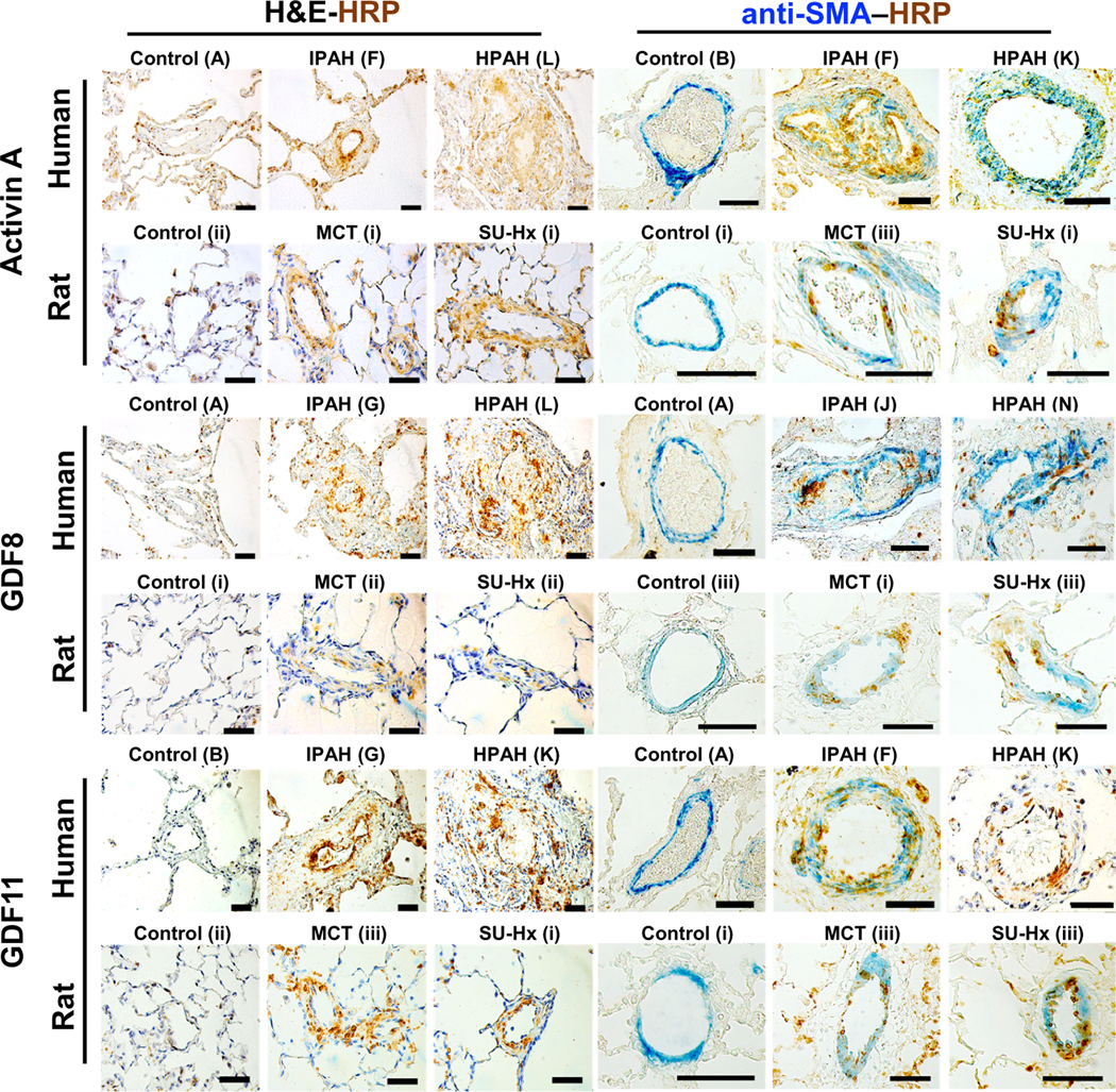
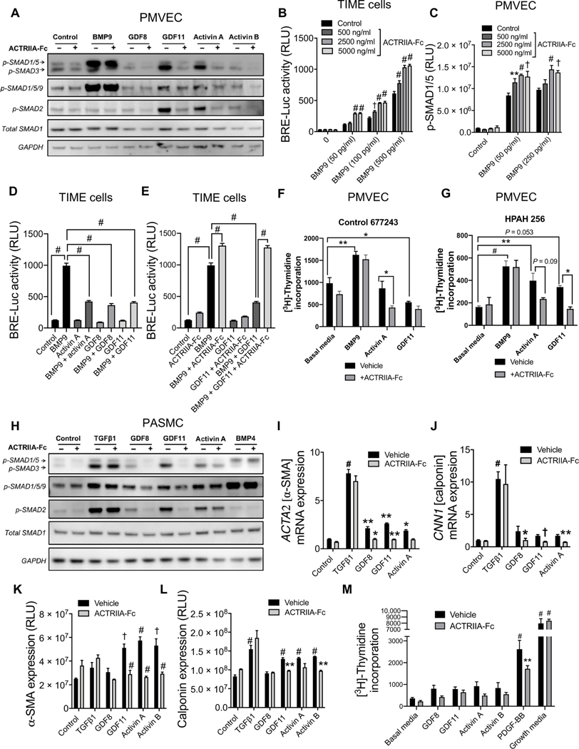
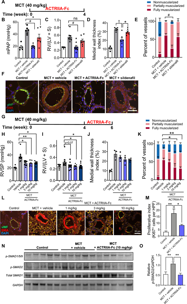
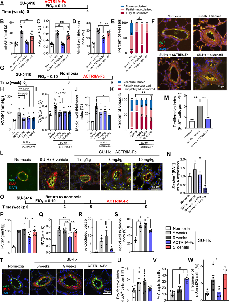
Human genetics, biomarker, and animal studies implicate loss of function in bone morphogenetic protein (BMP) signaling and maladaptive transforming growth factor-β (TGFβ) signaling as drivers of pulmonary arterial hypertension (PAH). Although sharing common receptors and effectors with BMP/TGFβ, the function of activin and growth and differentiation factor (GDF) ligands in PAH are less well defined. Increased expression of GDF8, GDF11, and activin A was detected in lung lesions from humans with PAH and experimental rodent models of pulmonary hypertension (PH). ACTRIIA-Fc, a potent GDF8/11 and activin ligand trap, was used to test the roles of these ligands in animal and cellular models of PH. By blocking GDF8/11- and activin-mediated SMAD2/3 activation in vascular cells, ACTRIIA-Fc attenuated proliferation of pulmonary arterial smooth muscle cells and pulmonary microvascular endothelial cells. In several experimental models of PH, prophylactic administration of ACTRIIA-Fc markedly improved hemodynamics, right ventricular (RV) hypertrophy, RV function, and arteriolar remodeling. When administered after the establishment of hemodynamically severe PH in a vasculoproliferative model, ACTRIIA-Fc was more effective than vasodilator in attenuating PH and arteriolar remodeling. Potent antiremodeling effects of ACTRIIA-Fc were associated with inhibition of SMAD2/3 activation and downstream transcriptional activity, inhibition of proliferation, and enhancement of apoptosis in the vascular wall. ACTRIIA-Fc reveals an unexpectedly prominent role of GDF8, GDF11, and activin as drivers of pulmonary vascular disease and represents a therapeutic strategy for restoring the balance between SMAD1/5/9 and SMAD2/3 signaling in PAH.


Figures

Fig. 1.
Fig. 1.. ACTRIIA ligands activin A, GDF8, and GDF11 are expressed in the intima and media of small arterioles in human PAH and rat PH.
Immunohistochemical staining for activin A, GDF8, and GDF11 along with α-SMA costain performed on paraffin-embedded lung sections from healthy controls and patients with IPAH or HPAH (n = 5 each) and lung sections from control, MCT-exposed, or SU-Hx–exposed rats (n = 3 each). Horseradish peroxidase (HRP)–conjugated secondary antibody (3,3′-diaminobenzidine staining, as indicated by label HRP in brown) and α-smooth muscle actin (α-SMA) AP-conjugated primary antibody (as indicated by label SMA in blue; right) are shown. Scale bars, 50 μm.
Fig. 2.
Fig. 2.. ACTRIIA-Fc modulates signaling, gene regulation, and proliferation of human pulmonary vascular cells.
(A) Expression of phosphorylated SMAD1/5/9 and SMAD2/3 in human pulmonary micro-vascular endothelial cells (PMVECs) exposed to BMP9 (100 pg/ml), GDF8 (50 ng/ml), GDF11 (50 ng/ml), activin A (50 ng/ml), or activin B (50 ng/ ml) for 30 min with or without ACTRIIA-Fc (2500 ng/ml), analyzed by Western blot. n = 3 replicates; N = 3 independent experiments. (B) Luciferase activity in telomerase immortalized human microvascular endothelial (TIME) cells transfected with BMP-responsive element transcriptional reporter (BRE-Luc) and exposed to varying concentrations of BMP9 with or without ACTRIIA-Fc (n = 3; N = 3; RLU, relative luminescence units). (C) Activation of SMAD1/5 in human PMVECs treated with BMP9 (below the EC50 250 ng/ml) with or without varying concentrations of ACTRIIA-Fc, analyzed by In-Cell Western. Data represent means ± SEM. *P < 0.05, **P < 0.01, †P < 0.001, and #P < 0.0001 compared to untreated controls in each group, two-way ANOVA with Dunnett’s test for multiple comparisons (n = 3; N = 3). (D and E) BMP-mediated transcriptional activity in TIME cells treated with BMP9, GDF8, GDF11, and activin A with or without ACTRIIA-Fc (n = 6; N = 3). (F and G) Proliferation of control- and PAH-derived PMVECs in response to BMP9, activin A, and GDF11 with or without ACTRIIA-Fc. (H) Expression of phosphorylated SMAD1/5/9 and SMAD2/3 in human pulmonary artery smooth muscle cells (PASMCs) exposed to TGFβ1 (1 ng/ml), GDF8 (50 ng/ml), GDF11 (50 ng/ml), activin A (50 ng/ml), or BMP4 (50 ng/ml) for 30 min with or without ACTRIIA-Fc (2500 ng/ml), analyzed by Western blot. (I and J) Expression of α-SMA and calponin mRNA at 24 hours after stimulation with TGFβ1 (1 ng/ml), GDF8 (50 ng/ml), GDF11 (50 ng/ml), or activin A (50 ng/ml) in human PASMCs with or without ACTRIIA-Fc (n = 3 to 6). (K and L) Expression of α-SMA and calponin protein in PASMCs at 72 hours after stimulation with TGFβ1, GDF8, GDF11, activin A, or activin B with or without ACTRIIA-Fc, as assessed by In-Cell Western (n = 4; N = 3). (M) Proliferation in PASMCs exposed to GDF8 (100 ng/ml), GDF11 (100 ng/ml), activin A (100 ng/ml), activin B (100 ng/ml), PDGF-BB (1 ng/ml), or complete media for 24 hours with or without ACTRIIA-Fc (10,000 ng/ml) as measured by 3H-thymidine incorporation. (n = 5; N = 3). Data represent means ± SEM. *P < 0.05, **P < 0.01, †P < 0.001, and #P < 0.0001 compared to vehicle controls, one-way ANOVA with Sidak’s test for multiple comparisons.
Fig. 3.
Fig. 3.. ACTRIIA-Fc attenuates PH and vascular remodeling in MCT-exposed rats.
(A) Treatment timing of adult rats after vehicle or MCT (40 mg/ kg, sc) with ACTRIIA-Fc (15 mg/kg, ip, twice weekly), sildenafil [30 mg/kg, orally, twice daily], or vehicle (10 mM TBS) for 4 weeks starting 1 day after MCT. (B) Mean pulmonary artery pressure (mPAP) and (C) RV hypertrophy (RVH) in rats treated with ACTRIIA-Fc or sildenafil compared to vehicle (n = 5 to 9 per group). Data represent means ± SEM. *P < 0.05, **P < 0.01, #P < 0.0001, and not significant (ns; P > 0.05) as indicated, one-way ANOVA with Sidak’s post test for mPAP and Kruskal-Wallis test with Dunn’s post test for RVH. (D) Medial wall thickness index and (E) the percentage of fully muscularized vessels in rats treated with ACTRIIA-Fc or sildenafil compared to vehicle (diameter, 10 to 50 μm). Values are shown as means ± SEM. n = 3 to 6 per group (30 to 50 vessels counted per sample). *P < 0.05, **P < 0.01, †P < 0.001, and #P < 0.0001 as indicated, one-way and two-way ANOVA with Tukey’s and Dunnett’s test for multiple comparisons. (F) Immunofluorescence images of pulmonary arteriole medial hypertrophy in lung sections from rats with MCT-induced PH treated with vehicle, ACTRIIA-Fc, or sildenafil. vWF, von Willebrand factor; SMA, α-SMA; DAPI, 4′,6-diamidino-2-phenylindole (nuclear stain). (G) Treatment timing of rats with established PH, with ACTRIIA-Fc (1, 3, and 10 mg/kg, ip, twice weekly) or isotype control with vehicle (mIgG2a, 10 mg/kg, ip, twice weekly) starting on day 28 after MCT. (H) RV systolic pressure (RVSP) and (I) RVH in rats treated with control IgG or ACTRIIA-Fc measured among surviving animals at 42 days (n = 6 to 10 per group). Values are shown as means ± SEM. *P < 0.05 and **P < 0.01, Kruskal-Wallis test with Dunn’s post test for multiple comparisons. (J) Medial wall thickness and (K) percentage of muscularized vessels in rats with established PH treated with vehicle IgG or ACTRIIA-Fc compared to control. Values are shown as means ± SEM. n = 3 to 6 per group (30 to 50 vessels counted per sample). *P < 0.05, **P < 0.01, †P < 0.001, and #P < 0.0001 as indicated, two-way ANOVA with Dunnett’s test for multiple comparisons. (L) Immunofluorescence images of MCT-induced medial hypertrophy in pulmonary arterioles from rats with established PH treated with vehicle IgG or ACTRIIA-Fc. (M) Ki67-positive cells in MCT-PH lungs treated with vehicle IgG or ACTRIIA-Fc. Values are shown as means ± SEM. n = 3 to 6 per group [20 random high-powered fields (HPF) containing >30 to 50 vessels counted per sample]. (N) Western blot and (O) densitometric analysis of phosphorylation of SMAD2 in lung lysates from MCT-exposed rats treated with vehicle IgG or ACTRIIA-Fc in comparison to control rats. Data represent means ± SEM. n = 3 to 4. *P < 0.05 and **P < 0.01, one-way ANOVA with Tukey’s test for multiple comparisons. Scale bars, 50 μm.
Fig. 4.
Fig. 4.. ACTRIIA-Fc ameliorates PH and vascular remodeling in SU-Hx–exposed rats.
(A) Treatment timing for SU-Hx PH. Male adult rats were injected with vehicle or VEGFR1/2 inhibitor SUGEN5416 (200 mg/kg, sc), subjected to normoxia (Nx) or normobaric hypoxia (FiO2 = 0.10) for 4 weeks, and received ACTRIIA-Fc (10 mg/kg, ip, twice weekly), sildenafil (60 mg/kg, twice daily, orally), or vehicle (10 mM TBS) for 4 weeks starting 1 day after SUGEN5416. (B) mPAP and (C) RVH in Nx-exposed and SU-Hx–exposed rats treated with ACTRIIA-Fc or sildenafil as compared to TBS vehicle–treated rats (n = 5 to 10 per group). Values are shown as means ± SEM. **P < 0.01 and #P < 0.0001, as indicated, one-way ANOVA with Dunnett’s multiple comparisons test. (D) Medial wall thickness and (E) the percentage of muscularized vessels (diameter, 10 to 50 μm) in rats treated with sildenafil or ACTRIIA-Fc as in (B). Values are shown as means ± SEM. n = 4 to 6 per group (30 to 50 vessels counted per sample). *P < 0.05, **P < 0.01, †P < 0.001, and #P < 0.0001 as indicated, one-way or two-way ANOVA with Tukey’s or Dunnett’s test for multiple comparisons. (F) Immunofluorescence images of medial hypertrophy of pulmonary arterioles in Nx-exposed and SU-Hx–exposed rats treated with vehicle, ACTRIIA-Fc, or sildenafil. (G) Treatment timing for established PH in SU-Hx model. Male adult rats received a single injection of SUGEN5416 (20 mg/kg, sc) and were subjected to normobaric hypoxia (FiO2 = 0.10) for 3 weeks, followed by 3 weeks of normoxia during which rats received varying doses of ACTRIIA-Fc (1, 3, or 10 mg/kg, ip, twice weekly) or isotype control (mIgG2a; 10 mg/kg, ip, twice weekly). (H) RVSP and (I) RVH in rats treated as in (G) (n = 5 to 8 per group). *P < 0.05, one-way ANOVA with Holm- Sidak’s test for multiple comparisons. (J and K) The highest dose of ACTRIIA-Fc attenuated medial wall thickening and (K) the percent-* age of muscularized vessels in rats treated as in (G). *P < 0.05 and **P < 0.01 as indicated, one-way and two-way ANOVA with Tukey’s and Dunnett’s tests for multiple comparisons. Values are shown as means ± SEM. n = 4 to 8 per group (30 to 50 vessels counted per sample). (L) Immunofluorescence images of medial hypertrophy and neointimal lesion formation in pulmonary arterioles from rats treated as in (G). (M) Quantification of proliferation index in small vessels and perivascular tissues of SU-Hx rats treated with isotype control or high dose of ACTRIIA-Fc. **P < 0.01, one-way ANOVA with Tukey’s test for multiple comparisons. Values are shown as means ± SEM. n = 3 to 4 per group (20 random high-powered fields of view quantified per sample). (N) Pai-1 mRNA expression via quantitative RT-PCR in lung tissues from rats with SU-Hx treated with ACTRIIA-Fc or control. *P < 0.05, one-way ANOVA with Tukey’s test for multiple comparisons. Values are shown as means ± SEM. n = 3 to 4 per group. (O) Treatment timing for rats with severe obliterative PH. SU-Hx rats were allowed to progress for 2 weeks under normoxia after 3 weeks of SU-Hx (20 mg/kg, sc; FiO2 = 0.10), followed by 4 weeks of treatment with vehicle, ACTRIIA-Fc, or sildenafil. (P) RVSP and (Q) RVH in untreated rats after 5 and 9 weeks or rats treated with ACTRIIA-Fc or sildenafil from weeks 6 to 9 (n = 3 to 8). *P < 0.05 and **P < 0.01, one-way ANOVA with Tukey’s test for multiple comparisons. (R) Proportion of occluded vessels (n = 3 to 4) and (S) vascular wall thickness index (n = 3 to 4 and 30 to 50 vessels counted per sample) in rats as in (P). (T) Immunofluorescence images of intimal and medial remodeling in pulmonary arterioles in SU-Hx rats with or without ACTRIIA-Fc treatment. (U) Proliferative index in small vessels and perivascular tissues in SU-Hx rats with or without ACTRIIA-Fc (P = 0.19 ACTRIIA-Fc compared to week 9, n = 4 to 6, 20 random high-powered fields of view quantified per sample). (V) Percentage of apoptotic vascular cells (TUNEL+ vessel-associated cells) in ACTRIIA-Fc–treated rats as compared to untreated rats at 5 and 9 weeks after SU-Hx treatment (n = 4 to 6). (W) Percentage of p-SMAD2/3+ cells in the luminal wall and media of remodeled vessels in ACTRIIA-Fc–, sildenafil-, or vehicle-treated SU-Hx rats at 9 weeks (n = 4 to 5). Values for (P) to (W) are shown as means ± SEM. *P < 0.05, **P < 0.01, †P < 0.001, and #P < 0.0001 as indicated, one-way ANOVA with Tukey’s test for multiple comparisons. Scale bars, 50 μm.

References

    1. Simonneau G, Montani D, Celermajer DS, Denton CP, Gatzoulis MA, Krowka M, Williams PG, Souza R, Haemodynamic definitions and updated clinical classification of pulmonary hypertension. Eur. Respir. J 53, 1801913 (2019). - PMC - PubMed
    1. Benza RL, Miller DP, Barst RJ, Badesch DB, Frost AE, McGoon MD, An evaluation of long-term survival from time of diagnosis in pulmonary arterial hypertension from the REVEAL Registry. Chest 142, 448–456 (2012). - PubMed
    1. Soubrier F, Chung WK, Machado R, Grünig E, Aldred M, Geraci M, Loyd JE, Elliott CG, Trembath RC, Newman JH, Humbert M, Genetics and genomics of pulmonary arterial hypertension. J. Am. Coll. Cardiol 62, D13–D21 (2013). - PubMed
    1. Gräf S, Haimel M, Bleda M, Hadinnapola C, Southgate L, Li W, Hodgson J, Liu B, Salmon RM, Southwood M, Machado RD, Martin JM, Treacy CM, Yates K, Daugherty LC, Shamardina O, Whitehorn D, Holden S, Aldred M, Bogaard HJ, Church C, Coghlan G, Condliffe R, Corris PA, Danesino C, Eyries M, Gall H, Ghio S, Ghofrani H-A, Gibbs JSR, Girerd B, Houweling AC, Howard L, Humbert M, Kiely DG, Kovacs G, MacKenzie Ross RV, Moledina S, Montani D, Newnham M, Olschewski A, Olschewski H, Peacock AJ, Pepke-Zaba J, Prokopenko I, Rhodes CJ, Scelsi L, Seeger W, Soubrier F, Stein DF, Suntharalingam J, Swietlik EM, Toshner MR, van Heel DA, Noordegraaf AV, Waisfisz Q, Wharton J, Wort SJ, Ouwehand WH, Soranzo N, Lawrie A, Upton PD, Wilkins MR, Trembath RC, Morrell NW, Identification of rare sequence variation underlying heritable pulmonary arterial hypertension. Nat. Commun 9, 1416 (2018). - PMC - PubMed
    1. Thomas M, Docx C, Holmes AM, Beach S, Duggan N, England K, Leblanc C, Lebret C, Schindler F, Raza F, Walker C, Crosby A, Davies RJ, Morrell NW, Budd DC, Activin-like kinase 5 (ALK5) mediates abnormal proliferation of vascular smooth muscle cells from patients with familial pulmonary arterial hypertension and is involved in the progression of experimental pulmonary arterial hypertension induced by monocrotaline. Am. J. Pathol 174, 380–389 (2009). - PMC - PubMed

Publication types