

Case Reports
doi: 10.1016/j.ajog.2020.05.023.
Epub 2020 May 13.
Visualization of severe acute respiratory syndrome coronavirus 2 invading the human placenta using electron microscopy
Affiliations
- PMID: 32405074
- PMCID: PMC7219376
- DOI: 10.1016/j.ajog.2020.05.023
Item in Clipboard
Case Reports
Visualization of severe acute respiratory syndrome coronavirus 2 invading the human placenta using electron microscopy
Am J Obstet Gynecol.
2020 Aug.
No abstract available
Figures
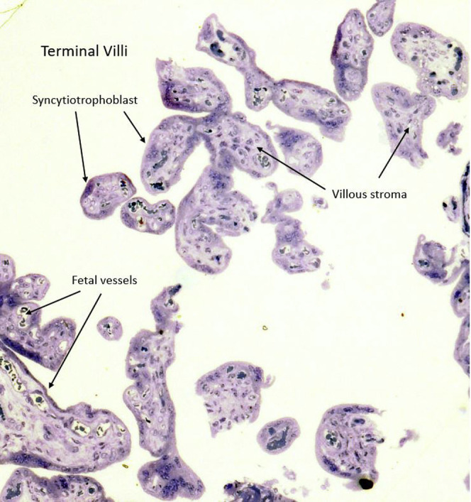
Placental thick section at 1 micron stained with toluidine blue showing the terminal villi containing fetal blood vessels (10×) This area was used for transmission electron microscopy. Algarroba. Visualization of SARS-CoV-2 invading the human placenta using electron microscopy. Am J Obstet Gynecol 2020.
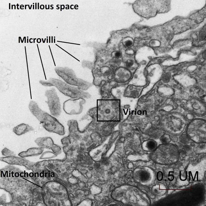
Transmission electron microscopy of a visible single virion invading a syncytiotrophoblast (30,000×) Algarroba. Visualization of SARS-CoV-2 invading the human placenta using electron microscopy. Am J Obstet Gynecol 2020.
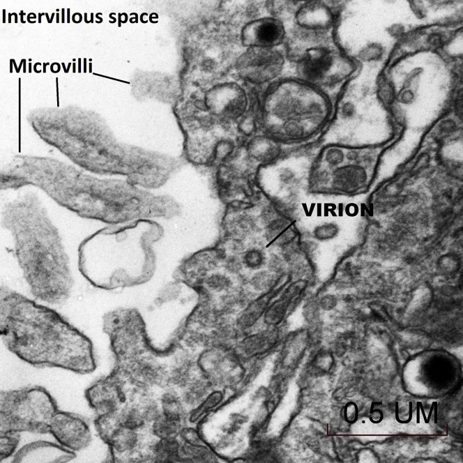
Transmission electron microscopy of a visible single virion invading a syncytiotrophoblast at a higher magnification (50,000×) Algarroba. Visualization of SARS-CoV-2 invading the human placenta using electron microscopy. Am J Obstet Gynecol 2020.
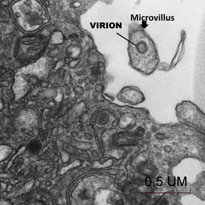
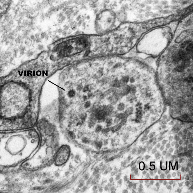
Transmission electron microscopy of a single virion visualized in a syncytiotrophoblast microvillus (50,000×) Algarroba. Visualization of SARS-CoV-2 invading the human placenta using electron microscopy. Am J Obstet Gynecol 2020.
Transmission electron microscopy of the trophoblastic layer in the mesenchymal core of the terminal villus where a single virion was visible, likely in the cell processes of fibroblasts (50,000×) Algarroba. Visualization of SARS-CoV-2 invading the human placenta using electron microscopy. Am J Obstet Gynecol 2020.
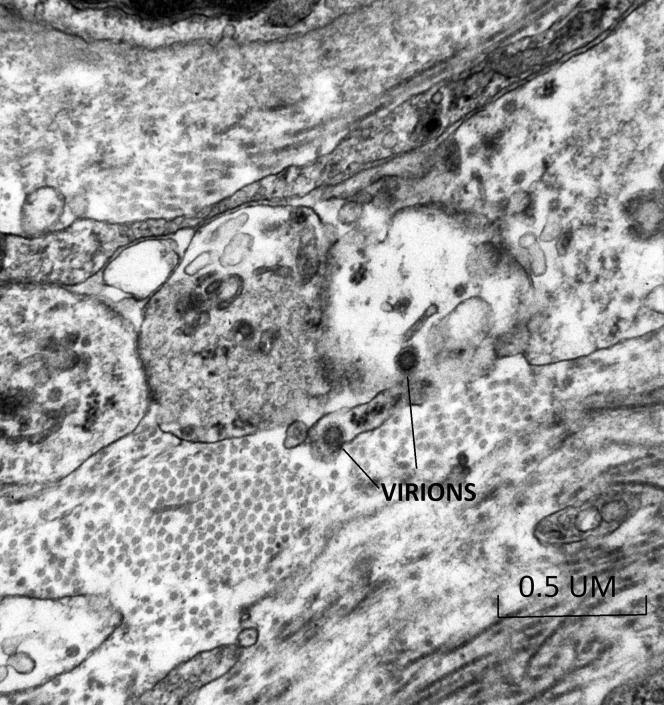
Transmission electron microscopy of the trophoblastic layer in the mesenchymal core of the terminal villus where 2 virions were visible, likely in the cell processes of fibroblasts (50,000×) Algarroba. Visualization of SARS-CoV-2 invading the human placenta using electron microscopy. Am J Obstet Gynecol 2020.
Comment in
-
Reply.Am J Obstet Gynecol. 2020 Nov;223(5):786-788. doi: 10.1016/j.ajog.2020.06.017. Epub 2020 Jun 9. Am J Obstet Gynecol. 2020. PMID: 32531214 Free PMC article. No abstract available.
-
Alternative interpretation to the findings reported in visualization of severe acute respiratory syndrome coronavirus 2 invading the human placenta using electron microscopy.Am J Obstet Gynecol. 2020 Nov;223(5):785-786. doi: 10.1016/j.ajog.2020.06.016. Epub 2020 Jun 10. Am J Obstet Gynecol. 2020. PMID: 32533928 Free PMC article. No abstract available.
-
Confirmatory evidence of the visualization of severe acute respiratory syndrome coronavirus 2 invading the human placenta using electron microscopy.Am J Obstet Gynecol. 2020 Dec;223(6):953-954. doi: 10.1016/j.ajog.2020.08.106. Epub 2020 Aug 28. Am J Obstet Gynecol. 2020. PMID: 32866527 Free PMC article. No abstract available.
References
-
- Valdés G., Neves L.A., Anton L., et al. Distribution of angiotensin-(1–7) and ACE2 in human placentas of normal and pathological pregnancies. Placenta. 2006;27:200–207. - PubMed
Publication types
MeSH terms
LinkOut - more resources
Full Text Sources
Medical
