

Review
doi: 10.1016/j.jcot.2020.03.010.
Epub 2020 Mar 28.
Ankle stability in ankle fracture
Affiliations
- PMID: 32405195
- PMCID: PMC7211817
- DOI: 10.1016/j.jcot.2020.03.010
Item in Clipboard
Review
Ankle stability in ankle fracture
J Clin Orthop Trauma.
2020 May-Jun.
Erratum in
-
Erratum regarding previously published articles.J Clin Orthop Trauma. 2020 Nov-Dec;11(6):1169-1171. doi: 10.1016/j.jcot.2020.09.032. Epub 2020 Sep 26. J Clin Orthop Trauma. 2020. PMID: 33013141 Free PMC article.
-
Erratum regarding missing Declaration of Competing Interest statements in previously published articles.J Clin Orthop Trauma. 2020 Nov-Dec;11(6):1178. doi: 10.1016/j.jcot.2020.10.026. Epub 2020 Oct 15. J Clin Orthop Trauma. 2020. PMID: 33078052 Free PMC article.
-
Erratum regarding previously published articles.J Clin Orthop Trauma. 2020 Nov-Dec;11(6):1172-1174. doi: 10.1016/j.jcot.2020.10.044. Epub 2020 Oct 23. J Clin Orthop Trauma. 2020. PMID: 33192025 Free PMC article.
-
Erratum regarding previously published articles.J Clin Orthop Trauma. 2021 Oct;21:101561. doi: 10.1016/j.jcot.2021.101561. Epub 2021 Aug 5. J Clin Orthop Trauma. 2021. PMID: 34381302 Free PMC article.
Abstract
Restoration of normal ankle kinematics should be the all-encompassing ethos in the approach to management of ankle fractures. To do this, the ligamentous stabilisers must also form part of its assessment and definitive management and be considered during index fracture fixation surgery. This article is a review of the anatomy, mechanics and clinical testing of instability in ankle fractures.
Keywords: Ankle fracture; Ankle instability; Fibular; Ligaments; Malleolar.
© 2020 Delhi Orthopedic Association. All rights reserved.
Figures

3D image of ankle skeleton with a movement sphere overlay showing the possible translation in the sagittal and frontal planes and rotation in the sagittal, frontal and horizontal planes.

3D image of ankle skeleton showing sagittal plane translation and rotation.

D image of ankle skeleton showing frontal plane translation and rotation.

3D image of ankle skeleton showing horizontal plane rotation.

3D image of ankle skeleton with schematic representation of the supporting ligaments (ATFL – anterior talofibular ligament, PTFL – posterior talofibular ligament, AITFL – anterior inferior tibiofibular ligament, PITFL – posterior inferior tibiofibular ligament, CFL – calcaneofibular ligament).
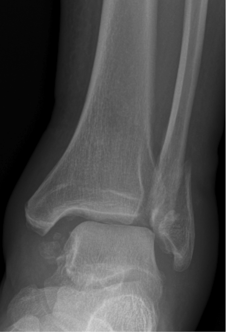
Anteroposterior radiograph of left ankle showing medical clear space greater than 5 mm with displaced fibular fracture.

Intraoperative loss of tibiofibular overlap as a result of an internal rotation test on a type 1 Mason and Molloy posterior Malleolar fracture following fibular fixation (first image). This overlap is corrected on syndesmotic fixation and retesting showed normal tibiofibular overlap on repeat screening (second image).
References
-
- Rasmussen O. Stability of the ankle joint. Analysis of the function and traumatology of the ankle ligaments. Acta Orthop Scand Suppl. 1985;211:1–75. - PubMed
-
- Grimston S.K., Nigg B.M., Hanley D.A., Engsberg J.R. Differences in ankle joint complex range of motion as a function of age. Foot Ankle. 1993;14(4):215–222. - PubMed
-
- Stauffer R.N., Chao E.Y., Brewster R.C. Force and motion analysis of the normal, diseased, and prosthetic ankle joint. Clin Orthop Relat Res. 1977;127:189–196. - PubMed
Publication types
LinkOut - more resources
Full Text Sources
