

Subtalar fusion and exostectomy in calcaneus malunion: How we do it
- PMID: 32405217
- PMCID: PMC7211907
- DOI: 10.1016/j.jcot.2020.03.011
Subtalar fusion and exostectomy in calcaneus malunion: How we do it
Erratum in
-
Erratum regarding previously published articles.J Clin Orthop Trauma. 2020 Nov-Dec;11(6):1169-1171. doi: 10.1016/j.jcot.2020.09.032. Epub 2020 Sep 26. J Clin Orthop Trauma. 2020. PMID: 33013141 Free PMC article.
-
Erratum regarding missing Declaration of Competing Interest statements in previously published articles.J Clin Orthop Trauma. 2020 Nov-Dec;11(6):1177. doi: 10.1016/j.jcot.2020.10.025. Epub 2020 Oct 15. J Clin Orthop Trauma. 2020. PMID: 33078051 Free PMC article.
-
Erratum regarding previously published articles.J Clin Orthop Trauma. 2020 Nov-Dec;11(6):1172-1174. doi: 10.1016/j.jcot.2020.10.044. Epub 2020 Oct 23. J Clin Orthop Trauma. 2020. PMID: 33192025 Free PMC article.
-
Erratum regarding previously published articles.J Clin Orthop Trauma. 2021 Aug 5;21:101560. doi: 10.1016/j.jcot.2021.101560. eCollection 2021 Oct. J Clin Orthop Trauma. 2021. PMID: 34414073 Free PMC article.
Abstract
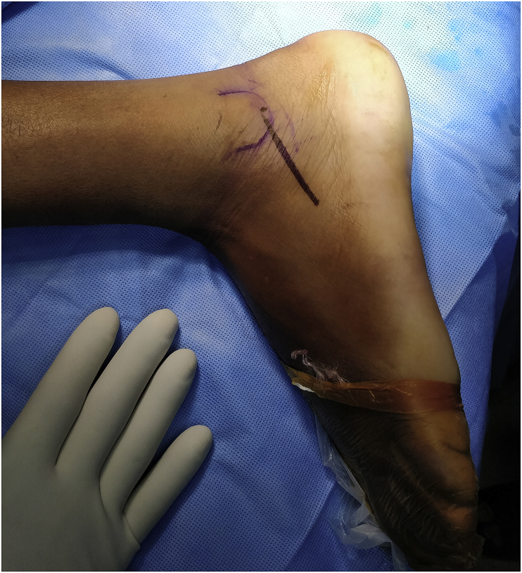
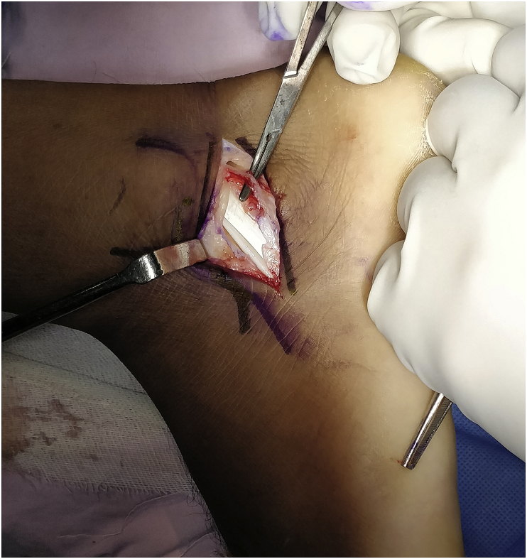
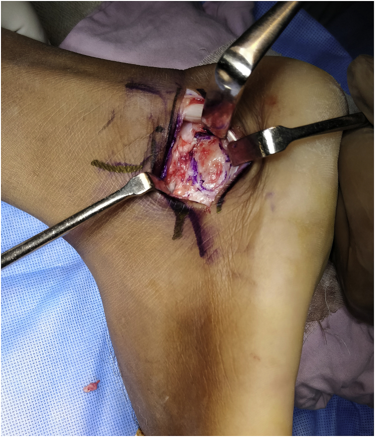
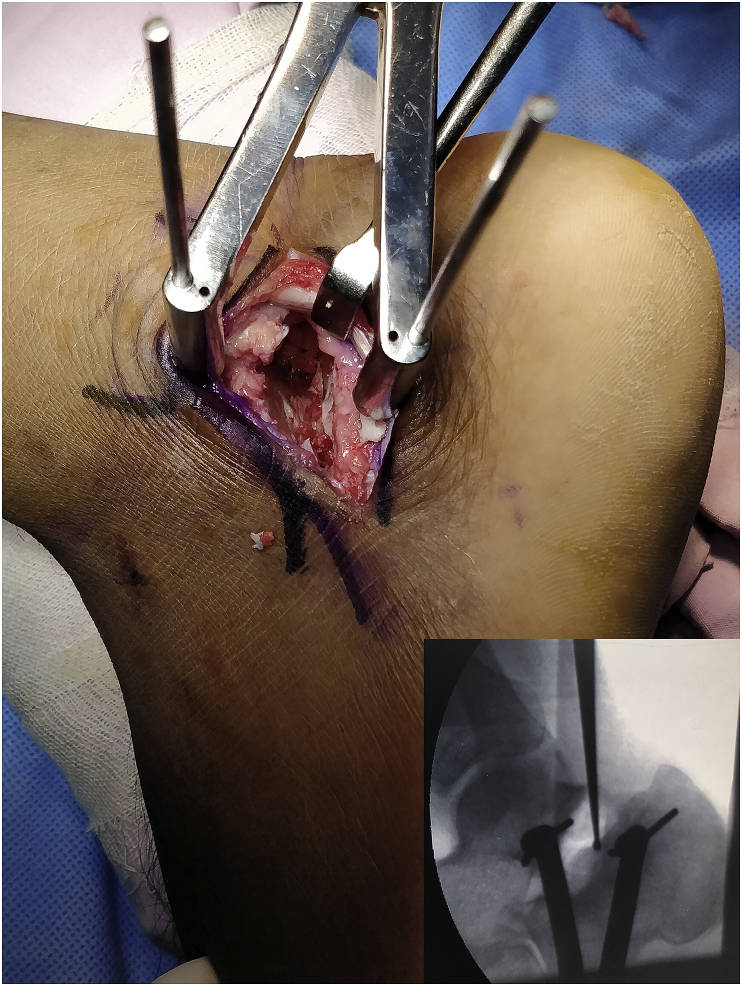
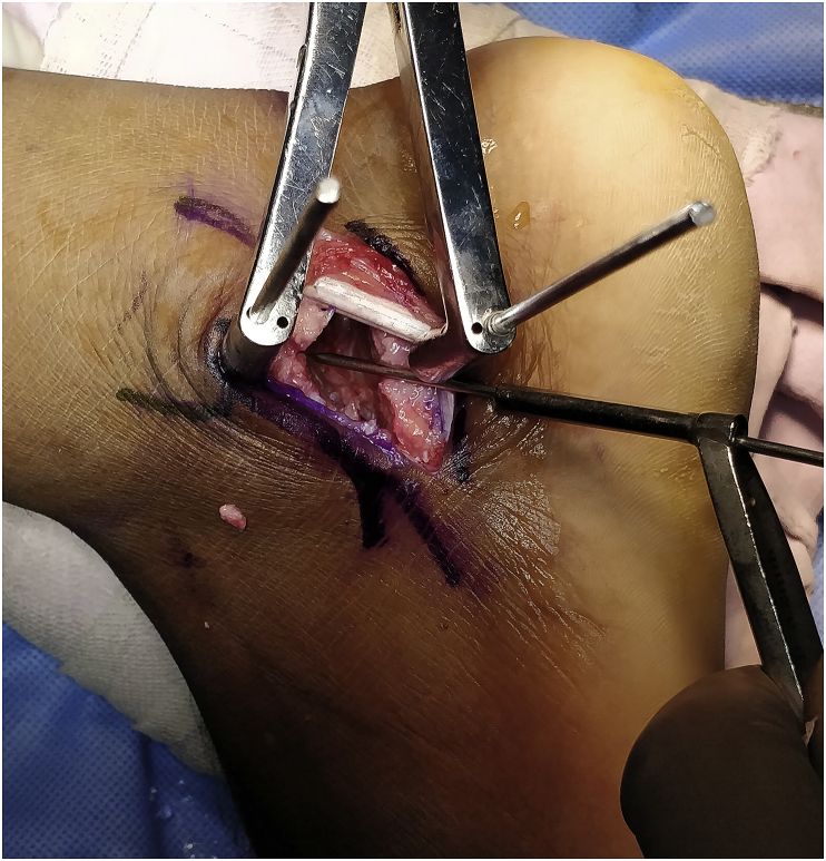
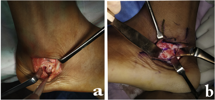
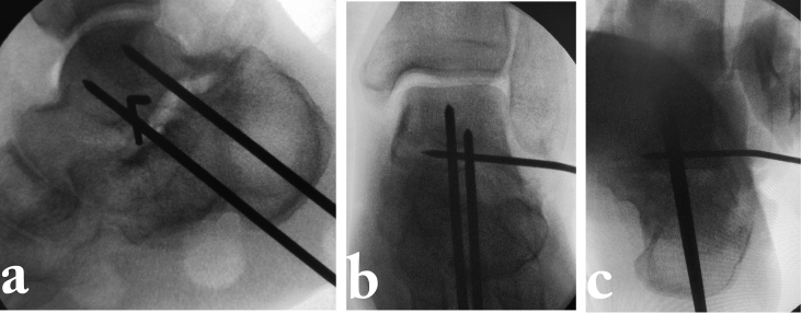
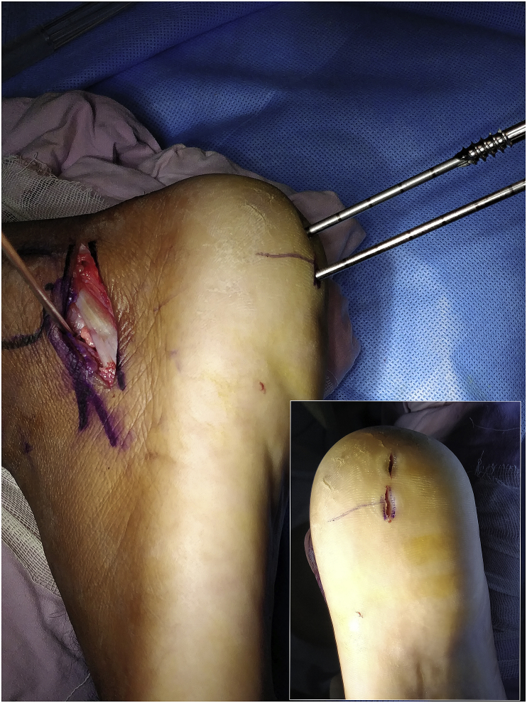
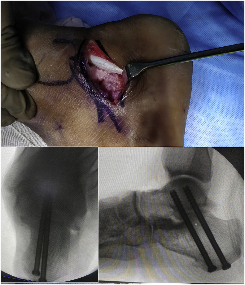
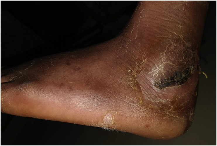
Calcaneus malunion is a common sequela to calcaneal fractures and is a cause of pain and discomfort. Multiple approaches have been described to address the subtalar joint and the lateral wall. Type 2 malunion is the most commonly encountered problem, and is usually addressed by the sinus tarsi approach. This has some limitations, as exposure for lateral wall excision beneath the peroneal tendons maybe a problem. We have slightly modified the sinus tarsi approach by a more horizontal skin incision, which may even be extended proximally by 1-2 cm; this allows access to the lateral wall on either side of the peroneal tendons. The approach is described in detail.
Keywords: Calcaneus malunion; Sub fibular impingement-sinus tarsi approach; Subtalar fusion.
© 2020.
Figures
References
-
- Myerson M.S., Quill G.E. Late complications of fractures of the calcaneus. J Bone Joint Surg. 1993;75A(3):331–341. - PubMed
-
- Stephens H.M., Sanders R. Calcaneal malunions: results of a prognostic computed tomography classification system. Foot Ankle Int. 1996 Jul;17(7):395–401. - PubMed
-
- Greisberg J., Sangeorzan B. Hindfoot arthrodesis. J Am Acad Orthop Surg. 2007 Jan;15(1):65–71. - PubMed
-
- Vulcano E., Ellington J.K., Myerson M.S. The spectrum of indications for subtalar joint arthrodesis. Foot Ankle Clin. 2015 Jun;20(2):293–310. Epub 2015 Apr 11. - PubMed
Publication types
LinkOut - more resources
Full Text Sources
