Editorial
. 2020 Jul;13(4):844-887.
doi: 10.1111/1751-7915.13576. Epub 2020 May 14.

Visualizing the invisible: class excursions to ignite children's enthusiasm for microbes

Affiliations
Editorial

Visualizing the invisible: class excursions to ignite children's enthusiasm for microbes

Terry J McGenity et al. Microb Biotechnol. 2020 Jul.

Abstract

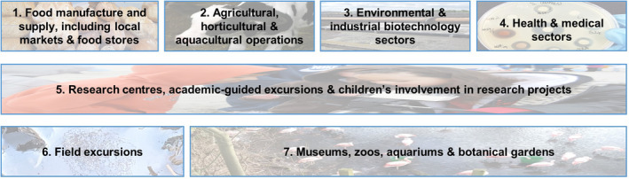
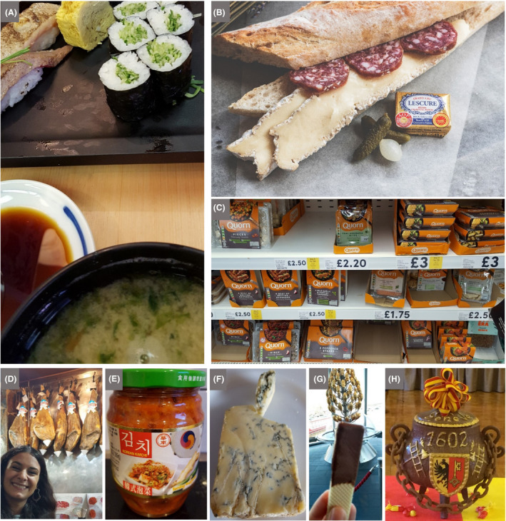
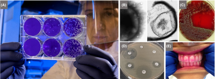
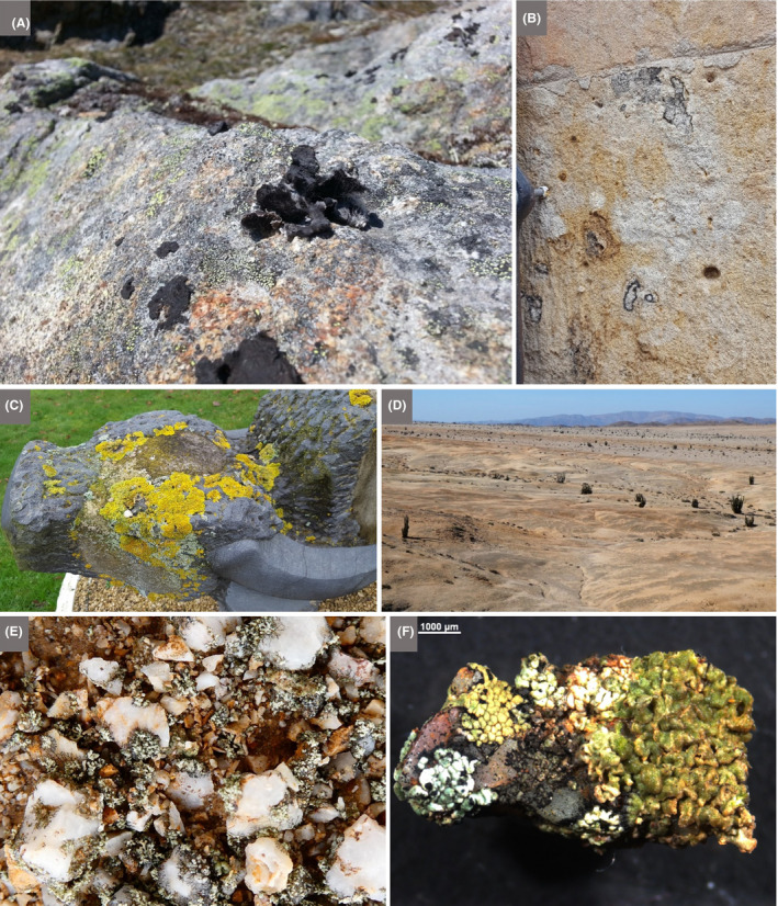
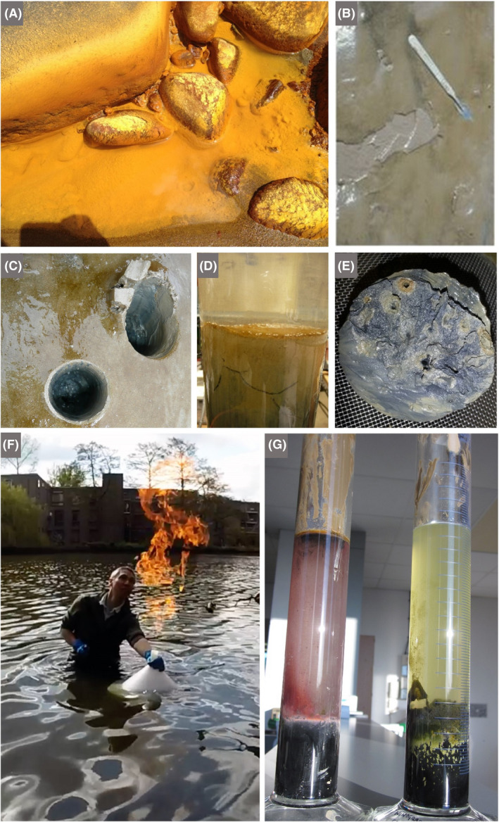
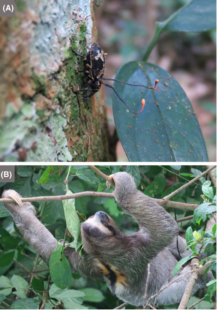
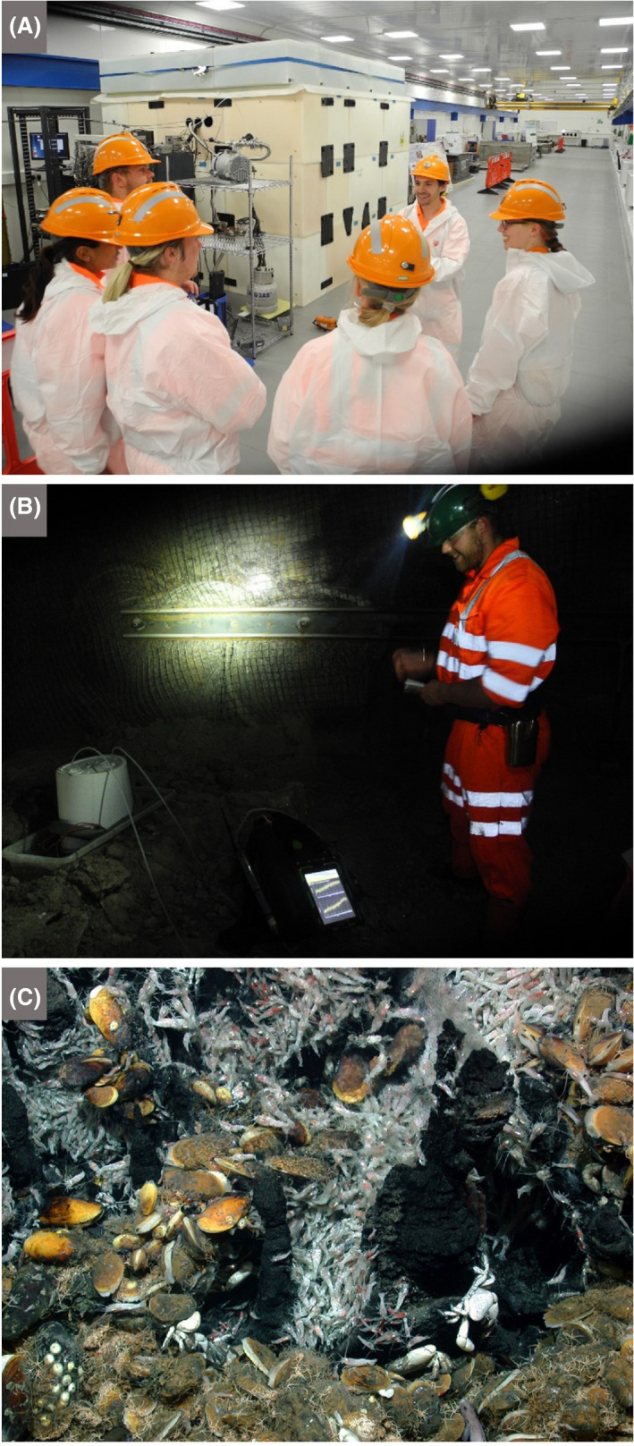
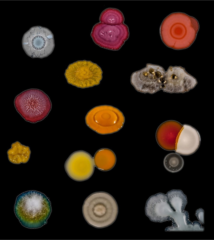
We have recently argued that, because microbes have pervasive - often vital - influences on our lives, and that therefore their roles must be taken into account in many of the decisions we face, society must become microbiology-literate, through the introduction of relevant microbiology topics in school curricula (Timmis et al. 2019. Environ Microbiol 21: 1513-1528). The current coronavirus pandemic is a stark example of why microbiology literacy is such a crucial enabler of informed policy decisions, particularly those involving preparedness of public-health systems for disease outbreaks and pandemics. However, a significant barrier to attaining widespread appreciation of microbial contributions to our well-being and that of the planet is the fact that microbes are seldom visible: most people are only peripherally aware of them, except when they fall ill with an infection. And it is disease, rather than all of the positive activities mediated by microbes, that colours public perception of 'germs' and endows them with their poor image. It is imperative to render microbes visible, to give them life and form for children (and adults), and to counter prevalent misconceptions, through exposure to imagination-capturing images of microbes and examples of their beneficial outputs, accompanied by a balanced narrative. This will engender automatic mental associations between everyday information inputs, as well as visual, olfactory and tactile experiences, on the one hand, and the responsible microbes/microbial communities, on the other hand. Such associations, in turn, will promote awareness of microbes and of the many positive and vital consequences of their actions, and facilitate and encourage incorporation of such consequences into relevant decision-making processes. While teaching microbiology topics in primary and secondary school is key to this objective, a strategic programme to expose children directly and personally to natural and managed microbial processes, and the results of their actions, through carefully planned class excursions to local venues, can be instrumental in bringing microbes to life for children and, collaterally, their families. In order to encourage the embedding of microbiology-centric class excursions in current curricula, we suggest and illustrate here some possibilities relating to the topics of food (a favourite pre-occupation of most children), agriculture (together with horticulture and aquaculture), health and medicine, the environment and biotechnology. And, although not all of the microbially relevant infrastructure will be within reach of schools, there is usually access to a market, local food store, wastewater treatment plant, farm, surface water body, etc., all of which can provide opportunities to explore microbiology in action. If children sometimes consider the present to be mundane, even boring, they are usually excited with both the past and the future so, where possible, visits to local museums (the past) and research institutions advancing knowledge frontiers (the future) are strongly recommended, as is a tapping into the natural enthusiasm of local researchers to leverage the educational value of excursions and virtual excursions. Children are also fascinated by the unknown, so, paradoxically, the invisibility of microbes makes them especially fascinating objects for visualization and exploration. In outlining some of the options for microbiology excursions, providing suggestions for discussion topics and considering their educational value, we strive to extend the vistas of current class excursions and to: (i) inspire teachers and school managers to incorporate more microbiology excursions into curricula; (ii) encourage microbiologists to support school excursions and generally get involved in bringing microbes to life for children; (iii) urge leaders of organizations (biopharma, food industries, universities, etc.) to give school outreach activities a more prominent place in their mission portfolios, and (iv) convey to policymakers the benefits of providing schools with funds, materials and flexibility for educational endeavours beyond the classroom.


Conflict of interest statement

None declared.

Figures

Fig. 1
Fig. 1
Mushrooms go hand in hand with folklore creatures in the playground of the Geneva Botanical Garden. Photograph by Kenneth Timmis
Fig. 2
Fig. 2
Exploring hypersaline environments and extremely halophilic microbes. Their vivid colours, extreme adaptations, biotechnological applications and ease of growth make halophiles very attractive to children. The manufacture of sea salt, with its pivotal role in food preservation, helped regions and civilizations to flourish. Thus, the interconnectedness of microbes, food and history can be explored by visiting hypersaline environments or when buying sea salt and salted foods from local shops A. Satellite view of the salterns in Bhavnagar, India, showing vivid red, orange and green ponds in which seawater evaporates leaving behind salt (field of view is ~ 10 km2). The colours are due to the pigments of the halophilic microbes in the brines of different salinity. The red/orange pigments of haloarchaea and Dunaliella salina (see D) encourage the absorption of solar radiation leading to increased rates of evaporation, resulting in more rapid salt production. B. A teacher and her class wade into Great Salt Lake (Utah, USA) to collect samples to study under field microscopes. C. A student investigates the biofilms that form stromatolite‐like structures in Great Salt Lake, impressive calcium carbonate deposits precipitated by the actions of cyanobacteria. D. Microscopic image from the hypersaline Lake Tyrrell, Australia (salinity> 20% w/v), in which we can tentatively identify the eukaryotic chlorophyte, Dunaliella salina (grown commercially for the carotenoid, β‐carotene, which is widely used as a natural food colorant as well as a precursor to vitamin A), living alongside the haloarchaeon, Haloquadratum walsbyi, which has flat square‐shaped cells with gas vesicles that allow flotation to the surface, most likely to acquire oxygen (scale bar is 5 μm). E. Gypsum crust from the bottom of a shallow saltern pond (~20% w/v salinity) in Eilat, Israel, showing layered microbial communities of phototrophic microbes. The orange‐brown upper layer is most likely dominated by unicellular cyanobacteria; the green layer by filamentous cyanobacteria; and the purple layer by anoxygenic purple sulfur bacteria. These microbes use light as a source of energy, and their layering is explained by differential tolerance to ultraviolet light and oxygen, and their capacity to use light of different wavelengths in photosynthesis. The anoxygenic purple sulfur bacteria are adapted to use light at the far‐red end of the spectrum, which is less attenuated than visible light in sediments, and they benefit by living in close proximity to anaerobic sulfate‐reducing bacteria (grey layer directly beneath the purple layer), which produce hydrogen sulfide. This compound is split to form oxidized sulfur in a similar way to which the oxygenic phototrophs, like cyanobacteria, produce oxygen from water. Such layering of microbial communities can trigger discussions about chemistry (the electron donors and products), physics (diffusion of oxygen and hydrogen sulfide, and the electromagnetic spectrum), geology (microbial fossils in gypsum) and ecological concepts (interspecies interactions and niche partitioning). F. A laboratory‐made salt (halite) crystal coloured red due to the presence of haloarchaea trapped inside small pockets of brine within the crystal (the central crystal is about 0.5 by 0.5 cm). This is a survival strategy used by haloarchaea to avoid desiccation. They remain viable inside the halite, and evidence suggests that some haloarchaea can survive over geological time inside buried halite. Some haloarchaea play a major role in hydrolysing biopolymers in salty environments, such as those used in the production of fish sauces. When we consume the sauces or sea salt, we consume haloarchaea! G. During an excursion, students can collect samples from which they can inoculate media (in this case to grow extreme halophiles) after returning to the classroom, bringing the field‐collected microorganisms into the laboratory and further connecting students to their environment. Photograph A by NASA https://earthobservatory.nasa.gov/ . Photographs B, C and G by Great Salt Lake Institute. Photograph D by Mike Dyall‐Smith. Photograph E by Andreas Thywißen.
Fig. 3
Fig. 3
Excursion categories discussed in the text
Fig. 4
Fig. 4
A trip to the food store or a meal can become an adventure of microbiological discovery A. Breakfast at Tsukiji Market, Japan. The miso in miso soup is a traditional Japanese paste produced by fermenting salted soya beans with kōji, which is also made by fermentation of rice or barley with the fungus Aspergillus oryzae. Soy sauce is made by fermenting salted soya beans and wheat by a complex community of hydrolytic fungi and bacteria, as well as lactic acid bacteria. Sushi rice is prepared with a sweet rice wine vinegar (mirin) to give it a delicate, sweet but sharp flavour. Vinegar can be prepared from almost any sugary solution by an alcoholic fermentation followed by an acetic fermentation. In Japan, vinegar traditionally derives from rice and kōji‐fermented rice. B. Salami and Brie baguette with pickles. Nearly all the foods in this image require microbes for their production: Bakers’ yeast (Saccharomyces cerevisiae) makes the dough rise for the bread; Penicillium species and lactic acid bacteria are among the microbes involved in both salami and Brie production; acetic acid‐producing bacteria make the vinegar from alcoholic drinks for the pickles. C. The drive towards reducing meat in the diet has opened a market for a range of meat‐free products, leading to a resurgence in the sales of Quorn, which is made from filamentous mycelium of the soil fungus Fusarium venenatum. D. Dry‐cured hams in a bar in Salamanca, Spain. Spanish ham is traditionally cured in brine with sea salt, often with added sodium nitrate, to lower the water activity to an extent that prevents the growth of spoilage organisms. E. Kimchi, a staple food in Korea, is becoming popular worldwide. Cabbage, along with numerous variations of vegetables and spices, is salted and undergoes fermentation dominated by lactic acid bacteria. The health benefits of this tangy, salty and often spicy product are widely reported (Tamang et al., 2016). F. Stilton cheese, which, in addition to requiring lactic acid bacteria and other microbes in its manufacture, is inoculated with Penicillium roqueforti to produce the blue veins that contribute to the distinctive flavour. G. Chocolate from the Lindt factory in Cologne, Germany – an enjoyable way to learn about the history and microbiology of chocolate. H. Chocolate is moulded into a multitude of artistic forms that titillate the senses, especially of children. The example shown is that of the Geneva ‘marmite’, a chocolate representation of a soup cauldron containing marzipan vegetables, made each year for the festival of ‘Escalade’, the celebration of the defeat of an assault on the city in 1602. Legend has it that an old lady (Catherine Cheynel or Mère Royaume), who was making vegetable soup at night, spotted the attackers who were scaling the city walls, sounded the alarm and threw the hot soup cauldron at them. Incidentally, Marmite – the yeast extract spread putatively developed by a German scientist, Justus von Liebig, using leftover yeast from the brewing of beer in Burton upon Trent, UK – derives from the French name ‘marmite’, as it is sold in pots with a similar shape to the cauldron. Photographs A and C‐F by Terry McGenity, B by Jez Timms on Unsplash, G by Mairi McGenity and H by Kenneth Timmis.
Fig. 5
Fig. 5
The beneficial and deleterious effects of microbes can be investigated on an excursion to a farm, horticulture centre or aquaculture facility A. Depuration tanks holding Pacific rock oysters (Magallana gigas, formerly Crassostrea gigas) harvested from the Colne Estuary, UK, by Colchester Oyster Fisheries. The incoming water is treated with ultraviolet light to reduce its microbial load. This process provides time for the filter‐feeding oysters to flush out any pathogens that they may have accumulated while they were in the estuarine waters, before being sold. In addition to providing a potentially sustainable source of protein, bivalves are important ecosystem engineers. For example, they help to ameliorate eutrophication directly by taking up nitrogen and phosphorus for shell and tissue growth and indirectly by creating anoxic sites that encourage denitrifying bacteria that convert soluble nitrate to nitrogen gas (van der Schatte Olivier et al., 2018). B. Nodules housing nitrogen‐fixing bacteria on the roots of southern pea, also known as cowpea (Vigna unguiculata), an important food crop in the semiarid tropics. C. Peach tree (Prunus persica) with crown gall, a cancerous growth caused by the bacterial species Agrobacterium tumefaciens. D. Aspergillus flavus, an opportunistic pathogenic fungus, growing on maize. A. flavus is the main producer of aflatoxin B1, the most carcinogenic mycotoxin yet known (Gasperini et al., 2019). E. Visible microbiota on a 50‐year‐old mango tree (Kolkata, India). The various colours on the bark largely reflect different microbial communities (e.g. algae, fungi and lichens) colonizing those habitats to which they are best adapted. Key factors that influence microbial distribution include exposure to the sun, availability of water and secretions from the tree. Thus, trees present a living illustration of the microbial biosphere across a series of environmental gradients. F. Silage bales for year‐round animal feed are wrapped in plastic (pink and blue wrappers were used to raise awareness of breast cancer and prostate cancer, respectively) at Gåseberg Sheep Farm, Lysekil Municipality, Sweden. Before wrapping the hay in plastic, its moisture content is optimized and then checked regularly to maintain ideal conditions for lactic acid fermentation while diminishing the risk of spoilage. The consumption of oxygen by aerobic microbes, followed by the production of acidic conditions by lactic acid bacteria (as part of a diverse community of microbes), minimizes the growth of spoilage organisms. Lübeck and Lübeck (2019) provide a clear review of how lactic acid bacteria and other microbes convert green crops (grasses, legumes and the green part of crops like carrots) into a range of agricultural feed products. Photograph A by Alex Shakspeare, B by Dave Whitinger released under the GNU Free Documentation Licence, C by University of Georgia Plant Pathology, University of Georgia, Bugwood.org, licensed under a Creative Commons Attribution 3.0 Licence, D by Alessandra Marcon Gasperini, E by John E. Hallsworth and F by W.carter under the Creative Commons CC0 1.0 Universal Public Domain Dedication.
Fig. 6
Fig. 6
Wastewater treatment plants as an opportunity to see microbes as the ultimate recyclers A. Colchester wastewater recycling plant run by Anglian Water. The image shows part of the activated sludge treatment, whereby flocs are encouraged to form, due to the activity of myriad microbes, including the extracellular polymer‐producing bacterial species Zoogloea ramigera. The organic microbe‐rich flocs are allowed to settle (see B): some will be recycled to seed the activated sludge tanks, and the rest can be used to make biogas (see C). B. The right‐hand bottle illustrates how activated sludge settles, thus separating the solids and leaving behind reasonably clear liquid with reduced organic load. The left‐hand bottle shows incomplete settling due to a suboptimal activated‐sludge microbial community. C. The upstream part of the anaerobic digestion facility at Colchester wastewater recycling plant. The sludge from the plant and surrounding locales is subjected to heating, pasteurization and hydrolysis, before the anaerobic digestion step, driven by methanogenic Archaea. The resultant methane is used to generate electricity and can be stored in the biogas dome shown on the left. Photographs by Terry McGenity.
Fig. 7
Fig. 7
Diagnostic and public health laboratories allow students to investigate different ways of growing, detecting and viewing microbes, and learning about treatment of pathogens A. Viral plaque assay to quantify influenza virus. Dilutions of the sample are added to a human cell line and incubated. The purple stain shows cells that are not infected with the virus, while clear zones (plaques) indicate viral infection and cell lysis. B. Some laboratories may perform electron microscopy, giving fascinating insights into the shape, size and ultrastructure of Bacteria and viruses. These images are transmission electron micrographs of the bacterium, Clostridium difficile, being attacked by bacteriophages. Left, surface of an intact cell of C. difficile, showing large numbers of attached bacteriophages, most with empty hexagonal heads, representing bacteriophages that have injected their DNA into the bacterium. Right, part of a C. difficile cell that has burst open due to the proliferation of bacteriophages within, which produce enzymes that lyse the cell membrane. The height of both images is ~ 800 nm. C. Throat swab sample inoculated on a blood agar plate, showing colonies of Streptococcus pyogenes, the causative agent of tonsillitis and other diseases, identified by β‐haemolysis (the production of clear zones around bacterial colonies, resulting from lysis of red blood cells by the haemolysin produced by the pathogen). D. Staphylococcus aureus antibiotic sensitivity testing using the disc diffusion method. S. aureus is an example of a pathogen, frequently acquired in hospital, that has developed resistance to multiple antibiotics. A culture of the bacterium is spread on the agar plate, and discs containing different antibiotics are then placed on the plate. During incubation, a bacterial lawn develops. A zone of clearing in the lawn around a disc indicates that the bacterium is sensitive to the corresponding antibiotic, thereby contributing to diagnosis as well as informing treatment strategies. E. Staining teeth biofilms (plaque) with plaque‐disclosing tablets provides a visual and memorable way to drive home the importance of good dental hygiene to remove plaque and thus prevent dental caries and periodontal disease (note the different meanings of the term ‘plaque’ here and in A). Photograph A by CDC on Unsplash, B by Stefan Hyman with samples from Martha Clokie, University of Leicester, C by Aurélie Villedieu, D by Selwa Alsam and E is licensed under the Creative Commons Attribution‐ShareAlike 4.0 International licence.
Fig. 8
Fig. 8
Bioplastic degradation in compost A. Strips (20.8 x 4.5 cm) of polyhydroxybutyrate (left), a biodegradable plastic that can be made by many Bacteria and Archaea (and which serves the role of a carbon reserve in nature), and polypropylene (right), a non‐biodegradable plastic derived from petroleum. B. Garden compost into which the strips were placed, after securing them in nylon tights, which served as ‘litter bags’. C. The polyhydroxybutyrate (below) shows clear signs of biodegradation after 15 days incubation, whereas the polypropylene (above) remains unchanged. Photographs by Terry McGenity.
Fig. 9
Fig. 9
Living skins on rocks A. Diverse lichens, including black foliose forms, on a rock in western Greenland. B. Lichens and fungi, including black ring forms on a sandstone building, which together with microbial communities, contribute to the biodeterioration of Villamayor Sandstone, the main building stone of the UNESCO World Heritage city of Salamanca. The field of view is ~ 20 x 20 cm. C. Lichens on a stone cow, Milton Keynes, UK. D. The seemingly barren landscape of the National Park, Pan de Azùcar, of the coastal Atacama Desert is covered by tiny, white stones that have a diameter of ~ 6 mm (termed grit). The black patterns are caused by lichens, cyanobacteria, green algae and fungi that colonize the surface and inner structures of the polycrystalline grits. These organisms also concatenate the single grits forming biological soil crusts (termed grit crust; Jung et al., 2020). E. Close‐up of the grit crust after a fog event. Mainly lichens in their wetted stage are visible growing attached to grit stones. F. Single grit stone covered by various lichens together with microfungi (mainly Lichenothelia). Many examples of microbes as part of the urban landscape can be viewed at ‘This is Microgeography’ website. Photographs A–C by Terry McGenity and D‐F by Patrick Jung.
Fig. 10
Fig. 10
Blooming microbes A. Satellite image of a milky white bloom of the phototropic coccolithophore, Emiliania huxleyi, in the North Sea. This eukaryotic microalgal species has a calcium carbonate wall which, when the bloom collapses (due to death caused by virus attack and/or grazing), can sink to the seafloor. Thus, coccolithophores play a role in sequestering carbon from the atmosphere and transferring it to marine sediments. The greener bloom at the bottom of the image may be dominated by diatoms, which, owing to their glassy silica walls, can also be manifest in the rock record as diatomaceous earth. B. The White Cliffs of Dover, UK, made of chalk from the calcium carbonate walls of coccolithophores and foraminifera that settled to the sea floor in the Cretaceous period C. The giant bacterium, Thiomargarita namibiensis, can be seen in this sediment core from the Benguela Upwelling System of the coast of Namibia. It appears like white ‘strings of pearls’, i.e. a string of connected cells, which can be seen in the sediment. Individual cells are ~ 0.4 mm in diameter, and so, unusually for a bacterium, can be seen with the naked eye. It is non‐motile, and so relies on currents to deliver its terminal electron acceptor, nitrate, which it accumulates in a large vacuole. The sulfide, which provides its energy source, is abundant in the deeper anaerobic zone due to its production by sulfate‐reducing bacteria. There is also a thick, jelly‐like biofilm at the top of the sediment. Image width is ~ 5 cm. D. Fossilization in action. Giant microbialites that have formed over the last several thousand years due to microbial activity coupled with high carbonate concentrations in freshwater Laguna Bacalar in the Yucatán Peninsula of Mexico. Cyanobacteria are the dominant photosynthetic microbes, whose activity raises the local pH, which, together with activities of the associated heterotrophic microbial community, encourages carbonate precipitation. The sticky extracellular polymeric substances produced by the microbes also trap detrital grains. Microbialites can have accretionary layers (stromatolites) or a more clotted structure (thrombolites), and both are abundant in the rock record, with some dating back to ~ 3500 million years (predating the evolution of oxygenic photosynthesis, and thus raising questions about the microbes responsible for their formation). The layers or clots typically represent cyanobacterial biofilms or colonies respectively (Gischler et al., 2008). E. Banded Iron Formation at the Fortescue Falls, Australia, with stripes rich in iron (darker bands) and silica (lighter bands) formed from oxidized iron sinking to the seafloor, mainly in the Archaean Eon. The way in which the ferrous iron minerals were oxidized in the early Earth is still under debate. The mechanism may have been oxygenic photosynthesis by the ancestors of modern cyanobacteria or iron‐dependent photosynthesis, which neither requires nor produces oxygen (Robbins et al., 2019; Thompson et al., 2019). Photograph A by NASA ( https://earthobservatory.nasa.gov ), B by Immanuel Giel under the Creative Commons Attribution‐ShareAlike 3.0 Unported licence, C by Natalie Hicks, D by Etienne Low‐Décarie and E by Graeme Churchard under the Creative Commons Attribution 2.0 Generic licence.
Fig. 11
Fig. 11
Fungi A. ‘Fairy ring’ of an unidentified Basidiomycete fungus around a pine tree at the University of Warwick, UK, a surface manifestation of a mycelial network belowground. B. Colony of the halophilic, melanin‐rich black yeast/fungus Hortaea werneckii (field of view is ~ 1 cm2). C. Three giant puffball fruiting bodies (Calvatia gigantea) in Wivenhoe Woods, UK, each ~ 30 cm in diameter. The hymenium, i.e. the tissue layer from which spores form, is internal. A specimen of this size would release around five trillion spores (Li, 2011). D. Basidiomycete fruiting bodies on the rotting, wooden edging of a path. E. Basidiomycete fruiting bodies of the shaggy bracket (Inonotus hispidus), which has its hymenium housed in pores, are seen here on the trunk of a tree in northern England. Hispidin, a phenolic metabolite produced by this species, is an antioxidant with potential pharmaceutical applications. F. Fly agaric (Amanita muscaria) fruiting body in Wivenhoe Woods, UK. G. Amethyst Deceiver (Laccaria sp.) fruiting body in Wivenhoe Woods, UK. H. Stereoscopic image of the underside of a Basidiomycete fruiting body with the hymenium housed in gills (Hygrophorus sp.; radius is ~ 1 cm). I. Rust fungus (most likely Puccinia aristidae) on Chenopodium album in a garden in St. Saturnin‐les‐Avignon, France. The copious urediospores have dusted the stones behind the plant, colouring them orange. J. Fungal rhizomorphs of the saprophytic and root parasitic honey fungus (Armillaria mellea) on a rotting tree trunk in Broc, Switzerland. These structures are very resistant and are able to explore their environments on metre scales to scavenge for nutrients and then to translocate fluids bidirectionally. There are many excellent mycology teaching resources, notably ‘Mushrooms primary school activity pack’. Photographs A, C, D, E, F and G by Terry McGenity, B by Polona Zalar, H by Saskia Bindschedler, I by Cindy Morris and J by Andrea Lohberger.
Fig. 12
Fig. 12
Slime moulds A. Physarum polycephalum plasmodium (single‐celled multinucleate amoeba capable of rapid streaming) on rotting wood. B. Fruiting body (sporocarp) of Diachea radiata in leaf litter of the evergreen tree, Bellucia grossularioides, in regrowth forest near Yangambi in the Democratic Republic of the Congo. Excellent videos of these fascinating organisms can be seen on the internet. Photograph A by Frankenstoen, under the Creative Commons Attribution 2.5 Generic licence, and B by Myriam de Haan, Meise Botanic Garden.
Fig. 13
Fig. 13
Microbial manifestations of their phototrophic and reduction–oxidation activities A. Boulder and pebbles in Río Sucio (Dirty River) in Costa Rica are coloured orange due to the presence of schwertmannite, a mineral generated by the activity of iron‐oxidizing bacteria, such a Gallionella spp., which use reduced iron as a form of energy, oxidizing it to form rusty minerals (Arce‐Rodríguez et al., 2017). B. Peel of mudflat sediment showing a thick gelatinous biofilm rich in diatoms (silica‐walled eukaryotic phototrophic microalgae) on top of the clay‐rich sediment. Diatoms are the main primary producers in this ecosystem and, by the production of sticky extracellular polymeric substances, they stabilize the sediment, thus limiting coastal erosion. C. Cored sediment from Tillingham mudflat, UK (holes are ~ 8 cm diameter), showing the golden mats formed by diatoms on the surface, and the transition to grey‐black sediment at depth caused by anaerobic sulfate‐reducing bacteria releasing sulfide that reacts with iron to form black iron sulfide. D. Extracted sediment core from C, incubated in the light, showing how the diatoms and other phototrophic microbes produce bubbles of oxygen visible at the surface. E. A transverse slice of the sediment shown in D, illustrating how burrowing animals such as the ragworm (Hediste diversicolor) can introduce oxygen deeper into the sediment and thus encourage the activity of aerobic microbes, such as iron‐oxidizing microbes, leading to a ring of rust around the holes. F. Burning methane from the lake at the University of York, UK. The sediment was stirred to release methane bubbles (produced by anaerobic methanogenic Archaea), which were collected in a funnel and set alight. This phenomenon can occur without human assistance, with the resultant ghostly light engendering myths and legends worldwide, such as Will‐o'‐the‐Wisp and ignis fatuus. G. Winogradsky columns set up with cellulose and sediment from an Essex salt marsh. The left‐hand column is dominated by purple sulfur anoxygenic phototrophs, while the right‐hand column is dominated by green sulfur anoxygenic phototrophs, both of which are Bacteria that use light as an energy source, but instead of splitting water and generating oxygen (as in the diatoms in D) they split hydrogen sulfide. The sulfide is produced by anaerobic sulfate‐reducing bacteria found in the black sediment. Photograph A by Max Chavarría, B by Graham J.C. Underwood, C, D, E and G by Terry McGenity and F by Paul Shields and James Chong.
Fig. 14
Fig. 14
Decaying objects can be found in all sorts of environments and provide the basis for discussing the underlying microbiology of decay and nutrient recycling. For example, children can be asked who is eating this rotting boat on the bank of the tidal River Deben, near Woodbridge, UK, and how they do it, thereby introducing children to extracellular and intracellular enzymes. Children can be encouraged to consider where else they encounter rotting and recycling (e.g. leaves in the forest, food in the fridge, fences in the garden, waste in landfill sites), when it is beneficial/damaging and how it can be encouraged/prevented. Photograph by Kenneth Timmis
Fig. 15
Fig. 15
Algal blooms and harmful algae A. Bloom of the dinoflagellate, Prymnesium parvum on the Norfolk Broads, UK. This dinoflagellate is a photosynthetic eukaryotic microalga that produces a toxin, which can lead to significant economic damage. B. A few of the ~ 30,000 fish killed by the blooming dinoflagellate, Prymnesium parvum on the Norfolk Broads, UK, in April 2015. Photographs by Martin Rejzek.
Fig. 16
Fig. 16
Microbial exhibits A. The giant Escherichia coli sculpture created by Luke Jerram was the centrepiece of the Bacterial World Exhibition. The 28‐m‐long piece is 5 million times bigger than a real microbial cell and highlights some of its external structures such as flagella and pili. B. ‘The Hunt for New Antimicrobials’ created by Anna Dumitriu in collaboration with Maggie Smith and Nicholas Read was part of the BioArt and Bacteria exhibition. This framed piece is composed of silk, sterilized wild‐type and genetically modified Streptomyces bacteria, wood, card and glass. Both exhibits shown in A and B were on display at the Oxford Museum of Natural History, UK. Redfern et al. (2020) provide more examples of exhibits that aim to highlight the issue of antibiotic resistance. C. The Fermentophone, designed by Joshua Rosenstock, consists of jars of fermenting fruits and vegetables producing CO2 bubbles, which are picked up by submerged microphones and converted to different sounds, creating a relaxing and aesthetically pleasing, as well as edible, display. Moreover, visitors can make their own ferments and add them to the living exhibit, shown here in action at Harvard Museum of Natural History, USA. D. The Broad Street Pump replica outside the John Snow Public House in London, UK, is an example of microbiology history – where the field of epidemiology was founded. By meticulous detective work, the Yorkshireman, John Snow, identified the pump as the source of a cholera outbreak in 1854, saving lives in the short term by the removal of the pump’s handle and in the long term by recognizing the role of sewage as the source of the waterborne disease. There are famous microbiologists and microbiological stories associated with many towns, which teachers can include in an excursion to enliven the subject. Photograph A by André Antunes, B by Carol Verheecke‐Vaessen, C by Joshua Rosenstock and D by Mairi McGenity.
Fig. 17
Fig. 17
Unusual and visible animal‐microbial interactions A. An adult weevil on a lichen‐covered tree (in Alta Floresta, Mato Grosso State in the southern section of the Brazilian Amazon) infected with the fungus Ophiocordyceps curculionum. The objects that look like antennae with orange/red ends are actually fungal fruiting bodies. Ophiocordyceps species are parasites of beetles and other insects. The behaviour of the infected beetle is changed to make it land at a certain height (~1 m) on a tree trunk. When infecting ants they modify their host’s behaviour, turning them into ‘zombies’ that move along the vegetation, locking their mandibles into precise locations on the underside of leaves where fungal development is optimized. The infected, fixed ants are generally positioned above foraging trails so that spores from Ophiocordyceps are likely to infect other ants (Araújo and Hughes, 2019). B. A male brown‐throated three‐toed sloth with green fur. Sloths’ hair has unique transverse cracks that retain moisture and thus support the hydroponic growth of a diverse range of eukaryotic microalgae and cyanobacteria. The highly digestible and lipid‐rich microalgae are eaten by sloths, supplementing their poor diet which otherwise consists exclusively of leaves (Pauli et al., 2014). Photographs by Margaret Adams.
Fig. 18
Fig. 18
Remote field locations and research facilities can be brought to life for children, e.g. by using live feeds to schools A. Students visiting the STFC Boulby Underground Laboratory, where the focus is low background radiation science, including the search for Dark Matter. In addition, the laboratory contains clean rooms in which the concentration of airborne particles (including microbes) is minimized, together with a deep underground astrobiology facility. B. Students measured methane flux in salt‐saturated ponds constructed in the salt mine’s ‘Mars Yard’. C. Living community at hydrothermal seeps on the mid‐ocean ridge at a water depth of 3,030 metres. This diverse animal community relies on microbes, primarily Bacteria, which grow by using gases that emanate from hydrothermal vents. For example, Bacteria use sulfide (or hydrogen) to gain energy and carbon dioxide to produce biomass and organic matter that support the food web. Such microbes are often found living symbiotically in the gill chamber of the clams and shrimp. This type of symbiosis between animals and chemolithoautotrophic microbes is very common, not only in unusual environments like hydrothermal vents (where geothermally heated water and gases escape from fissures) and cold seeps (where the efflux is more diffuse and cooler than in hydrothermal vents), but also more normal environments like intertidal marine sediments (Dubilier et al., 2008). New techniques provide unprecedented visual insight at the micrometre scale into such host–microbe symbioses and their metabolic interactions (Geier et al., 2020). Photographs A and B by Terry McGenity and C by MARUM – Center for Marine Environmental Sciences, University of Bremen (CC‐BY 4.0).
Fig. 19
Fig. 19
An array of microbial colonies growing on agar plates, demonstrating different forms, textures and colours. Beautiful images of microbes and their habitats, along with interesting context, can be found in the book called ‘Life at the Edge of Sight’ (Chimileski and Kolter, 2017). Photograph by Scott Chimileski and Roberto Kolter
Fig. 20
Fig. 20
Agar Art. The top three entries in the American Society for Microbiology (ASM) Agar Art Kids 2019 competition A. ‘Circle of Life’ by Kate Lin (Age 11) using E. coli MM294 pGFPuv (green), pYellow (yellow), pCherri (purple) and pTang (Pink) growing on LB Agar + Ampicillin, with support from Cold Spring Harbor Laboratory DNA Learning Center. B. ‘The Honey Bee’ by Manal Faisal Khan (Age 5) using E. coli growing on Nutrient Agar. C. ‘Fall’ by Lilu Good‐Martinez (Age 10) using Methylobacterium extorquens growing on minimal medium with methanol as a carbon source. Congratulations to the entrants named above and thanks to them and the ASM (https://asm.org/Press-Releases/2019/November-1/ASM-s-5th-Agar-Art-Contest-Showcases-the-Beauty-of) for these images. The ‘Microbial Art’ website and the June 2017 edition of SfAM’s magazine, Microbiologist, provide many examples of the interplay between art/fashion and microbiology, and Park (2012) provides practical tips for creating microbial art.
Fig. 21
Fig. 21
Classroom activity making lupin‐based tempeh. Tempeh is a fermented Indonesian food, usually made from soya beans. Here, the soya beans have been replaced with lupin beans, e.g. from Lupinus angustifolius. The fungus, Rhizopus oryzae, makes a white mycelial network knitting together the lupin grits into sliceable tempeh, ready for marinating or frying. These slices contain vitamin B12 thanks to the use of a co‐culture of the Rhizopus oryzae with the vitamin B12‐producing food‐grade bacterium Propionibacterium freudenreichii, thus making this specific type of lupin tempeh an excellent replacement of meat (Wolkers‐Rooijackers et al., 2018). Children can inspect the spores of Rhizopus oryzae before using them to inoculate the soaked lupin grits. It takes 3 days of incubation before the ‘tempeh burgers’ are ready for cooking. Photograph by Martha Endika
Fig. 22
Fig. 22
New and diverse imaging techniques make microbes more attractive to broadcasters and publishers A. Fluorescence micrograph of a coral polyp, showing autofluorescence of the coral tissue (green) and the photosynthetic Symbiodinium algae living inside (red). To image these photosensitive corals, a custom light‐sheet microscope was made. This image won the 2019 Nikon Small World in Motion Competition, and method details can be found in Laissue et al. (2019). B. Fluorescence micrograph of the diatom Thalassiosira nanae. Chloroplasts are red, neutral lipids green, DNA blue and the cell wall white. Fluorescence microscopy provides three‐dimensional information of individual components, enabling the measurement of their volumes. For more information, see Chansawang et al. (2016). C. Fluorescence in situ hybridization image of coral reef biofilms using confocal microscopy. Samples were hybridized with the Cy5‐labelled Bacteria‐specific probe (EUB338), the Cy3‐labelled GAM42a (for Gammaproteobacteria), the fluorescein‐labelled ALF1b (for Alphaproteobacteria) and the Cy3‐labelled Arch915 (for Archaea). Cells which appear magenta are Gammaproteobacteria, cyan cells are Alphaproteobacteria, blue cells are other Bacteria, and red cells are Archaea. Large autofluorescent algal filaments can also be observed interspersed with the smaller bacterial and archaeal cells. The scale bar is 100 µm. D. Helium ion microscopy image showing T4 phage infecting E. coli. Some of the attached phage have contracted tails indicating that they have injected their DNA into the host. The bacterial cells are ~ 0.5 µm wide, equivalent in size to the small dots seen in C. For more information, see Leppänen et al. (2017). Time‐lapse imaging and videos of live organisms (e.g. A), rotatable images (e.g. C), and realistic graphics coupled with high‐resolution imaging to demonstrate relative position and very fine detail from nanometre to micrometre scales (brilliantly exemplified by using cryo‐electron microscopy to illustrate cell surface structures in Caulobacter crescentus by von Kügelgen et al. (2019)), all add an extra dimension that can help to attract children to the wonder of microbes, their structures, activities and interactions. Goodsell et al. (2020) review the plethora of techniques that are allowing cellular machines, compartments and macromolecules to be visualized, and they consider the important issue of the use of artistic licence, particularly to stimulate interest and to educate the non‐technical audience. Photographs A and B by Philippe Laissue, C by Nicole Webster, D by Miika Leppänen (permission from Wiley).

Comment on

Similar articles

  • A concept for international societally relevant microbiology education and microbiology knowledge promulgation in society.
    Timmis K, Hallsworth JE, McGenity TJ, Armstrong R, Colom MF, Karahan ZC, Chavarría M, Bernal P, Boyd ES, Ramos JL, Kaltenpoth M, Pruzzo C, Clarke G, López-Garcia P, Yakimov MM, Perlmutter J, Greening C, Eloe-Fadrosh E, Verstraete W, Nunes OC, Kotsyurbenko O, Nikel PI, Scavone P, Häggblom MM, Lavigne R, Le Roux F, Timmis JK, Parro V, Michán C, García JL, Casadevall A, Payne SM, Frey J, Koren O, Prosser JI, Lahti L, Lal R, Anand S, Sood U, Offre P, Bryce CC, Mswaka AY, Jores J, Kaçar B, Blank LM, Maaßen N, Pope PB, Banciu HL, Armitage J, Lee SY, Wang F, Makhalanyane TP, Gilbert JA, Wood TK, Vasiljevic B, Soberón M, Udaondo Z, Rojo F, Tamang JP, Giraud T, Ropars J, Ezeji T, Müller V, Danbara H, Averhoff B, Sessitsch A, Partida-Martínez LP, Huang W, Molin S, Junier P, Amils R, Wu XL, Ron E, Erten H, de Martinis ECP, Rapoport A, Öpik M, Pokatong WDR, Stairs C, Amoozegar MA, Serna JG. Timmis K, et al. Microb Biotechnol. 2024 May;17(5):e14456. doi: 10.1111/1751-7915.14456. Microb Biotechnol. 2024. PMID: 38801001 Free PMC article.
  • Microbiology in the 21st Century: Where Are We and Where Are We Going? This report is based on a colloquium sponsored by the American Academy of Microbiology held September 5–7, 2003, in Charleston, South Carolina.
    [No authors listed] [No authors listed] Washington (DC): American Society for Microbiology; 2004. Washington (DC): American Society for Microbiology; 2004. PMID: 32809308 Free Books & Documents. Review.
  • The future of Cochrane Neonatal.
    Soll RF, Ovelman C, McGuire W. Soll RF, et al. Early Hum Dev. 2020 Nov;150:105191. doi: 10.1016/j.earlhumdev.2020.105191. Epub 2020 Sep 12. Early Hum Dev. 2020. PMID: 33036834
  • The urgent need for microbiology literacy in society: children as educators.
    Timmis K, Timmis J, Jebok F. Timmis K, et al. Microb Biotechnol. 2020 Sep;13(5):1300-1303. doi: 10.1111/1751-7915.13619. Epub 2020 Jul 10. Microb Biotechnol. 2020. PMID: 32649058 Free PMC article.
  • The Minderoo-Monaco Commission on Plastics and Human Health.
    Landrigan PJ, Raps H, Cropper M, Bald C, Brunner M, Canonizado EM, Charles D, Chiles TC, Donohue MJ, Enck J, Fenichel P, Fleming LE, Ferrier-Pages C, Fordham R, Gozt A, Griffin C, Hahn ME, Haryanto B, Hixson R, Ianelli H, James BD, Kumar P, Laborde A, Law KL, Martin K, Mu J, Mulders Y, Mustapha A, Niu J, Pahl S, Park Y, Pedrotti ML, Pitt JA, Ruchirawat M, Seewoo BJ, Spring M, Stegeman JJ, Suk W, Symeonides C, Takada H, Thompson RC, Vicini A, Wang Z, Whitman E, Wirth D, Wolff M, Yousuf AK, Dunlop S. Landrigan PJ, et al. Ann Glob Health. 2023 Mar 21;89(1):23. doi: 10.5334/aogh.4056. eCollection 2023. Ann Glob Health. 2023. PMID: 36969097 Free PMC article. Review.

Cited by

References

    1. Adkins, S.J. , Rock, R.K. , and Morris, J.J. (2018) Interdisciplinary STEM education reform: dishing out art in a microbiology laboratory. FEMS Microbiol Lett 365 10.1093/femsle/fnx245 - DOI - PubMed
    1. Anderson, D.C. , and Hairston, R.V. (1999) The Winogradsky column and biofilms models for teaching nutrient cycling and succession in an ecosystem. Am Biol Teacher 61: 453–459.
    1. Antunes, A. , Stackebrandt, E. , and Lima, N. (2016) Fuelling the bio‐economy: European culture collections and microbiology education and training. Trends Microbiol 24: 77–79. - PubMed
    1. Araújo, J.P.M. , and Hughes, D.P. (2019) Zombie‐ant fungi emerged from non‐manipulating, beetle‐infecting ancestors. Curr Biol 29: 3735–3738.e2. - PubMed
    1. Arce‐Rodríguez, A. , Puente‐Sánchez, F. , Avendaño, R. , Libby, E. , Rojas, L. , Cambronero, J.C. , et al (2017) Pristine but metal‐rich Río Sucio (Dirty River) is dominated by Gallionella and other iron‐sulfur oxidizing microbes. Extremophiles 21: 235–243. - PubMed