. 2020 Nov;84(5):2512-2522.
doi: 10.1002/mrm.28310. Epub 2020 May 13.

Improved velocity-selective-inversion arterial spin labeling for cerebral blood flow mapping with 3D acquisition

Affiliations

Improved velocity-selective-inversion arterial spin labeling for cerebral blood flow mapping with 3D acquisition

Dapeng Liu et al. Magn Reson Med. 2020 Nov.

Abstract

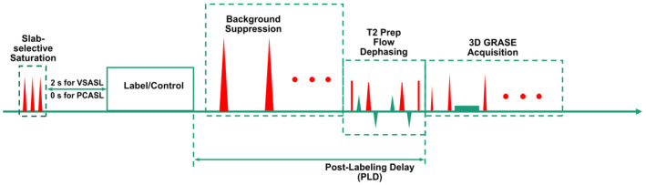
Purpose: To further optimize the velocity-selective arterial spin labeling (VSASL) sequence utilizing a Fourier-transform based velocity-selective inversion (FT-VSI) pulse train, and to evaluate its utility for 3D mapping of cerebral blood flow (CBF) with a gradient- and spin-echo (GRASE) readout.

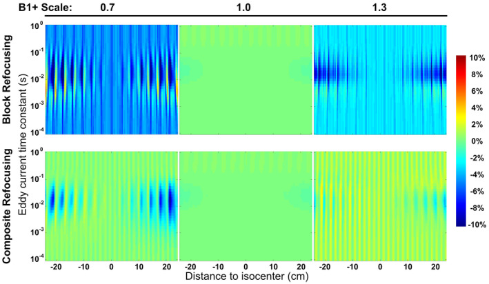
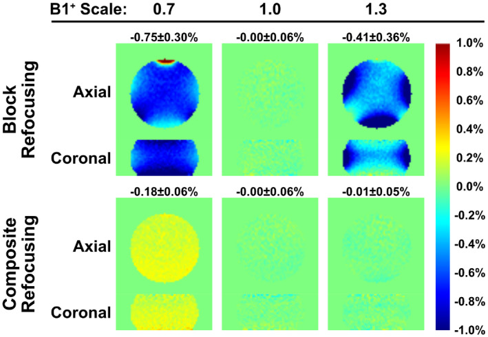
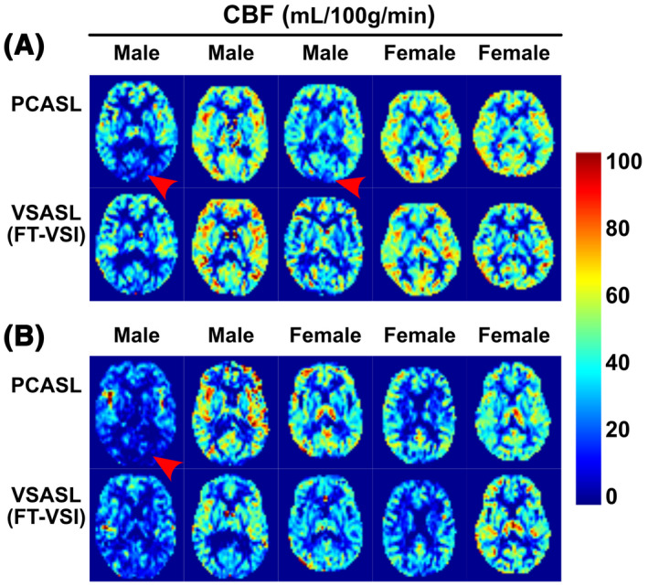
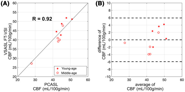
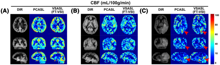
Methods: First, numerical simulations and phantom experiments were done to test the susceptibility to eddy currents and B1 field inhomogeneities for FT-VSI pulse trains with block and composite refocusing pulses. Second, the choices of the post-labeling delay (PLD) for FT-VSI prepared 3D VSASL were evaluated for the sensitivity to perfusion signal. The study was conducted among a young-age and a middle-age group at 3T. Both signal-to-noise ratio (SNR) and CBF were quantitatively compared with pseudo-continuous ASL (PCASL). The optimized 3D VSI-ASL was also qualitatively compared with PCASL in a whole-brain coverage among two healthy volunteers and a brain tumor patient.

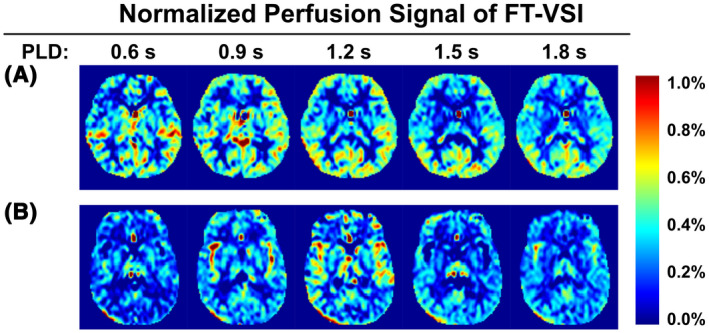
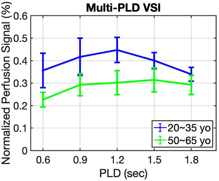
Results: The simulations and phantom test showed that composite refocusing pulses are more robust to both eddy-currents and B1 field inhomogeneities than block pulses. 3D VSASL images with FT-VSI preparation were acquired over a range of PLDs and PLD = 1.2 s was selected for its higher perfusion signal. FT-VSI labeling produced quantitative CBF maps with 27% higher SNR in gray matter compared to PCASL. 3D whole-brain CBF mapping using VSI-ASL were comparable to the corresponding PCASL results.

Conclusion: FT-VSI with 3D-GRASE readout was successfully implemented and showed higher sensitivity to perfusion signal than PCASL for both young and middle-aged healthy volunteers.

Keywords: 3D GRASE acquisition; arterial spin labeling; cerebral blood flow; velocity-selective inversion.


Figures

FIGURE 1
FIGURE 1
Pulse sequence diagram of the ASL sequences used in this study, with FT‐VSI or PCASL label/control pulse trains, combined with slab‐selective saturation pulses post acquisition, spatially selective BGS pulses during the PLD, and a T2‐prep module with flow‐dephasing gradients for suppressing large‐vessel signal followed by 3D GRASE acquisition
FIGURE 2
FIGURE 2
Simulated subtraction errors of static spins following a FT‐VSI pulse train applying block (first row) and composite (second row) refocusing pulses, with EC effects of different time constants at varying distances to isocenter at B1+ scales of 0.7 (left column), 1.0 (middle column), and 1.3 (right column). Large errors from incorrect B1+ settings were reduced substantially when switching from block to composite refocusing pulses
FIGURE 3
FIGURE 3
Normalized subtracted signal of the phantom experiment using FT‐VSI based VSASL with PLD of 1.2 s and 3D GRASE acquisition. In the VSI pulse trains, block (first row) and composite (second row) refocusing pulses were applied, at B1+ scales of 0.7 (left column), 1.0 (middle column), and 1.3 (right column). Incorrect B1+ settings yielded considerable false signal for block refocusing pulses, which were reduced substantially when using composite refocusing pulses, similar to the findings of numerical simulation results in Figure 2
FIGURE 4
FIGURE 4
Normalized perfusion signal images using FT‐VSI preparations acquired with PLDs from 0.6 s to 1.8 s with examples (one slice) from one female subject (31 yo) of the young‐age group (A) and one male subject (52 yo) of the middle‐age group (B)
FIGURE 5
FIGURE 5
Normalized perfusion signal in gray matter using FT‐VSI preparations as function of PLD averaged across the young‐age group (blue) and the middle‐aged group (green)
FIGURE 6
FIGURE 6
Quantitative CBF maps (one slice) using PCASL and FT‐VSI prepared VSASL of each participant from the young‐age group (A) and the middle‐age group (B). Note that the PCASL results of several subjects show CBF underestimation in the occipital lobes (red arrowhead), most likely related to its sensitivity to long transit time delay or inefficient labeling. In contrast, VSASL results do not display such artifacts
FIGURE 7
FIGURE 7
Correlation (A) and agreement (B) plots of the averaged individual CBF values measured between FT‐VSI prepared VSASL and PCASL of gray matter ROIs from subjects of the young (stars) and the middle‐age (open circles) groups. For the correlation plots (A), the black dashed lines show the unity line y = x. The Bland‐Altman plot are shown in (B), in which the difference of the two methods are plotted against their mean, with the middle of the three horizontal dashed lines indicating the mean difference of all subjects, and the top and bottom dashed lines indicating the mean difference ± two times of the standard deviation of their differences
FIGURE 8
FIGURE 8
CBF maps of PCASL and FT‐VSI based VSASL along axial, sagittal and coronal views of a female subject (31 yo) (A), a male subject (42 yo) (B), and a male patient (61 yo) with recurrent glioblastoma in the left occipital lobe (C, red arrowhead). The perfusion signal across brain regions are consistent between PCASL and VSI‐ASL, and their contrast closely resemble the gray‐matter‐only anatomical images by DIR

References

    1. Alsop DC, Detre JA, Golay X, et al. Recommended implementation of arterial spin‐labeled perfusion MRI for clinical applications: A consensus of the ISMRM perfusion study group and the European consortium for ASL in dementia. Magn Reson Med. 2015;73:102‐116. - PMC - PubMed
    1. Ye FQ, Frank JA, Weinberger DR, McLaughlin AC. Noise reduction in 3D perfusion imaging by attenuating the static signal in arterial spin tagging (ASSIST). Magn Reson Med. 2000;44:92‐100. - PubMed
    1. Alsop DC, Detre JA. Reduced transit‐time sensitivity in noninvasive magnetic resonance imaging of human cerebral blood flow. J Cereb Blood Flow Metab. 1996;16:1236‐1249. - PubMed
    1. Dai W, Robson PM, Shankaranarayanan A, Alsop DC. Reduced resolution transit delay prescan for quantitative continuous arterial spin labeling perfusion imaging. Magn Reson Med. 2012;67:1252‐1265. - PMC - PubMed
    1. MacIntosh BJ, Filippini N, Chappell MA, Woolrich MW, Mackay CE, Jezzard P. Assessment of arterial arrival times derived from multiple inversion time pulsed arterial spin labeling MRI. Magn Reson Med. 2010;63:641‐647. - PubMed

Publication types