. 2020 Jun 19;48(11):5967-5985.
doi: 10.1093/nar/gkaa377.

PvrA is a novel regulator that contributes to Pseudomonas aeruginosa pathogenesis by controlling bacterial utilization of long chain fatty acids

Affiliations

PvrA is a novel regulator that contributes to Pseudomonas aeruginosa pathogenesis by controlling bacterial utilization of long chain fatty acids

Xiaolei Pan et al. Nucleic Acids Res. .

Abstract

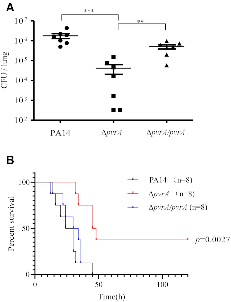
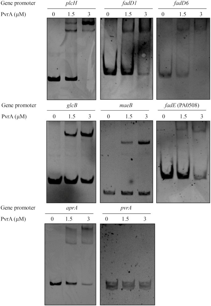
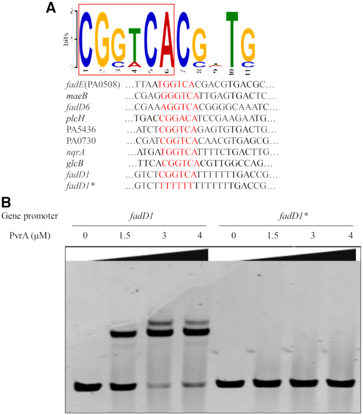
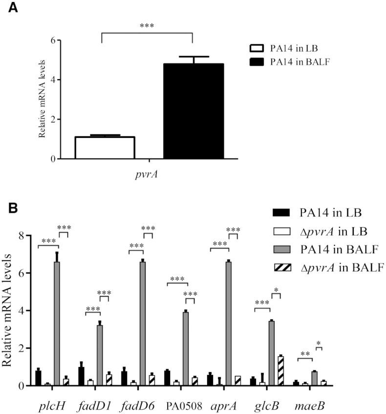
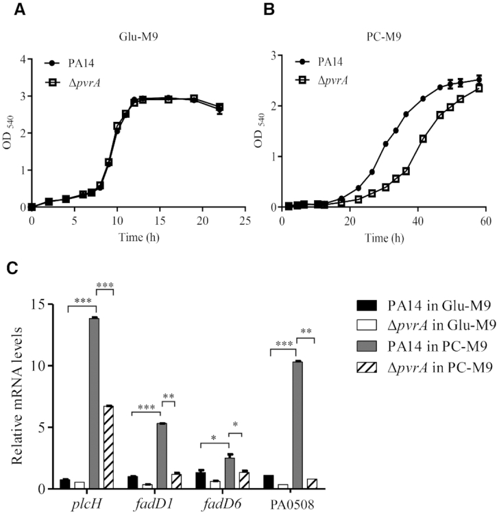
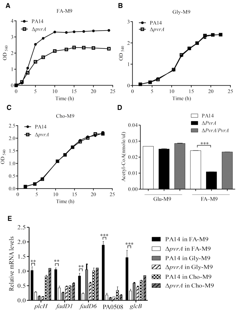
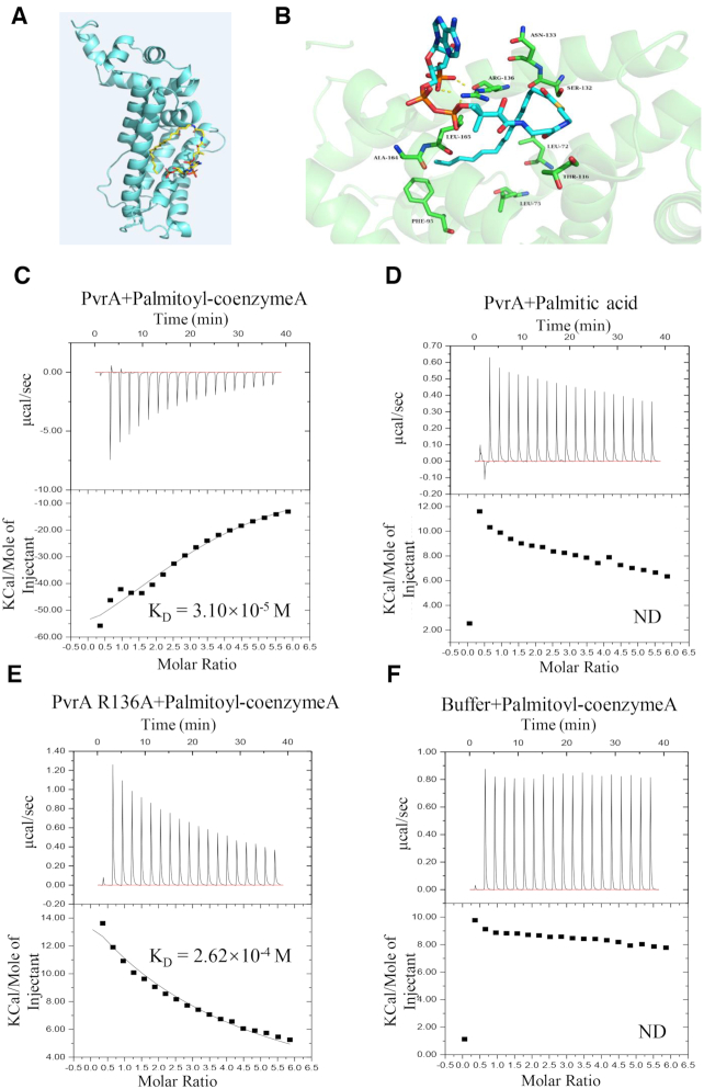
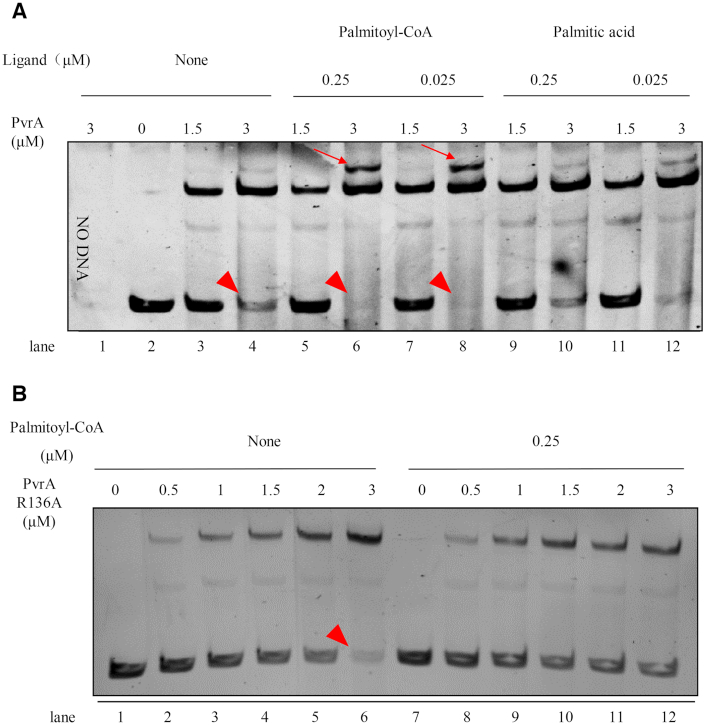
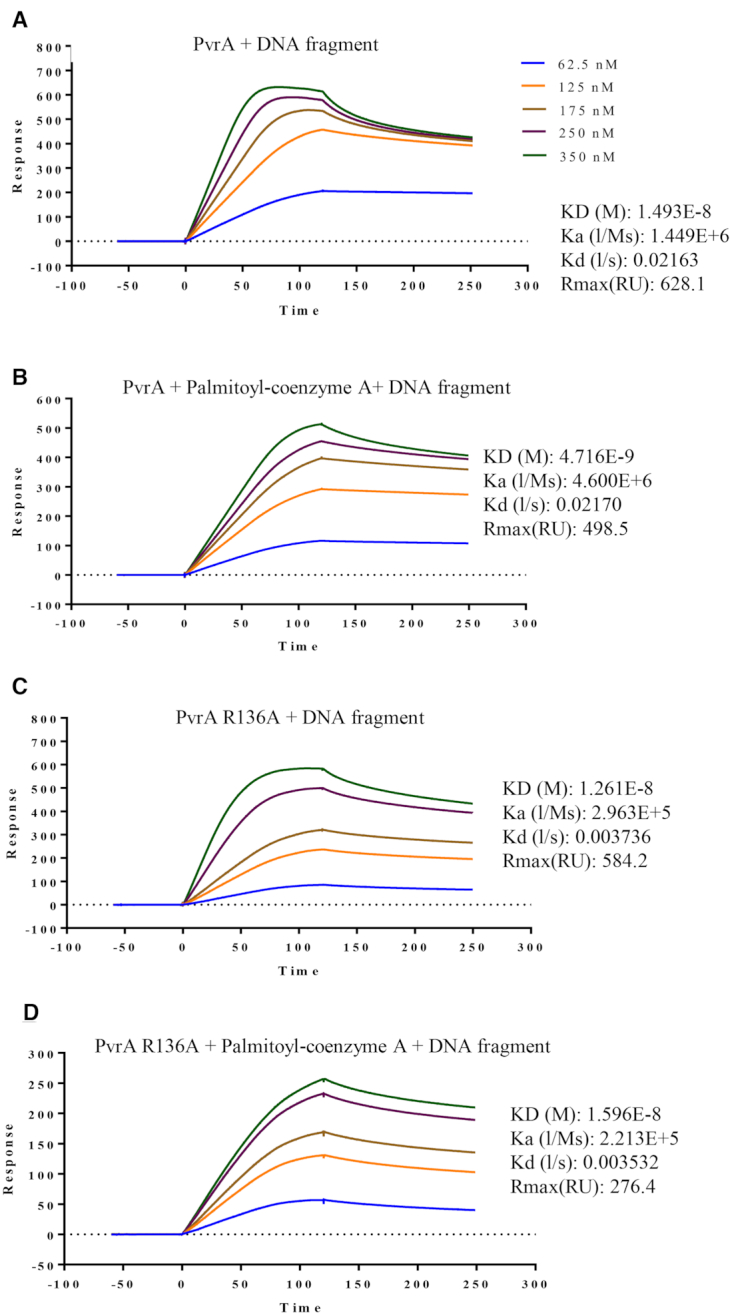
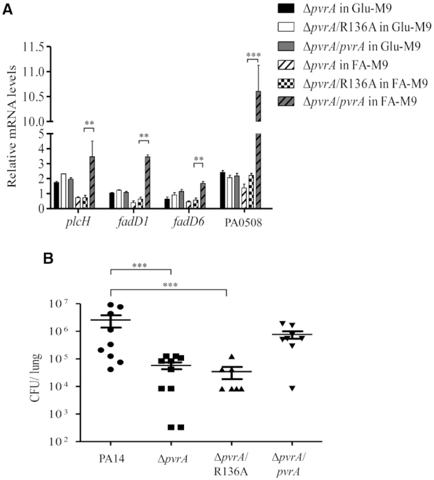
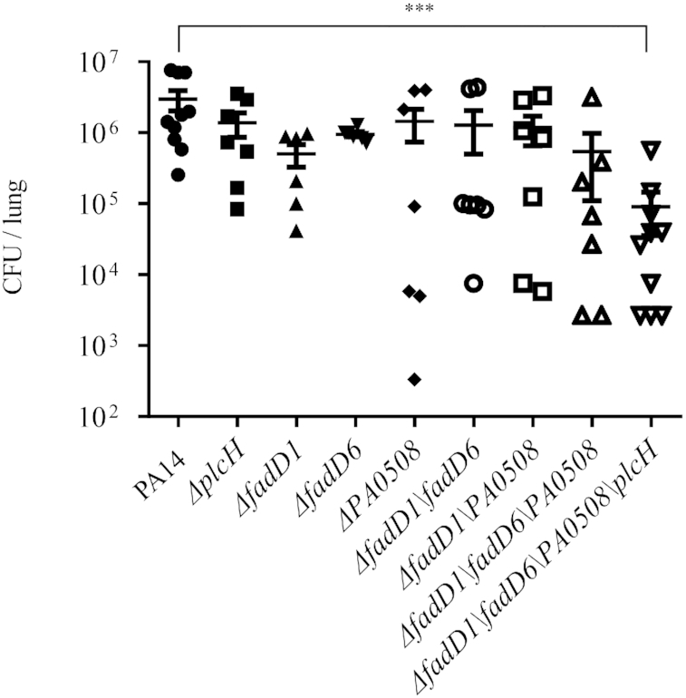
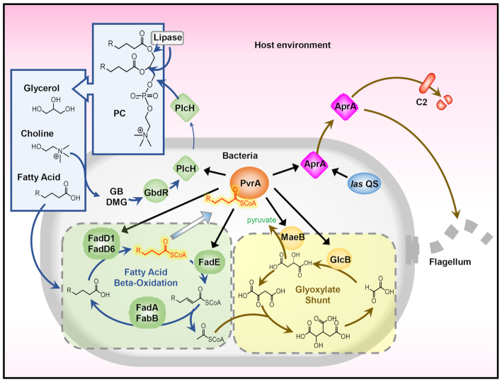
During infection of a host, Pseudomonas aeruginosa orchestrates global gene expression to adapt to the host environment and counter the immune attacks. P. aeruginosa harbours hundreds of regulatory genes that play essential roles in controlling gene expression. However, their contributions to the bacterial pathogenesis remain largely unknown. In this study, we analysed the transcriptomic profile of P. aeruginosa cells isolated from lungs of infected mice and examined the roles of upregulated regulatory genes in bacterial virulence. Mutation of a novel regulatory gene pvrA (PA2957) attenuated the bacterial virulence in an acute pneumonia model. Chromatin immunoprecipitation (ChIP)-Seq and genetic analyses revealed that PvrA directly regulates genes involved in phosphatidylcholine utilization and fatty acid catabolism. Mutation of the pvrA resulted in defective bacterial growth when phosphatidylcholine or palmitic acid was used as the sole carbon source. We further demonstrated that palmitoyl coenzyme A is a ligand for the PvrA, enhancing the binding affinity of PvrA to its target promoters. An arginine residue at position 136 was found to be essential for PvrA to bind palmitoyl coenzyme A. Overall, our results revealed a novel regulatory pathway that controls genes involved in phosphatidylcholine and fatty acid utilization and contributes to the bacterial virulence.


Figures

Figure 1.
Figure 1.
Mutation of pvrA reduces the virulence of PA14. (A). Bacterial colonization in the murine acute pneumonia model. Each mouse was infected intranasally with 4 × 10⁶ CFU of wild type PA14, ΔpvrA or the complemented strain (ΔpvrA/pvrA). 12 hours post infection, mice were sacrificed and bacterial loads in the lungs were determined. Bars represent medians, and error bars represent standard deviations. *P < 0.05; ***P < 0.001 by Student's t-test. (B). Survival rates of infected mice. Mice were infected intranasally with 6 × 10⁶ CFU of wild type PA14, the ΔpvrA mutant or the complemented strain (ΔpvrA/pvrA). The mice were monitored for 5 days. P values were calculated by the log-rank test.
Figure 2.
Figure 2.
Binding of PvrA to the promoter regions of genes involved in PC utilization and fatty acid metabolism. Interaction between PvrA and its target DNA was examined by EMSA. Increasing amount of the purified PvrA protein was incubated with the promoter regions of plcH, fadD1, fadD6, PA0508, aprA, glcB, maeB and pvrA. The mixtures were electrophoresed on an agarose gel and the bands were visualized under UV light following ethidium bromide stain. Data represent results from three independent experiments.
Figure 3.
Figure 3.
Identification of the potential PvrA binding motif. (A). Potential PvrA binding motif was identified by MEME from the ChIP-seq peak regions. Representative sequences bound by PvrA in the EMSAs are listed below. The conserved sequence is shown in red. (B). In the EMSA, indicated amount of the purified PvrA protein was incubated with the fadD1 promoter region or the same fragment with the predicted binding motif deleted (fadD1*). The bands were visualized by staining with EB.
Figure 4.
Figure 4.
Role of PvrA in gene regulation during infection. Wild type PA14 and the ΔpvrA mutant were grown in LB to an OD600 of 1.0. A portion of the bacteria were subjected to RNA isolation. The remaining bacteria were washed and resuspended in PBS. Mice were infected with the bacteria intranasally. The bacteria were isolated from BALF 6 hpi. The mRNA levels of indicated genes were determined by real time PCR with rpsL (encoding the 16S rRNA) as the internal control. (A). Relative mRNA levels of pvrA of PA14 in BALF and LB medium. (B). Relative mRNA levels of plcH, fadD1, fadD6, PA0508, aprA, glcB, maeB in BALF or LB. *P < 0.05; **P < 0.01; ***P < 0.001 by Student's t-test. Data represent the mean ± standard deviation from three samples.
Figure 5.
Figure 5.
PvrA contributes to the bacterial utilization of PC. Same amount of wild type PA14 and the ΔpvrA mutant were inoculated in Glu-M9 (A) and PC-M9 (B). The bacterial growth was monitored by measuring OD540. (C) PA14 and the ΔpvrA mutant were inoculated into Glu-M9 or PC-M9 and grown to an OD600 of 1.0. The relative mRNA levels of plcH, fadD1, fadD6 and PA0508 were determined by real time PCR. rpsL was used as the internal control. *P < 0.05; **P < 0.01; ***P < 0.001 by Student's t-test. Data represent the mean ± standard deviation from three samples.
Figure 6.
Figure 6.
Growth analyses and genes expression of PA14 and the ΔpvrA mutant on palmitic acid, glycerine and choline. Same amount of wild type PA14 and the ΔpvrA mutants were inoculated in FA-M9 (A), Gly-M9 (B) or Cho-M9 (C). The bacterial growth was monitored by measuring OD540. (D). PA14, the ΔpvrA mutant and the complemented strain were grown in Gly-M9 or FA-M9 for 10 h. The intracellular acetyl-CoA concentration was determined and normalized to the corresponding total protein concentration. (E). PA14 and the ΔpvrA mutant were grown in FA-M9, Gly-M9 or Cho-M9 for 10 h. The relative mRNA levels of fadD1, fadD6, PA0508, plcH, glcB were determined by real time PCR. *P < 0.05; **P < 0.01; ***P < 0.001 by Student's t-test. Data represent the mean ± standard deviation from three samples.
Figure 7.
Figure 7.
Interaction between PvrA and palmitoyl-coenzyme A. (A) Model structure of the interaction between palmitoyl-coenzyme A and PvrA by the MetaPocket program. (B) Binding modes of palmitoyl-coenzyme A in the PvrA binding pocket. Dotted lines represent hydrogen-bonds between palmitoyl-coenzyme A and the arginine residue at the 136 position of PvrA. (C–F) Direct binding of palmitoyl-coenzyme A to the soluble PvrA was measured by isothermal titration microcalorimetry (ITC). Isothermal titration microcalorimetry of palmitoyl-coenzyme A (C) or palmitic acid (D) (0.3 mM) binding to 0.03 mM PvrA protein at 25°C. Palmitoyl-coenzyme A exhibited binding affinity of KD = 3.10 × 10⁻⁵ M to PvrA; No detectable binding of palmitic acid to PvrA was exhibited. (E) ITC analysis revealed the affinity between PvrA R136A mutant and palmitoyl-coenzyme A was KD = 2.62 × 10⁻⁴ M. (F). Interaction between the buffer and palmitoyl-coenzyme A was analysed as negative control. Affinity and molar ratio are indicated. ND: No detectable binding.
Figure 8.
Figure 8.
Palmitoyl-coenzyme A enhances the binding of PvrA to the target DNA fragment. The binding of wild type PvrA (A) or the mutated PvrA (R136A) (B) to the fadD1 promoter was examined by EMSA. The fadD1 promoter fragment was incubated with increasing concentrations of PvrA or the mutated PvrA (R136A) with or without the indicated concentrations of palmitoyl-coenzyme A. The DNA bands were visualized by EB staining. The free probes are indicated by arrowheads and the super shifts are indicated by arrows.
Figure 9.
Figure 9.
Surface Plasmon Resonance (SPR) analyses of binding of PvrA to the fadD1 promoter fragment. The PvrA or PvrA(R136A) protein at the indicated concentrations were injected over the immobilized DNA fragment of the fadD1 promoter region (30 bp). (A, B) The binding affinities of PvrA to the DNA probe in the absence (A) or presence (B) of palmitoyl-coenzyme A. (C, D) The binding affinities of PvrA(R136A) to the DNA probe in the absence (C) or presence (D) of palmitoyl-coenzyme A. ka, association rate constant; kd, dissociation rate constant; KD, equilibrium dissociation constant. The concentrations of the proteins and values of ka, kd and KD are indicated in the inset to the figures.
Figure 10.
Figure 10.
Role of the R136 in the function of PvrA. (A) The ΔpvrA mutant and the mutant carrying a wild type pvrA or the R136A mutant allele were grown in Glu-M9 and FA-M9. The relative mRNA levels of plcH, fadD1, fadD6 and PA0508 were determined by real time PCR. rpsL was used as the internal control. (B) Mice were infected intranasally with 4 × 10⁶ CFU of the indicated strains. 12 hpi, the mice were sacrificed and bacterial loads in the lungs were determined by plating. Data represents the mean ± standard deviation from three samples. **P < 0.01, ***P < 0.001 by Student's t-test.
Figure 11.
Figure 11.
Roles of the PvrA regulated genes in the bacterial virulence. The indicated strains were grown in LB to an OD600 of 1.0. Mice were infected intranasally with 4 × 10⁶ CFU of each strain. The mice were sacrificed 12 hpi and the bacterial loads in the lungs were determined by plating. Bars represent medians, and error bars represent standard deviations. ***P < 0.001 by Student's t-test.
Figure 12.
Figure 12.
Proposed model of PvrA mediated regulatory pathways. During infection, PC is degraded by lipases and PlcH, resulting in glycerol, choline and fatty acids. Glycine betaine (GB) and dimethylglycine (DMG) derived from choline are sensed by GbdR, which directly upregulates plcH (57–59). The fatty acids were converted into fatty acyl-CoA. PvrA binds to the fatty acyl-CoA and activates the expression of plcH, fadD1, fadD6, PA0508, aprA, maeB, and glcB. FadD1, FadD6 and PA0508 are key enzymes in the β-oxidation pathway through which a long-chain acyl-CoA molecule is broken down to acetyl-CoA molecules. The las quorum sensing system regulates the expression of aprA (76,77). AprA degrades self flagellin and host complement component C2 to evade recognition by the immune system (50,51). MaeB and GlcB are key enzymes in the glyoxylate shunt which plays an important role in utilizing fatty acids in bacteria (61).

Similar articles

Cited by

References

    1. Lyczak J.B., Cannon C.L., Pier G.B.. Establishment of Pseudomonas aeruginosa infection: lessons from a versatile opportunist. Microbes Infect. 2000; 2:1051–1060. - PubMed
    1. Skaar E.P., Madhani H.D.. The battle for iron between bacterial pathogens and their vertebrate hosts. PLoS Pathog. 2010; 6:e1000949. - PMC - PubMed
    1. Balasubramanian D., Schneper L., Kumari H., Mathee K.. A dynamic and intricate regulatory network determines Pseudomonas aeruginosa virulence. Nucleic Acids Res. 2013; 41:1–20. - PMC - PubMed
    1. Huang H., Shao X., Xie Y., Wang T., Zhang Y., Wang X., Deng X.. An integrated genomic regulatory network of virulence-related transcriptional factors in Pseudomonas aeruginosa. Nat. Commun. 2019; 10:2931. - PMC - PubMed
    1. Winsor G.L., Griffiths E.J., Raymond L., Dhillon B.K., Shay J.A., Brinkman F.S.L.. Enhanced annotations and features for comparing thousands of Pseudomonas genomes in the Pseudomonas genome database. Nucleic Acids Res. 2016; 44:D646–D653. - PMC - PubMed

Publication types

MeSH terms