

Stapled Peptide Inhibitors of Autophagy Adapter LC3B
- PMID: 32406996
- PMCID: PMC7872222
- DOI: 10.1002/cbic.202000212
Stapled Peptide Inhibitors of Autophagy Adapter LC3B
Abstract
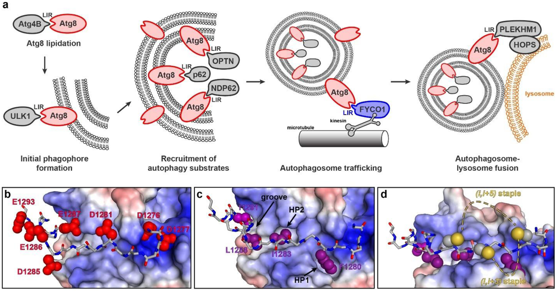
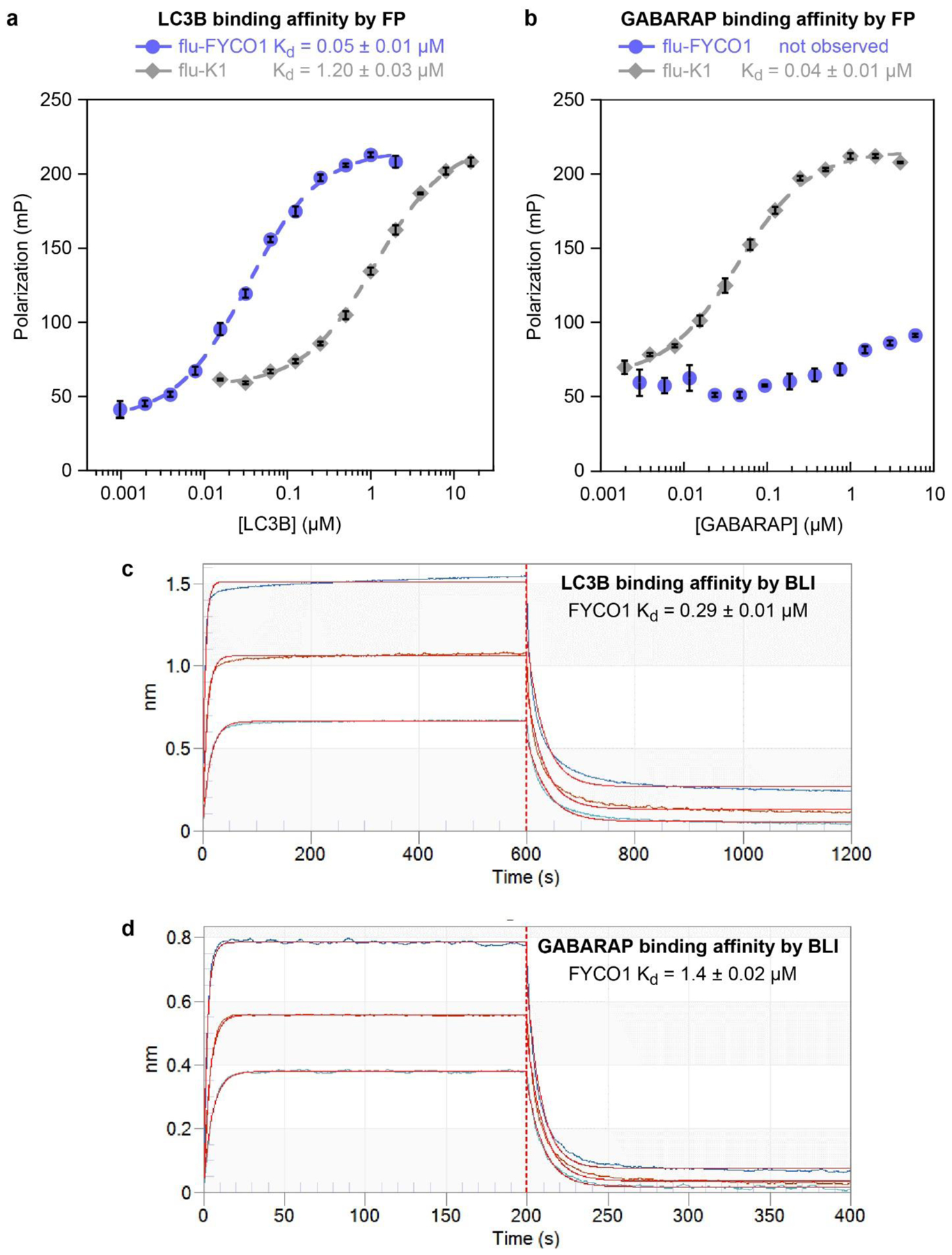
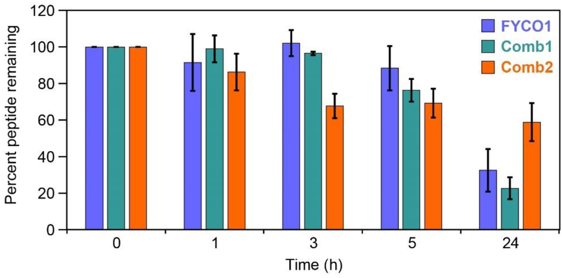
A growing body of evidence suggests that autophagy inhibition enhances the effectiveness of chemotherapy, especially in difficult-to-treat cancers. Existing autophagy inhibitors are primarily lysosomotropic agents. More specific autophagy inhibitors are highly sought-after. The microtubule-associated protein 1A/1B light chain 3B protein, LC3B, is an adapter protein that mediates key protein-protein interactions at several points in autophagy pathways. In this work, we used a known peptide ligand as a starting point to develop improved LC3B inhibitors. We obtained structure-activity relationships that quantify the binding contributions of peptide termini, individual charged residues, and hydrophobic interactions. Based on these data, we used artificial amino acids and diversity-oriented stapling to improve affinity and resistance to biological degradation, while maintaining or improving LC3B affinity and selectivity. These peptides represent the highest-affinity LC3B-selective ligands reported to date, and they will be useful tools for further elucidation of LC3B's role in autophagy and in cancer.
Keywords: LC3B; autophagy; cancer; protein-protein interactions; stapled peptides.
© 2020 Wiley-VCH Verlag GmbH & Co. KGaA, Weinheim.
Conflict of interest statement
Conflicts of interest
There are no conflicts of interest to declare.
Figures
Similar articles
-
FYCO1 Peptide Analogs: Design and Characterization of Autophagy Inhibitors as Co-Adjuvants in Taxane Chemotherapy of Prostate Cancer.Int J Mol Sci. 2025 Jun 3;26(11):5365. doi: 10.3390/ijms26115365. Int J Mol Sci. 2025. PMID: 40508175 Free PMC article.
-
LC3B: A microtubule-associated protein influences disease progression and prognosis.Cytokine Growth Factor Rev. 2025 Feb;81:16-26. doi: 10.1016/j.cytogfr.2024.11.006. Epub 2024 Dec 12. Cytokine Growth Factor Rev. 2025. PMID: 39701849 Review.
-
Diversity-Oriented Stapling Yields Intrinsically Cell-Penetrant Inducers of Autophagy.J Am Chem Soc. 2017 Jun 14;139(23):7792-7802. doi: 10.1021/jacs.7b01698. Epub 2017 May 9. J Am Chem Soc. 2017. PMID: 28414223 Free PMC article.
-
Demonstrating Ligandability of the LC3A and LC3B Adapter Interface.J Med Chem. 2021 Apr 8;64(7):3720-3746. doi: 10.1021/acs.jmedchem.0c01564. Epub 2021 Mar 26. J Med Chem. 2021. PMID: 33769048
-
Design and structure of peptide and peptidomimetic antagonists of protein-protein interaction.Curr Protein Pept Sci. 2005 Apr;6(2):151-69. doi: 10.2174/1389203053545462. Curr Protein Pept Sci. 2005. PMID: 15853652 Review.
Cited by
-
Stapled β-Hairpins Featuring 4-Mercaptoproline.J Am Chem Soc. 2021 Sep 22;143(37):15039-15044. doi: 10.1021/jacs.1c04378. Epub 2021 Sep 13. J Am Chem Soc. 2021. PMID: 34516087 Free PMC article.
-
Computational Prediction of Cyclic Peptide Structural Ensembles and Application to the Design of Keap1 Binders.J Chem Inf Model. 2023 Nov 13;63(21):6925-6937. doi: 10.1021/acs.jcim.3c01337. Epub 2023 Nov 2. J Chem Inf Model. 2023. PMID: 37917529 Free PMC article.
-
Comparing Cell Penetration of Biotherapeutics across Human Cell Lines.ACS Chem Biol. 2024 Jun 21;19(6):1351-1365. doi: 10.1021/acschembio.4c00211. Epub 2024 Jun 5. ACS Chem Biol. 2024. PMID: 38836425 Free PMC article.
-
Design of Protein Segments and Peptides for Binding to Protein Targets.Biodes Res. 2022 Apr 15;2022:9783197. doi: 10.34133/2022/9783197. eCollection 2022. Biodes Res. 2022. PMID: 37850124 Free PMC article. Review.
-
Exploring Arylidene-Indolinone Ligands of Autophagy Proteins LC3B and GABARAP.ACS Med Chem Lett. 2025 Jan 6;16(2):271-277. doi: 10.1021/acsmedchemlett.4c00517. eCollection 2025 Feb 13. ACS Med Chem Lett. 2025. PMID: 39967642
References
Publication types
MeSH terms
Substances
Grants and funding
LinkOut - more resources
Full Text Sources
