

Clinical and Chest Radiography Features Determine Patient Outcomes in Young and Middle-aged Adults with COVID-19
- PMID: 32407255
- PMCID: PMC7507999
- DOI: 10.1148/radiol.2020201754
Clinical and Chest Radiography Features Determine Patient Outcomes in Young and Middle-aged Adults with COVID-19
Abstract
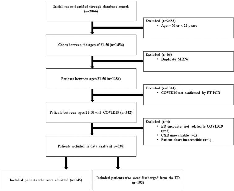
Background Chest radiography has not been validated for its prognostic utility in evaluating patients with coronavirus disease 2019 (COVID-19). Purpose To analyze the prognostic value of a chest radiograph severity scoring system for younger (nonelderly) patients with COVID-19 at initial presentation to the emergency department (ED); outcomes of interest included hospitalization, intubation, prolonged stay, sepsis, and death. Materials and Methods In this retrospective study, patients between the ages of 21 and 50 years who presented to the ED of an urban multicenter health system from March 10 to March 26, 2020, with COVID-19 confirmation on real-time reverse transcriptase polymerase chain reaction were identified. Each patient's ED chest radiograph was divided into six zones and examined for opacities by two cardiothoracic radiologists, and scores were collated into a total concordant lung zone severity score. Clinical and laboratory variables were collected. Multivariable logistic regression was used to evaluate the relationship between clinical parameters, chest radiograph scores, and patient outcomes. Results The study included 338 patients: 210 men (62%), with median age of 39 years (interquartile range, 31-45 years). After adjustment for demographics and comorbidities, independent predictors of hospital admission (n = 145, 43%) were chest radiograph severity score of 2 or more (odds ratio, 6.2; 95% confidence interval [CI]: 3.5, 11; P < .001) and obesity (odds ratio, 2.4 [95% CI: 1.1, 5.4] or morbid obesity). Among patients who were admitted, a chest radiograph score of 3 or more was an independent predictor of intubation (n = 28) (odds ratio, 4.7; 95% CI: 1.8, 13; P = .002) as was hospital site. No significant difference was found in primary outcomes across race and ethnicity or those with a history of tobacco use, asthma, or diabetes mellitus type II. Conclusion For patients aged 21-50 years with coronavirus disease 2019 presenting to the emergency department, a chest radiograph severity score was predictive of risk for hospital admission and intubation. © RSNA, 2020 Online supplemental material is available for this article.
Figures
![Images show examples of the chest severity score. Yellow circles indicate regions of the lung with visible opacities. A, Chest radiograph in a 26-year-old man with no past medical history other than obesity (body mass index [BMI] of 38 kg/m2) who was admitted for coronavirus disease 2019 (COVID-19) requiring oxygen supplementation via nasal cannula. He initially tested negative for COVID-19 via nasopharyngeal swab, but later tested positive for antibodies to severe acute respiratory syndrome coronavirus 2. Portable chest radiograph shows hazy opacities in right lower lung zone, left middle lung zone, and left upper lung zone; total score of 3. B, Chest radiograph in a 23-year-old man with no past medical history who tested positive for COVID-19 via reverse transcription polymerase chain reaction and was subsequently discharged from emergency department with home care and isolation precautions. Portable chest radiograph shows hazy opacities (arrows) in right and left peripheral lower lung zone; total score of 2. C, Chest radiograph in a 32-year-old overweight (BMI of 30 kg/m2) COVID-19–positive man with history of childhood asthma who was subsequently admitted and intubated in intensive care unit for 3 days. Portable chest radiograph shows opacities in all three right lung zones and in left middle and lower lung zones; total score of 5.](https://cdn.ncbi.nlm.nih.gov/pmc/blobs/858e/7507999/8442e21cf685/radiol.2020201754.fig2.jpg)
Comment in
-
Chest Radiography Features Help to Predict a Favorable Outcome in Patients with Coronavirus Disease 2019.Radiology. 2020 Oct;297(1):E238. doi: 10.1148/radiol.2020202326. Epub 2020 Jun 2. Radiology. 2020. PMID: 32484419 Free PMC article. No abstract available.
References
-
- COVID-19 dashboard by the Center for Systems Science and Engineering (CCSE). Johns Hopkins Coronavirus Resource Center. https://coronavirus.jhu.edu/map.html. Accessed April 30, 2020.
-
- American College of Radiology. ACR recommendations for the use of chest radiography and computed tomography (CT) for suspected COVID-19 infection. https://www.acr.org/Advocacy-and-Economics/ACR-Position-Statements/Recom.... Published March 11, 2020. Updated March 22, 2020. Accessed April 30, 2020.
MeSH terms
LinkOut - more resources
Full Text Sources
Other Literature Sources
