

Case Reports
doi: 10.1148/radiol.2020201753.
Epub 2020 May 14.
COVID-19-associated Leukoencephalopathy
Affiliations
- PMID: 32407258
- PMCID: PMC7437492
- DOI: 10.1148/radiol.2020201753
Item in Clipboard
Case Reports
COVID-19-associated Leukoencephalopathy
Radiology.
2020 Sep.
No abstract available
Figures
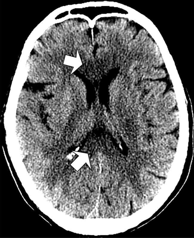
Unenhanced CT image of head demonstrates confluent hypoattenuation in genu (anterior) and splenium (posterior) of corpus callosum (arrows).
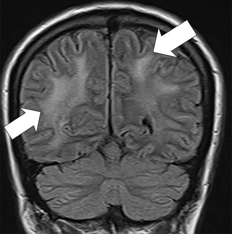
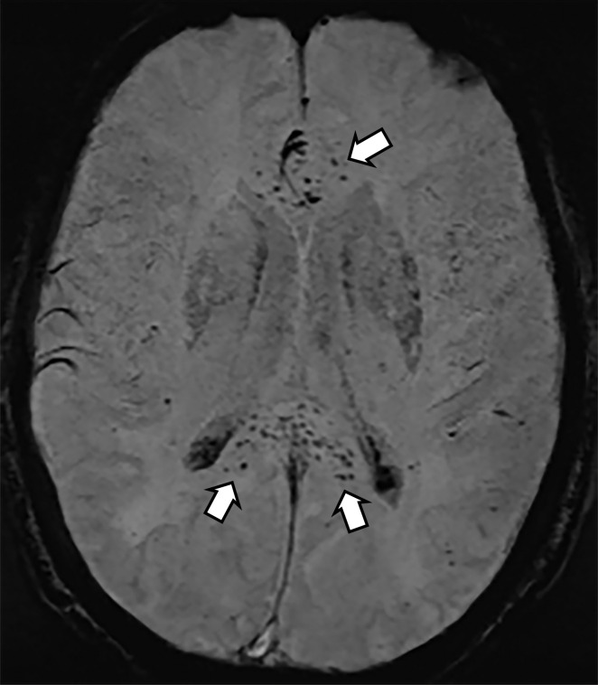
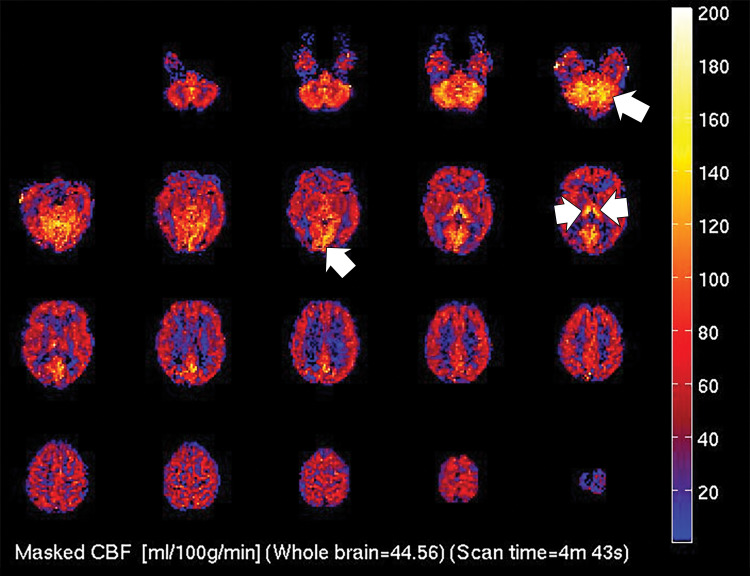
(a) T2-weighted fluid-attenuated inversion recovery MRI scan of brain shows extensive posterior greater than anterior white matter hyperintensity (arrows) without mass effect. (b) Susceptibility-weighted image demonstrates a cluster of microhemorrhages in corpus callosum (arrows). (c) Arterial-spin-labeling perfusion images are suggestive of posterior circulation hyperperfusion (arrows). CBF = cerebral blood flow.
(a) T2-weighted fluid-attenuated inversion recovery MRI scan of brain shows extensive posterior greater than anterior white matter hyperintensity (arrows) without mass effect. (b) Susceptibility-weighted image demonstrates a cluster of microhemorrhages in corpus callosum (arrows). (c) Arterial-spin-labeling perfusion images are suggestive of posterior circulation hyperperfusion (arrows). CBF = cerebral blood flow.
(a) T2-weighted fluid-attenuated inversion recovery MRI scan of brain shows extensive posterior greater than anterior white matter hyperintensity (arrows) without mass effect. (b) Susceptibility-weighted image demonstrates a cluster of microhemorrhages in corpus callosum (arrows). (c) Arterial-spin-labeling perfusion images are suggestive of posterior circulation hyperperfusion (arrows). CBF = cerebral blood flow.
References
-
- Pinto PS, Taipa R, Moreira B, Correia C, Melo-Pires M. Acute hemorrhagic leukoencephalitis with severe brainstem and spinal cord involvement: MRI features with neuropathological confirmation. J Magn Reson Imaging 2011;33(4):957–961. - PubMed
-
- Abdel Razek AA, Alvarez H, Bagg S, Refaat S, Castillo M. Imaging spectrum of CNS vasculitis. RadioGraphics 2014;34(4):873–894. - PubMed
-
- Moore JB, June CH. Cytokine release syndrome in severe COVID-19. Science 2020;368(6490):473–474. - PubMed
Publication types
MeSH terms
LinkOut - more resources
Full Text Sources
