. 2020 Aug 3;130(8):4104-4117.
doi: 10.1172/JCI122237.

Elevated circulating amyloid concentrations in obesity and diabetes promote vascular dysfunction

Affiliations

Elevated circulating amyloid concentrations in obesity and diabetes promote vascular dysfunction

Paul J Meakin et al. J Clin Invest. .

Abstract

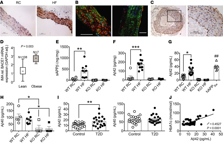
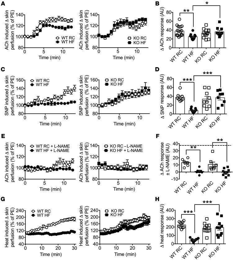
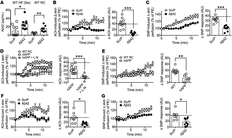
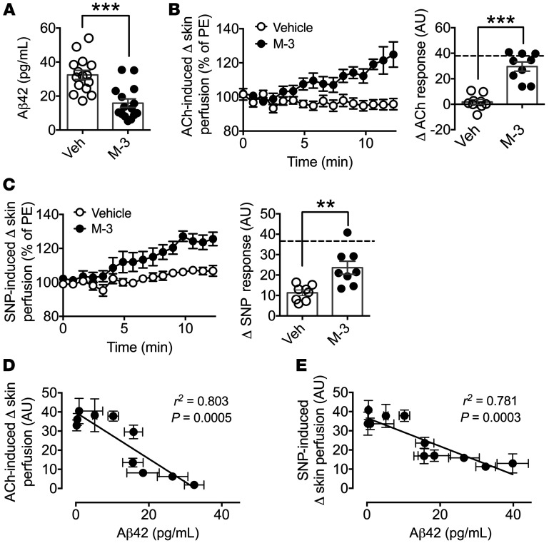
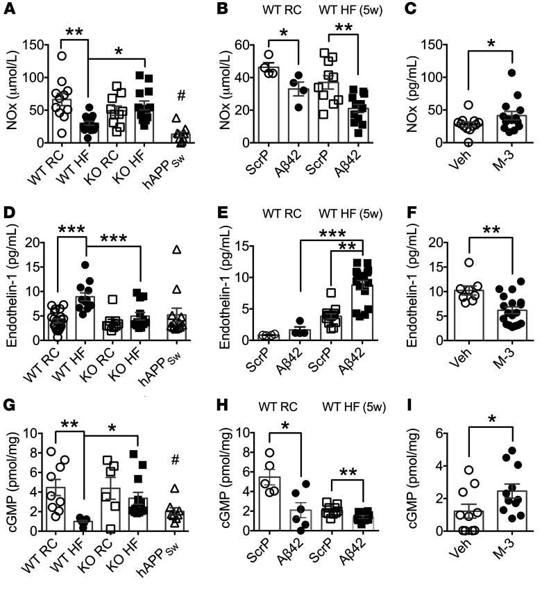
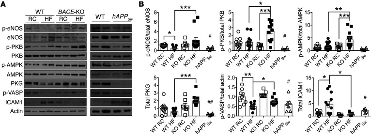
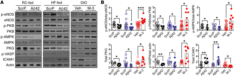
Diabetes, obesity, and Alzheimer's disease (AD) are associated with vascular complications and impaired nitric oxide (NO) production. Furthermore, increased β-site amyloid precursor protein-cleaving (APP-cleaving) enzyme 1 (BACE1), APP, and β-amyloid (Aβ) are linked with vascular disease development and increased BACE1 and Aβ accompany hyperglycemia and hyperlipidemia. However, the causal relationship between obesity and diabetes, increased Aβ, and vascular dysfunction is unclear. We report that diet-induced obesity (DIO) in mice increased plasma and vascular Aβ42 that correlated with decreased NO bioavailability, endothelial dysfunction, and increased blood pressure. Genetic or pharmacological reduction of BACE1 activity and Aβ42 prevented and reversed, respectively, these outcomes. In contrast, expression of human mutant APP in mice or Aβ42 infusion into control diet-fed mice to mimic obese levels impaired NO production, vascular relaxation, and raised blood pressure. In humans, increased plasma Aβ42 correlated with diabetes and endothelial dysfunction. Mechanistically, higher Aβ42 reduced endothelial NO synthase (eNOS), cyclic GMP (cGMP), and protein kinase G (PKG) activity independently of diet, whereas endothelin-1 was increased by diet and Aβ42. Lowering Aβ42 reversed the DIO deficit in the eNOS/cGMP/PKG pathway and decreased endothelin-1. Our findings suggest that BACE1 inhibitors may have therapeutic value in the treatment of vascular disease associated with diabetes.

Keywords: Endocrinology; Nitric oxide; Obesity; Vascular Biology; endothelial cells.


Conflict of interest statement

Conflict of interest: The authors have declared that no conflict of interest exists.

Figures

Figure 1
Figure 1. BACE1 vascular expression and activity and plasma levels of Aβ in mice and humans.
(A) Immunohistochemical staining for BACE1 (brown) showing representative sections from aortas of RC-fed and HF-fed (DIO) WT mice. (B) Confocal images of DIO mouse aortas stained for BACE1 (green) and SMA (left) or CD31 (right), respectively (red). Scale bars: 50 μm. (C) Immunohistochemical staining for BACE1 in human nonatherosclerotic temporal artery, with a section (rectangle) at higher magnification. (D) BACE1 mRNA expression in internal mammary arteries from lean and obese individuals (Mann-Whitney U test). HF feeding increases BACE1 activity in WT mouse aorta, as determined by sAPPβ (E) and Aβ42 (F) levels, with BACE1-KO aortas showing negligible peptide levels (n = 4–12). (G) Plasma Aβ42 levels are increased in DIO mice to RC-fed hAPPSw mouse levels, with insignificant plasma Aβ42 in RC- or HF-fed BACE1-KO mice (n = 6–14). (H) Plasma Aβ40 levels of RC-fed and DIO mice and RC- and HF-fed BACE1-KO mice (n = 6–14). (I) Plasma Aβ42 and Aβ40 levels in control (n = 20) and obese individuals with type 2 diabetes (T2D) (n = 20). (J) Linear regression between plasma Aβ42 and HbA1c in control and obese individuals with T2D (P < 0.001). Data presented as means ± SEM for all figures except D, where SD given. *P < 0.05; **P < 0.01; ##P < 0.01; ***P < 0.001 by 2-way ANOVA with Tukey’s multiple-comparisons test (EH) or 2-tailed unpaired Student’s t test (G and I).
Figure 2
Figure 2. Loss of BACE1 prevents HF feeding–induced endothelial and vascular smooth muscle dysfunction.
(A) Endothelium-dependent microvascular responses induced by ACh in RC-fed WT, DIO (20-week HF-fed) mice, and RC- and HF-fed BACE1-KO mice (n = 10–21). PE, phenylephrine. (B) Quantitative analysis of ACh response from A. (C) Endothelium-independent responses induced by SNP in RC-WT, DIO, and BACE1-KO mice (n = 8–9). (D) Quantitative analysis of SNP responses from C. (E) Microvascular responses induced by ACh in the presence of L-NAME in RC-fed WT, DIO, and RC- and HF-fed WT and BACE1-KO mice (n = 6–10). (F) Quantitative analysis of ACh responses from E. The broken line denotes the RC-fed WT ACh response. (G) Microvascular responses to localized heating in RC-fed WT, DIO, and RC- and HF-fed BACE1-KO mice (n = 9–11). (H) Quantitative analysis of heating responses from G. Data are means ± SEM. *P < 0.05; **P < 0.01; ***P < 0.001 by 2-way ANOVA with Tukey’s multiple-comparisons test.
Figure 3
Figure 3. Increased circulating Aβ42 impairs microvascular function.
(A) Plasma Aβ42 levels in HF-fed (5 weeks) and RC-fed WT mice after 4 weeks of Aβ42 or ScrP infusion (n = 7–10). (B) Endothelium-dependent microvascular responses induced by ACh in ScrP- and Aβ42-treated HF-fed (5 weeks) WT mice (n = 9). Quantitative analysis of ACh responses for ScrP- and Aβ42-treated mice. The broken line denotes the RC-fed WT ACh response. PE, phenylephrine. (C) Endothelium-independent responses induced by SNP in ScrP- and Aβ42-treated HF-fed (5 weeks) WT mice (n = 8–9). Quantitative analysis of SNP responses for ScrP- and Aβ42-treated mice. The broken line denotes the RC-fed WT SNP response. (D) Microvascular responses induced by ACh in RC-fed WT and hAPPSw mice and hAPPSw mice with L-NAME (L-N) (n = 6–16). Quantitative analysis of ACh responses for hAPPSw mice in the absence and presence of L-NAME. The broken line denotes the RC-fed WT ACh response. (E) Endothelium-independent responses induced by SNP in RC-fed WT and hAPPSw mice (n = 5–8). Quantitative analysis of SNP responses. (F) Endothelium-dependent microvascular responses induced by ACh in ScrP- and Aβ42-treated RC-fed WT mice (n = 7–9). Quantitative analysis of ACh responses for ScrP- and Aβ42-treated mice. The broken line denotes the RC-fed WT ACh response. (G) Endothelium-independent responses induced by SNP in ScrP- and Aβ42-treated RC-fed WT mice (n = 7–9). Quantitative analysis of SNP responses for ScrP- and Aβ42-treated mice. The broken line denotes the RC-fed WT SNP response. Data are means ± SEM. *P < 0.05; **P < 0.01; ***P < 0.001 by 2-tailed unpaired Student’s t test.
Figure 4
Figure 4. M-3 recovers microvascular function in DIO mice and plasma Aβ42 correlates with microvascular responsiveness.
(A) Plasma Aβ42 levels of DIO mice treated with M-3 or vehicle (n = 8). (B) Endothelium-dependent microvascular responses induced by ACh in vehicle- and M-3–treated DIO mice (n = 9). Quantitative analysis of ACh responses for vehicle- and M-3–treated mice. The broken line denotes the RC-fed WT ACh response. PE, phenylephrine. (C) Endothelium-independent responses induced by SNP in vehicle- and M-3–treated DIO mice (n = 8). Quantitative analysis of SNP responses for vehicle- and M-3–treated mice. The broken line denotes the RC-fed WT SNP response. Linear regression between plasma Aβ42 levels and ACh (D) and SNP (E) responses for experimental groups of mice. Data are means ± SEM. **P < 0.01, ***P < 0.001 by 2-tailed unpaired Student’s t test.
Figure 5
Figure 5. Plasma NOx, ET-1, and aortic cGMP are modified by BACE1 activity and Aβ42.
(A) Plasma NOx levels in RC-fed WT, hAPPSw, and BACE1-KO, DIO, and HF-fed BACE1-KO mice (n = 9–14). (B) Plasma NOx levels in RC-fed WT and HF-fed (5 weeks) mice after 4 weeks of Aβ42 or ScrP infusion (n = 4–12). (C) Plasma NOx levels in vehicle- and M-3–treated DIO mice (n = 12–14). (D) Plasma ET-1 levels in RC-fed WT, hAPPSw, and BACE1-KO, DIO, and HF-fed BACE1-KO mice (n = 9–21). (E) Plasma ET-1 levels in RC-fed WT and HF-fed (5 weeks) mice after 4 weeks of Aβ42 or ScrP infusion (n = 4–12). (F) Plasma ET-1 levels in vehicle- and M-3–treated DIO mice (n = 8–18). (G) Aortic cGMP levels in RC-fed WT, hAPPSw, and BACE1-KO, DIO, and HF-fed BACE1-KO mice (n = 6–12). (H) Aortic cGMP levels in HF-fed (5 weeks) mice after 4 weeks of Aβ42 or ScrP infusion (n = 9–10) and (I) in vehicle- and M-3–treated DIO mice (n = 10–11). Data are means ± SEM. *P < 0.05; **P < 0.01; ***P < 0.001 by 2-way ANOVA with Tukey’s multiple-comparisons test (A, B, D, E, G, and H) or 2-tailed unpaired Student’s t test (C, F, and I). #P < 0.05 vs. RC-fed WT (A and G).
Figure 6
Figure 6. Aortic NO signaling is modified in BACE1-KO and hAPPSw mice.
(A) Representative immunoblots of p-eNOS(Ser1177), eNOS, p-PKB(Ser473), PKB, p-AMPK(Thr172), AMPK, PKG, p-VASP(Ser239), ICAM1, and actin in aortas of RC-fed WT, DIO, and RC- and HF-fed BACE1-KO mice (left panel), and from aortas of RC-fed WT and RC-fed hAPPSw mice (right panel). (B) Ratio of signal intensities for p-eNOS to eNOS, p-PKB to PKB, p-AMPK to AMPK; and for p-VASP, PKG, and ICAM1 to actin (n = 6–15). Data are means ± SEM. *P < 0.05; **P < 0.01; ***P < 0.001 by Kruskal-Wallis test with Dunn’s multiple-comparisons test. #P < 0.05, ##P < 0.01 by Mann-Whitney U test (vs. RC-fed WT mice).
Figure 7
Figure 7. BACE1 activity and Aβ42 regulate aortic NO signaling.
(A) Representative immunoblots of p-eNOS(Ser1177), eNOS, p-PKB(Ser473), PKB, p-AMPK(Thr172), AMPK, PKG, p-VASP(Ser239), ICAM1, and actin in aortas of ScrP- and Aβ42-treated RC-fed (left panel) and HF-fed (5 weeks) WT mice (middle panel) and in aortas of vehicle- or M-3–treated DIO mice (right panel). (B) Ratio of signal intensities for p-eNOS to eNOS, p-PKB to PKB, p-AMPK to AMPK; and for PKG, p-VASP, and ICAM1 to actin (n = 5–14). Data are means ± SEM. *P < 0.05; **P < 0.01; ***P < 0.001 by Mann-Whitney U test.

Similar articles

Cited by

References

    1. de Bruijn RF, Ikram MA. Cardiovascular risk factors and future risk of Alzheimer’s disease. BMC Med. 2014;12:130. - PMC - PubMed
    1. Attems J, Jellinger KA. The overlap between vascular disease and Alzheimer’s disease--lessons from pathology. BMC Med. 2014;12:206. - PMC - PubMed
    1. Wiesmann M, Kiliaan AJ, Claassen JA. Vascular aspects of cognitive impairment and dementia. J Cereb Blood Flow Metab. 2013;33(11):1696–1706. doi: 10.1038/jcbfm.2013.159. - DOI - PMC - PubMed
    1. Cheng X, He P, Yao H, Dong Q, Li R, Shen Y. Occludin deficiency with BACE1 elevation in cerebral amyloid angiopathy. Neurology. 2014;82(19):1707–1715. doi: 10.1212/WNL.0000000000000403. - DOI - PMC - PubMed
    1. Attems J, Lintner F, Jellinger KA. Amyloid beta peptide 1-42 highly correlates with capillary cerebral amyloid angiopathy and Alzheimer disease pathology. Acta Neuropathol. 2004;107(4):283–291. doi: 10.1007/s00401-004-0822-6. - DOI - PubMed

Publication types

MeSH terms