. 2020 May 14;15(5):e0232991.
doi: 10.1371/journal.pone.0232991. eCollection 2020.

The role of the C2A domain of synaptotagmin 1 in asynchronous neurotransmitter release

Affiliations

The role of the C2A domain of synaptotagmin 1 in asynchronous neurotransmitter release

Mallory C Shields et al. PLoS One. .

Abstract

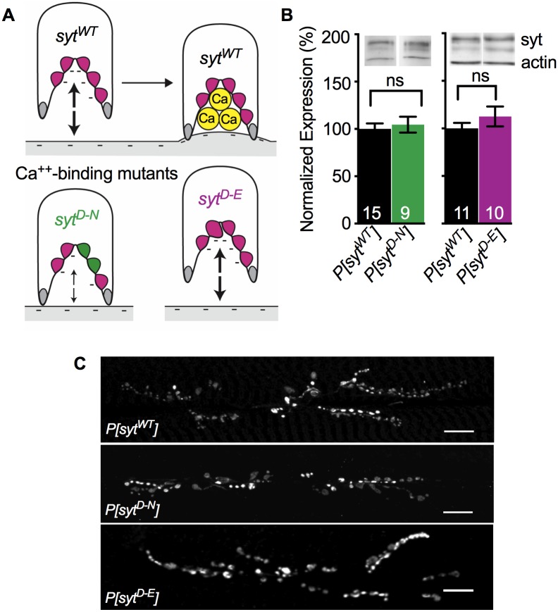
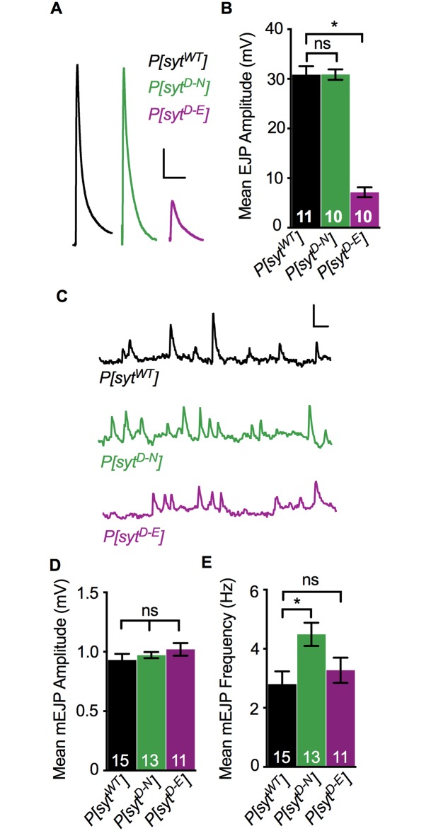
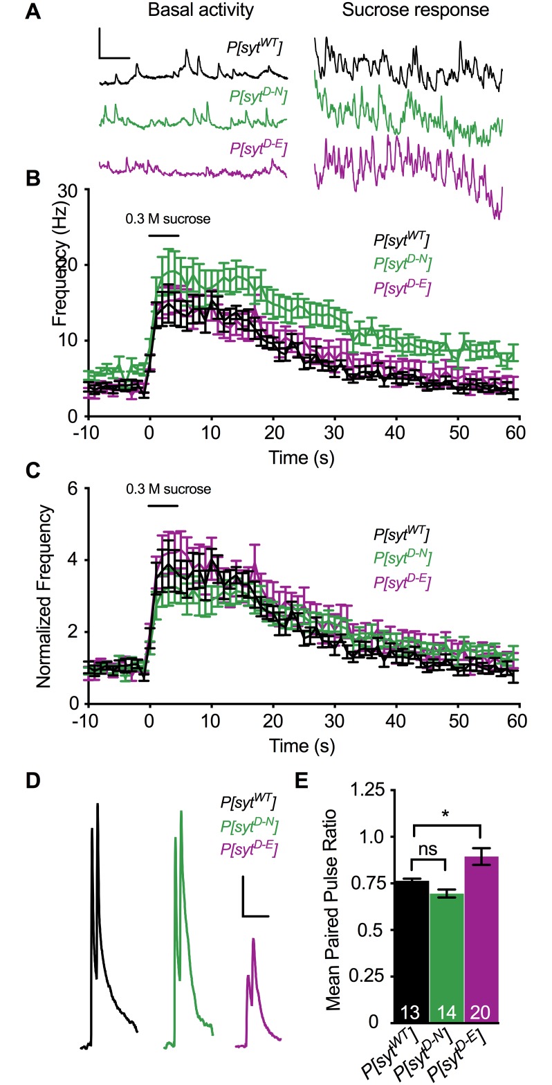
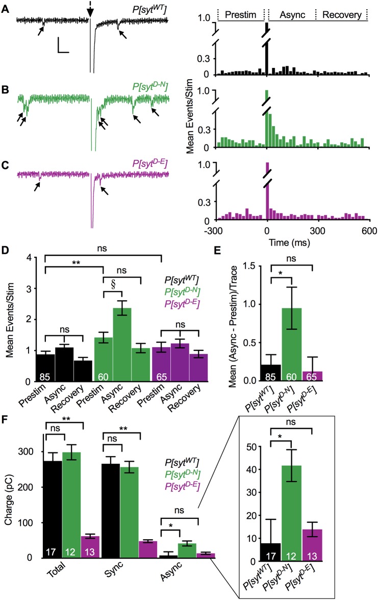
Following nerve stimulation, there are two distinct phases of Ca2+-dependent neurotransmitter release: a fast, synchronous release phase, and a prolonged, asynchronous release phase. Each of these phases is tightly regulated and mediated by distinct mechanisms. Synaptotagmin 1 is the major Ca2+ sensor that triggers fast, synchronous neurotransmitter release upon Ca2+ binding by its C2A and C2B domains. It has also been implicated in the inhibition of asynchronous neurotransmitter release, as blocking Ca2+ binding by the C2A domain of synaptotagmin 1 results in increased asynchronous release. However, the mutation used to block Ca2+ binding in the previous experiments (aspartate to asparagine mutations, sytD-N) had the unintended side effect of mimicking Ca2+ binding, raising the possibility that the increase in asynchronous release was directly caused by ostensibly constitutive Ca2+ binding. Thus, rather than modulating an asynchronous sensor, sytD-N may be mimicking one. To directly test the C2A inhibition hypothesis, we utilized an alternate C2A mutation that we designed to block Ca2+ binding without mimicking it (an aspartate to glutamate mutation, sytD-E). Analysis of both the original sytD-N mutation and our alternate sytD-E mutation at the Drosophila neuromuscular junction showed differential effects on asynchronous release, as well as on synchronous release and the frequency of spontaneous release. Importantly, we found that asynchronous release is not increased in the sytD-E mutant. Thus, our work provides new mechanistic insight into synaptotagmin 1 function during Ca2+-evoked synaptic transmission and demonstrates that Ca2+ binding by the C2A domain of synaptotagmin 1 does not inhibit asynchronous neurotransmitter release in vivo.


Conflict of interest statement

The authors have declared that no competing interests exist.

Figures

Fig 1
Fig 1. Ca2+ binding mutants expressed and localized correctly.
A. Schematic depiction of the C2A domain of wild type and mutant synaptotagmin and their postulated interactions with the negatively-charged presynaptic membrane. A. Top, The C2A Ca2+-binding pocket of wild type synaptotagmin (sytWT). Prior to Ca2+ entry (left), 5 negatively charged (magenta) aspartate residues repel the negatively-charged presynaptic membrane (large arrows). Ca2+ binding neutralizes the negative charge of the pocket (right), resulting in the penetration of the presynaptic membrane by hydrophobic residues (grey). A. Below, By replacing two aspartate residues with neutral (green) asparagines, the sytD-N mutation in C2A blocks Ca2+ binding and partially neutralizes the negative charge of the pocket. Importantly, this partial neutralization also decreases the electrostatic repulsion of the presynaptic membrane (small arrows), which may mimic Ca2+ binding. By replacing one aspartate residue deep in the Ca2+-binding pocket with a larger, negatively-charged glutamate residue (magenta, larger), the sytD-E mutation in C2A blocks Ca2+ binding by steric hindrance while maintaining electrostatic repulsion of the presynaptic membrane (large arrows). B. Above, Representative western blots showing expression levels of synaptotagmin and actin from individual larval CNSs of each genotype. B. Below, quantification of P[sytWT] vs. P[sytD-N] (left) and P[sytWT] vs. P[sytD-E] (right). All measurements were normalized to actin levels. Both mutant lines exhibited levels of synaptotagmin expression similar to control. ns = not significant. C. Anti-synaptotagmin labeling of third instar body wall musculature demonstrated that transgenic synaptotagmin is appropriately concentrated at the neuromuscular junction in all genotypes. Scale bars = 20 μm.
Fig 2
Fig 2. Ca2+ binding mutants had differential effects on evoked and spontaneous release.
A. Representative EJP traces from P[sytWT], P[sytD-N], and P[sytD-E]. Scale bars = 5 mV, 0.1 s. B. Mean EJP amplitude in P[sytD-N] was unimpaired, but in P[sytD-E], it was significantly decreased compared to control (*p < 0.0001). C. Representative mEJP traces from P[sytWT], P[sytD-N], and P[sytD-E] showing 3 consecutive seconds of spontaneous mEJPs. Scale bars = 1 mV, 0.2 s. D. Mean mEJP amplitude was similar among genotypes. E. Mean mEJP frequency was increased in P[sytD-N] (*p = 0.01) but unchanged in P[sytD-E] relative to P[sytWT]. Recorded in HL3.1 containing 1.0 mM Ca2+, all error bars depict SEM, and n’s within bars represent number of muscle fibers tested. ns = not significant.
Fig 3
Fig 3. Ca2+ binding mutants had no impact on the number of fusion competent vesicles, but had differential effects on release probably.
A. Representative traces of event frequency before, during, and after sucrose-stimulated neurotransmitter release from P[sytWT], P[sytD-N], and P[sytD-E]. Scale bars = 2 mV, 0.5 s. B. Mean event frequencies over time in response to a 5 s application of a hypertonic sucrose solution (n = 11 fibers for each genotype). C. Mean event frequencies over time normalized to the basal mEJP frequency prior to sucrose application. No statistically significant changes were found among genotypes during the sucrose response. The black bar above the traces in B,C represents the 5 s sucrose application. D. Representative paired pulse traces with a 20 ms interpulse interval from P[sytWT], P[sytD-N], and P[sytD-E]. Scale bars = 5 mV, 0.1 s. E. There was a significant increase in the paired pulse ratio in P[sytD-E] compared to control (*p < 0.0001). There was no significant change in paired pulse ratios between control and P[sytD-N]. Recorded in HL3.1 containing 1.0 mM Ca2+, error bars are SEM, and n’s within bars represent number of fibers tested. ns = not significant.
Fig 4
Fig 4. Asynchronous release was increased in the P[sytD-N] mutant but not the P[sytD-E] mutant.
A-C Left. Representative traces from P[sytWT], P[sytD-N], and P[sytD-E] recorded in HL3.1 containing 1.0 mM Ca2+ showing events between 280 ms before to 300 ms after stimulation (large dotted arrow). Stimulation artifact removed for clarity. Individual release events before and after the large, multi-quantal synchronous response are indicated (small arrows). Scale bars = 1 nA, 0.04 s. Right. Latency histograms. Data were parsed into 20 ms bins from 280 ms before to 580 ms after single stimulations and the mean number of events/stimulation was plotted in each bin for all genotypes. D. Mean events/stimulation during the 280 ms before stimulation (Prestim), the 20–300 ms after stimulation, asynchronous release period (Async), and the 300–580 ms after stimulation (Recovery) were graphed for each genotype. P[sytD-N] larvae exhibited a significant increase in asynchronous release compared to Prestim (§p = 0.001). Control and the P[sytD-E] mutant did not. No significant differences were found in any genotype when comparing Prestim and Recovery time periods. P[sytD-N] also exhibited a significant increase in mEJC frequency (Prestim) compared to control (**p = 0.01). The P[sytD-E] mutant did not. E. Compared to control, the P[sytD-N] mutant exhibited a significant increase in asynchronous release corrected for mEJC frequency [Mean (Async—Prestim)/Trace, *p = 0.04], but the P[sytD-E] mutant did not. F. Compared to control, the P[sytD-N] mutant displayed no change in charge transfer during the total stimulated response (Total, green vs black) or during the synchronous phase of release (Sync, green vs black), but did display significantly greater charge transfer during the asynchronous phase of release (Async, green vs black, *p = 0.01). Conversely, the P[sytD-E] mutant displayed no change in asynchronous release (Async, magenta vs black), but did display significantly less charge transfer during the total stimulated response and during the synchronous phase of release (Total and Sync respectively, magenta vs black **p < 0.0001). F Inset. Async data magnified for clarity. The n’s within bars represent number of fibers tested. All error bars represent SEM. ns = not significant.

References

    1. Goda Y, Stevens CF. Two components of transmitter release at a central synapse. Proceedings of the National Academy of Sciences of the United States of America. 1994;91(26):12942–6. Epub 1994/12/20. 10.1073/pnas.91.26.12942 . - DOI - PMC - PubMed
    1. Perin MS, Fried VA, Mignery GA, Jahn R, Sudhof TC. Phospholipid binding by a synaptic vesicle protein homologous to the regulatory region of protein kinase C. Nature. 1990;345(6272):260–3. Epub 1990/05/17. 10.1038/345260a0 . - DOI - PubMed
    1. Mackler JM, Drummond JA, Loewen CA, Robinson IM, Reist NE. The C(2)B Ca(2+)-binding motif of synaptotagmin is required for synaptic transmission in vivo. Nature. 2002;418(6895):340–4. Epub 2002/07/12. 10.1038/nature00846 . - DOI - PubMed
    1. Striegel AR, Biela LM, Evans CS, Wang Z, Delehoy JB, Sutton RB, et al. Calcium binding by synaptotagmin’s C2A domain is an essential element of the electrostatic switch that triggers synchronous synaptic transmission. The Journal of neuroscience: the official journal of the Society for Neuroscience. 2012;32(4):1253–60. Epub 2012/01/27. 10.1523/jneurosci.4652-11.2012 . - DOI - PMC - PubMed
    1. Geppert M, Goda Y, Hammer RE, Li C, Rosahl TW, Stevens CF, et al. Synaptotagmin I: a major Ca2+ sensor for transmitter release at a central synapse. Cell. 1994;79(4):717–27. Epub 1994/11/18. 10.1016/0092-8674(94)90556-8 . - DOI - PubMed

Publication types

MeSH terms

LinkOut - more resources