

Can changes in implant macrogeometry accelerate the osseointegration process?: An in vivo experimental biomechanical and histological evaluations
- PMID: 32407416
- PMCID: PMC7224560
- DOI: 10.1371/journal.pone.0233304
Can changes in implant macrogeometry accelerate the osseointegration process?: An in vivo experimental biomechanical and histological evaluations
Abstract
Objectives: The propose was to compare this new implant macrogeometry with a control implant with a conventional macrogeometry.
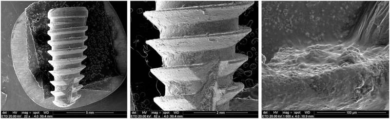
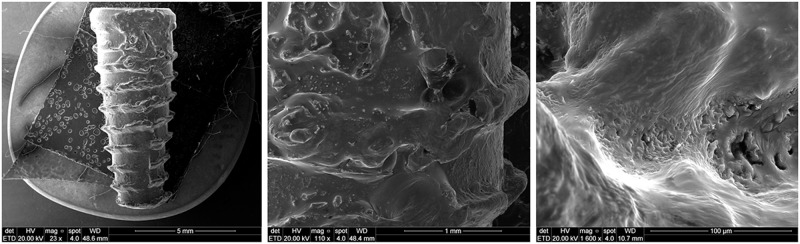
Materials and methods: Eighty-six conical implants were divided in two groups (n = 43 per group): group control (group CON) that were used conical implants with a conventional macrogeometry and, group test (group TEST) that were used implants with the new macrogeometry. The new implant macrogeometry show several circular healing cambers between the threads, distributed in the implant body. Three implants of each group were used to scanning electronic microscopy (SEM) analysis and, other eighty samples (n = 40 per group) were inserted the tibia of ten rabbit (n = 2 per tibia), determined by randomization. The animals were sacrificed (n = 5 per time) at 3-weeks (Time 1) and at 4-weeks after the implantations (Time 2). The biomechanical evaluation proposed was the measurement of the implant stability quotient (ISQ) and the removal torque values (RTv). The microscopical analysis was a histomorphometric measurement of the bone to implant contact (%BIC) and the SEM evaluation of the bone adhered on the removed implants.
Results: The results showed that the implants of the group TEST produced a significant enhancement in the osseointegration in comparison with the group CON. The ISQ and RTv tests showed superior values for the group TEST in the both measured times (3- and 4-weeks), with significant differences (p < 0.05). More residual bone in quantity and quality was observed in the samples of the group TEST on the surface of the removed implants. Moreover, the %BIC demonstrated an important increasing for the group TEST in both times, with statistical differences (in Time 1 p = 0.0103 and in Time 2 p < 0.0003).
Conclusions: Then, we can conclude that the alterations in the implant macrogeometry promote several benefits on the osseointegration process.
Conflict of interest statement
The authors have read the journal’s policy and have the following potential competing interests: SAG is a paid employee of Biotecnos. This does not alter our adherence to PLOS ONE policies on sharing data and materials. There are no patents, products in development or marketed products associated with this research to declare.
Figures


Similar articles
-
Comparison Between Micro- and Micro-Nano Surface Texturization in the Initial Osseointegration Process: An Experimental In Vitro and In Vivo Preclinical Study.Bioengineering (Basel). 2025 Feb 12;12(2):175. doi: 10.3390/bioengineering12020175. Bioengineering (Basel). 2025. PMID: 40001694 Free PMC article.
-
Comparison of two dental implant surface modifications on implants with same macrodesign: an experimental study in the pelvic sheep model.Clin Oral Implants Res. 2015 Aug;26(8):898-908. doi: 10.1111/clr.12411. Epub 2014 May 21. Clin Oral Implants Res. 2015. PMID: 24954017
-
Biomechanical and histological evaluation of four different implant macrogeometries in the early osseointegration process: An in vivo animal study.J Mech Behav Biomed Mater. 2022 Jan;125:104935. doi: 10.1016/j.jmbbm.2021.104935. Epub 2021 Oct 28. J Mech Behav Biomed Mater. 2022. PMID: 34736028
-
The osseointegration stimulatory effect of macrogeometry-modified implants: a study in the rabbit.Clin Oral Implants Res. 2014 Sep;25(9):1051-5. doi: 10.1111/clr.12212. Epub 2013 Jun 18. Clin Oral Implants Res. 2014. PMID: 23782296
-
Synergistic Effect of Implant Surface Physicochemical Modifications and Macrogeometry on the Early Stages of Osseointegration: An In Vivo Preclinical Study.J Biomed Mater Res B Appl Biomater. 2025 Apr;113(4):e35569. doi: 10.1002/jbm.b.35569. J Biomed Mater Res B Appl Biomater. 2025. PMID: 40156250
Cited by
-
Randomized Clinical Trial Comparing Insertion Torque and Implant Stability of Two Different Implant Macrogeometries in the Initial Periods of Osseointegration.Medicina (Kaunas). 2023 Jan 14;59(1):168. doi: 10.3390/medicina59010168. Medicina (Kaunas). 2023. PMID: 36676792 Free PMC article. Clinical Trial.
-
Comparison Between Micro- and Micro-Nano Surface Texturization in the Initial Osseointegration Process: An Experimental In Vitro and In Vivo Preclinical Study.Bioengineering (Basel). 2025 Feb 12;12(2):175. doi: 10.3390/bioengineering12020175. Bioengineering (Basel). 2025. PMID: 40001694 Free PMC article.
-
Effects of insertion torque values on the marginal bone loss of dental implants installed in sheep mandibles.Sci Rep. 2022 Jan 11;12(1):538. doi: 10.1038/s41598-021-04313-5. Sci Rep. 2022. PMID: 35017552 Free PMC article.
-
Effectiveness of biomolecule-based bioactive surfaces, on os-seointegration of titanium dental implants: A systematic review and meta-analysis of in vivo studies.Front Bioeng Biotechnol. 2022 Sep 26;10:986112. doi: 10.3389/fbioe.2022.986112. eCollection 2022. Front Bioeng Biotechnol. 2022. PMID: 36225604 Free PMC article.
-
Micro-Computed Tomography Analysis of Peri-Implant Bone Defects Exposed to a Peri-Implantitis Microcosm, with and without Bone Substitute, in a Rabbit Model: A Pilot Study.Bioengineering (Basel). 2024 Apr 19;11(4):397. doi: 10.3390/bioengineering11040397. Bioengineering (Basel). 2024. PMID: 38671818 Free PMC article.
References
MeSH terms
Substances
LinkOut - more resources
Full Text Sources
